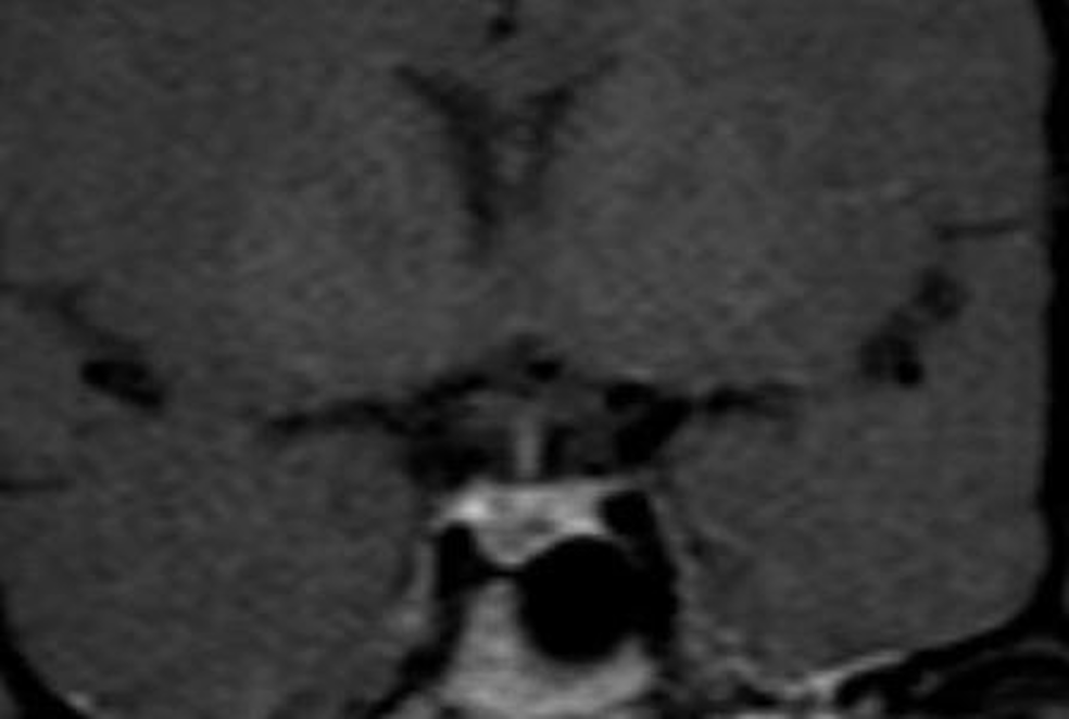
Figure 6. Coronal T1 weighted image of the pituitary gland immediately after contrast. There is a microadenoma in the right side of the gland. After contrast a dynamic acquisition shows an area of lesser enhancement indicative of a microadenoma.
Figure 6. Coronal T1 weighted image of the pituitary gland immediately after contrast. There is a microadenoma in the right side of the gland. After contrast a dynamic acquisition shows an area of lesser enhancement indicative of a microadenoma.