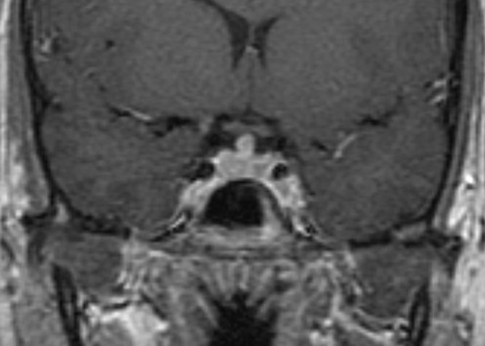
Figure 34. Coronal T1 weighted enhanced image of lymphocytic hypophysitis. There is slight enlargement and heterogeneity of the gland with thickening of the lower part of the stalk. The fossa is not enlarged.
Figure 34. Coronal T1 weighted enhanced image of lymphocytic hypophysitis. There is slight enlargement and heterogeneity of the gland with thickening of the lower part of the stalk. The fossa is not enlarged.