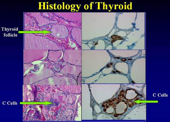
Figure 37. C cell staining with H&E and Immunohistochemical method. C cells are identified with difficulty in sections stained with hematoxylin and eosin, where they appear polygonal and with a granular weakly eosinophilic cytoplasm that is larger and paler than that of follicular cells. (Fig 1a,c,e) (See arrow). When compared with sections from the same patients using immunohistochemical staining, C cells as more readily identified part of the follicular epithelium and as isolated cluster between follicles