The best Clinical Endocrinology source in the world, and FREE.

Endotext requires free registration
Login or Register

Menu

Skip to content
  • Home / Sections
  • Clinical Guidelines
  • Webinar Case Studies
  • Editorial Team
  • Country Associates
  • About
  • Instructions for Authors

figure8

Published May 16, 2013 at 400 × 300 in Testicular Cancer: Pathogenesis, Diagnosis and Management with Focus on Endocrine Aspects.
← Previous Next →

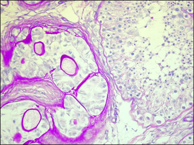
Figure 8.Large cell calcifying Sertoli cell tumor isolated from a 12-year-old boy. The neoplastic seminiferous tubules contain only large pale Sertoli cells and visible calcifications in the lumen (stained with PAS). Adjacent normal tubules show advanced spermatogenesis.

  • Agreement on Use
  • Supporters
  • Donations
  • Privacy Policy
  • Instructions for Authors (5/03/23)

Copyright © 2000-2025, MDText.com, Inc. All rights reserved.