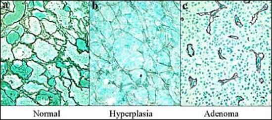
Figure 14. The reticulin stain is the most valuable tool to distinguish normal acinar architecture (a) from the expanded acini of hyperplasia (b) and to confirm total breakdown of the reticulin fiber network in adenomas (c).
Figure 14. The reticulin stain is the most valuable tool to distinguish normal acinar architecture (a) from the expanded acini of hyperplasia (b) and to confirm total breakdown of the reticulin fiber network in adenomas (c).