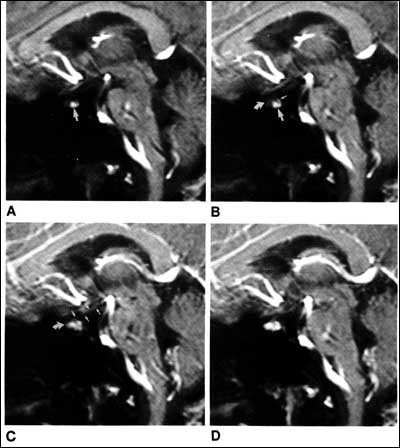
Fig. 9.(A-D) MRI of sequential sequences of the stalk and pituitary gland in saggital orientation following the intravenous administration of gadolineum. (A) Appearance prior to gadolineum. (B) Following gadolineum, the posterior pituitary is the first structure to show contrast enhancement. (C) This is followed by the pituitary stalk (arrow) and then finally (D) the anterior pituitary. (From Yuh et al, AJNR 15: 101-108, 1994.)