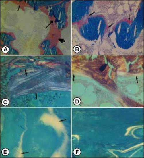
Figure 5. Photomicrographs of transilial bone biopsies of patients with rickets and osteomalacia (A,C and E) and siblings without bone disease (B,D and F) (Courtesy of Drs. D. Gazit and I.A. Bab).A and B: Modified Mason stain; magnification x130. Note in A: broad osteoid seams (arrow), osteoid trabeculae (heavy arrow) and irregular mineralization front (rectangular arrow).C and D: Polarized light; Von Kossa toluidine blue stain; magnification x360. Note in C: increased number of oseoid lamellae (arrows).E and F: Fluorescent photomicrograph, unstained; magnification x200. Note in E wide fluorescent bands (arrows), no double or single tetracycline labels and ground glass appearance.