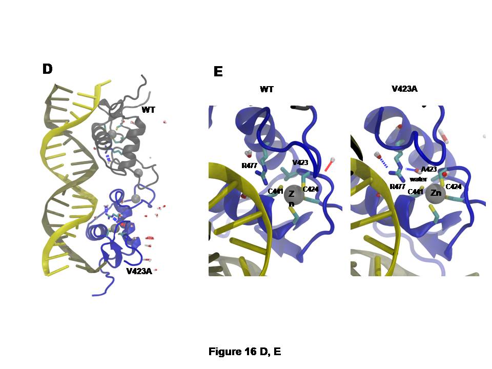
D and E: The V423A mutation alters the specific binding of the hGR DBD to GRE.
Replacement of valine (V) by alanine (A) at position 423 in the hGR DBD reduces the hydrophobic nature of the site and permits water to diffuse into the ion-binding region of the protein, where it is captured by hydrogen bonds to C424 and C441 among others (D). Water is almost never observed in this area in the wild type simulation. The structures in green indicate the wild type (WT) receptor, while those in blue represent the mutant receptor. Water molecules are indicated by capsule-like molecules in grey and red color.
The most significant changes in hydrogen bonding in this area occur to C424, C441, and R477 (E).
Modified from (315, 319).