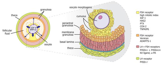
Figure 14. An oocyte morphogen gradient influences granulosa cell phenotypes. The model shown is based mainly on experimental evidence from rodent model systems. Protein morphogens including GDF-9 and BMP-15 are secreted by the oocyte, resulting in a concentration gradient that diminishes with distance from the oocyte. Because granulosa cell differentiation is dictated by the morphogen concentration, position in the follicle relative to the oocyte is a critical determinant of the final phenotype. Additional modulation of granulosa cell phenotype is accomplished by other factors from the follicle itself and from the endocrine system, including activin, inhibin, and steroid hormones. Granulosa cells differentiate into three distinct phenotypes based on position within the follicle – cumulus, periantral, and membrana granulosa cells. Each granulosa cell type exhibits a distinct response to FSH stimulation. Some examples of the many genes differentially expressed in the various follicle compartments are listed at the right. Abbreviations: Cox-2, cyclooxygenase 2; FSH, follicle-stimulating hormone; HAS2, hyaluronic acid synthase 2; IGF-I, insulin-like growth factor I; PTX, pentraxin; TNFAIP6, tumor necrosis factor-induced protein 6; LH, luteinizing hormone; P450SCC, P450 side chain cleavage; P450AROM, P450 aromatase; u-PA, urokinase-type plasminogen activator. (Revised from Erickson GF, Shimasaki S: The role of the oocyte in folliculogenesis. Trends Endocrinol Metab 11:193, 2000.)