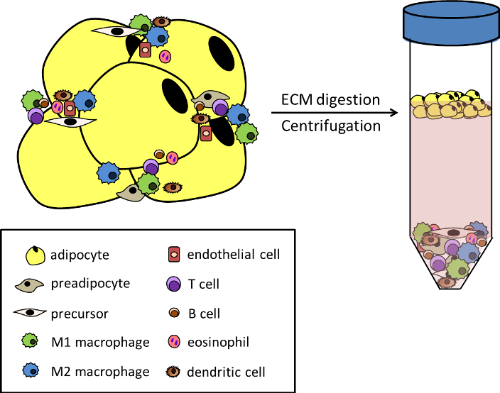
Figure 8. Constituents of adipose tissue (AT). Left: Along with mature, functional adipocytes and precursor cells, many cell types related to vasculature and immune function reside within AT. They perform both physiological and pathophysiological functions by communicating with the adipocytes via secreted factors and scavenging lipid from dying fat cells. The number and diversity of these cell types increases with developing obesity and metabolic dysfunction. Right: The non-adipocyte cells are collectively referred to as the stromal vascular fraction (SVF), and the SVF can be separated from lipid-containing adipocytes by digesting the extracellular matrix (ECM) and centrifuging the cellular mixture. The SVF will form a pellet at the bottom of the tube, while the adipocytes will float and form a visible lipid layer at the top of the aqueous medium. This separation technique is critical to studying the cellular composition of adipose tissue and gaining insight regarding the individual functions of these diverse and distinct cell types under physiological and pathophysiological conditions.