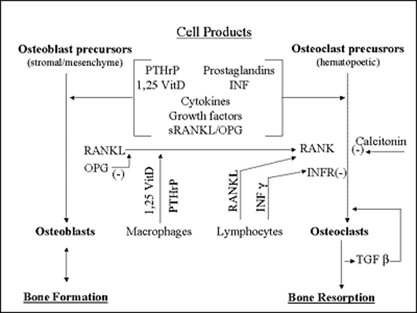
Figure 6. Schematic representation of the cellular and molecular mechanisms of the effects of OPG, RANK, and RANKL on skeletal metabolism. A variety of skeletal and non-skeletal cells can express several cell products [in brackets] that regulate the balance between osteoblastic bone formation (left) and osteoclastic bone resorption (right). They include PTHrP (parathyroid hormone related protein); 1, 25 Vit D (1, 25- dihydroxyvitamin D); prostaglandins, especially of the PGE2 series; cytokines, especially interleukin 1 (IL-1); growth factors, especially TGF beta; RANKL (receptor activator of nuclear factor kappa B ligand), a cell membrane-associated member of the tumor necrosis factor family of cytokines; soluble RANKL (sRANKL); and their cognate receptor, RANK; and OPG (osteoprotegerin), a soluble “decoy” receptor for RANKL. The latter group are also expressed by osteoblast precursors as they develop into osteoblasts in the osteoblastic cascade (left). In addition to OPG, the stimulation of osteoclastic bone resorption by RANKL is opposed by activation of the gamma interferon receptor (INFR) by gamma interferon (INF) production by activated lymphocytes and by the peptide hormone, calcitonin. The relative activity of the osteoclast stimulatory effects of RANKL and sRANKL and the inhibitory effects of OPG and INF determine the balance between bone resorption and formation. Arrows indicate a positive (stimulatory) effect except where indicated by the negative sign, (-). Several growth factors in addition to TGF beta reside in bone matrix and can be released upon resorption to exert their biological effects, often osteoclast stimulation. They include BMP (bone morphogenetic proteins, especially BMP-2); FGF (fibroblast growth factor); PDGF (platelet derived growth factor); and IGFs in (insulin like growth factors). Macrophages may fuse into giant cells and resorb bone. (see Acknowledgements).