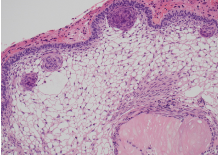
Figure 1B. Histology of adamantinomatous craniopharyngioma. Well-differentiated epithelium with peripheral palisading, nodular whorls, and pale, microcystic areas termed ‘stellate reticulum’, as well as pale eosinophlic ‘wet keratin’ nodule (right bottom); HE x200 magnification.