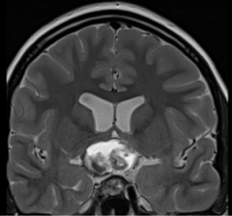
Figure 3F. MRI images of craniopharyngiomas. Coronal section showing a mixed solid and cystic craniopharyngioma with mixed signal intensities on T2-weighted imaging.
Figure 3F. MRI images of craniopharyngiomas. Coronal section showing a mixed solid and cystic craniopharyngioma with mixed signal intensities on T2-weighted imaging.