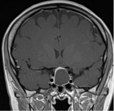
Figure 3A. MRI images of craniopharyngiomas. Coronal section showing cystic craniopharyngioma on post-contrast T1-weighted MRI. The cyst contents are isointense and the cyst rim enhances following contrast.
Figure 3A. MRI images of craniopharyngiomas. Coronal section showing cystic craniopharyngioma on post-contrast T1-weighted MRI. The cyst contents are isointense and the cyst rim enhances following contrast.