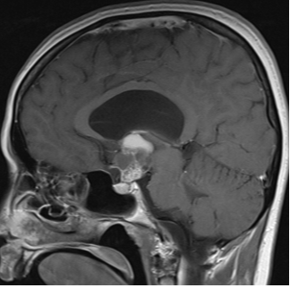
Figure 3E. MRI images of craniopharyngiomas. Sagittal section showing a craniopharyngioma with mixed solid and cystic components on post-contrast T1-weighted imaging.
Figure 3E. MRI images of craniopharyngiomas. Sagittal section showing a craniopharyngioma with mixed solid and cystic components on post-contrast T1-weighted imaging.