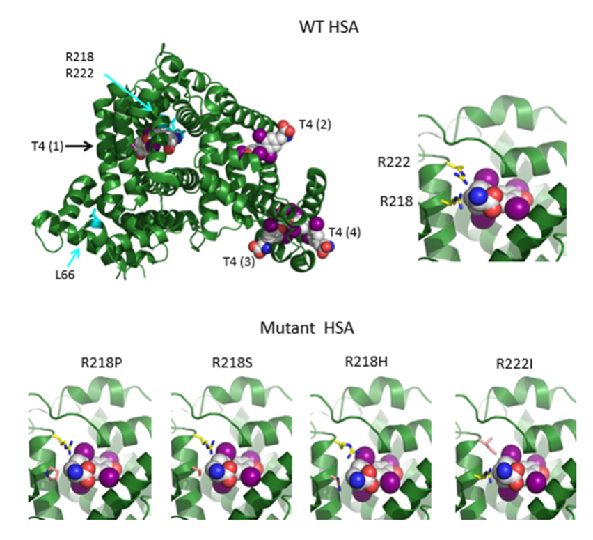
Figure 6. The structures of HSA in the presence of T4 as modeled on the structures 1BM0, 1HK1, 1HK3 in the Protein Data Bank (http://www.rcsb.org/pdb/home/home.do). Top panel shows on the left the entire WT HSA molecule (in green) with its four T4 binding sites [T4(1) to T4(4)] according to Petitpas et al (120) and to the right a close up of the binding pocket, T4 (1) containing arginine’s 218 and 222 along with the T4 molecule (carbons are in white, nitrogen’s in blue, oxygens in red and iodine in magenta). In the bottom panel are represented the structures of the T4 (1) binding pockets of the four mutant HSA showing, a better accommodation of T4 than in the WT HSA and thus, resulting in enhanced binding (From Erik Schoenmakers, University of Cambridge, UK).