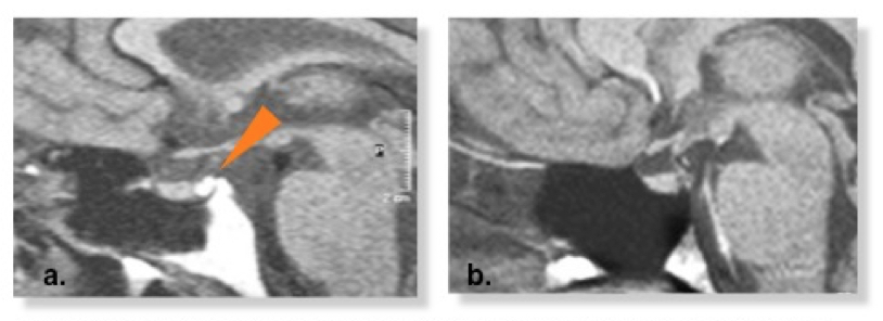
Figure 5. MRI appearance of the posterior pituitary in hypothalamic/cranial DI. Figure a. demonstrates a normal posterior pituitary ‘bright-spot’ T1-weighted MRI, highlighted by arrow. Figure b. demonstrates absence of the equivalent ‘bright-spot’ in a patient with idiopathic HDI.