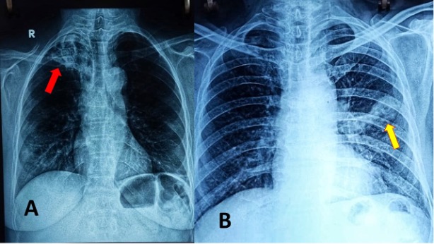
Figure 3. Chest Radiographs suggesting fibrocavitatory lesions. Post primary infections and reactivation of pulmonary TB are more likely to cavitate. They are most common in the posterior segments of the upper lobes (85%) as seen in Picture A. Red arrow pointing to the cavity. The other common site is the superior segment of the lower lobe (Picture B) Yellow arrow pointing to the cavity (Picture courtesy- Prof Mary John, Christian Medical College and Hospital, Ludhiana)