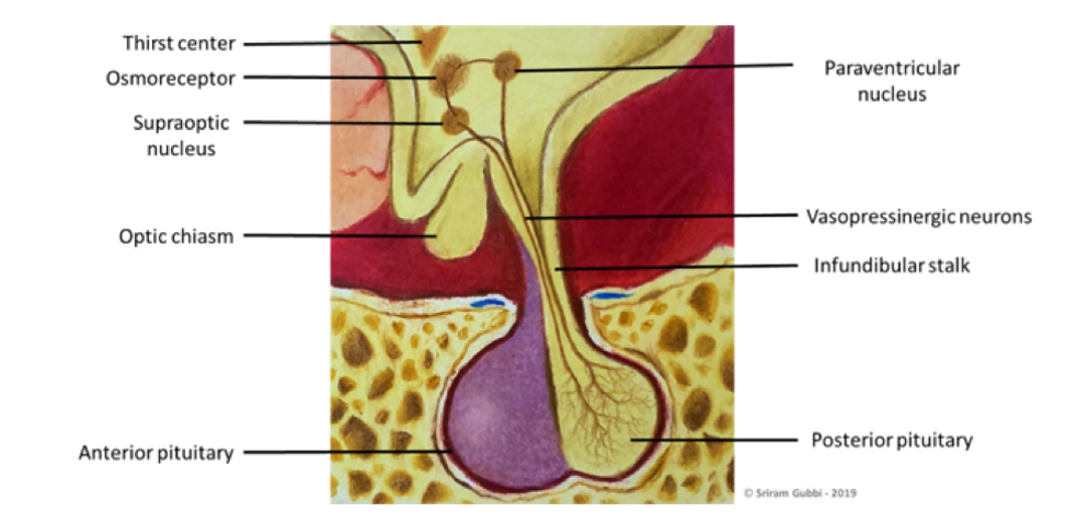
Figure 3. Anatomy of the pituitary gland and the hypothalamus. The pituitary gland comprises of two developmentally and functionally distinct parts: the anterior pituitary (adenohypophysis), derived from the Rathke’s cleft and the posterior pituitary (neurohypophysis). The gland is attached to the hypothalamus through the posterior pituitary via a stalk. The posterior pituitary along with the hypothalamus and the stalk forms the functional unit of infundibuloneurohypophysis. This diagram emphasizes on the neuronal network that supplies the posterior pituitary. The vasopressinergic neurons (magnocellular neurons) are present in the supraoptic and paraventricular nuclei of the hypothalamus. These neurons synthesize arginine vasopressin (AVP), its precursors and co-peptides, and their axons project into the posterior pituitary where the AVP is stored along with oxytocin (produced by the oxytotic neurons present in the same nuclei) in the axonal terminals. These hormones are transported to the posterior pituitary through the supraoptic-hypophyseal tract. Another set of neurons of the paraventricular nucleus (also called parvocellular neurons) project into other areas of the brain and spinal cord, some of which secrete AVP. Image courtesy: Sriram Gubbi, NIDDK, NIH.