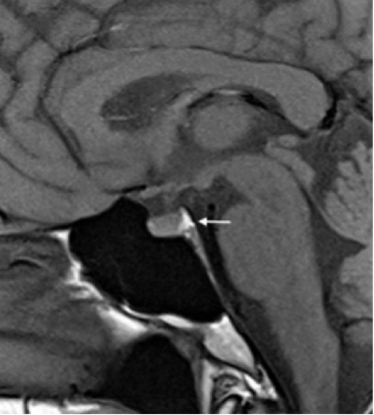
Figure 5. Magnetic resonance imaging (MRI) of the pituitary gland. The above image is a non-contrast T1 MRI image of a normal pituitary gland. The white arrow point towards the ‘bright spot’ seen in the posterior pituitary. This finding is a result of phospholipid-rich granules that store arginine vasopressin (AVP) and oxytocin. Image courtesy: NIDDK, NIH.