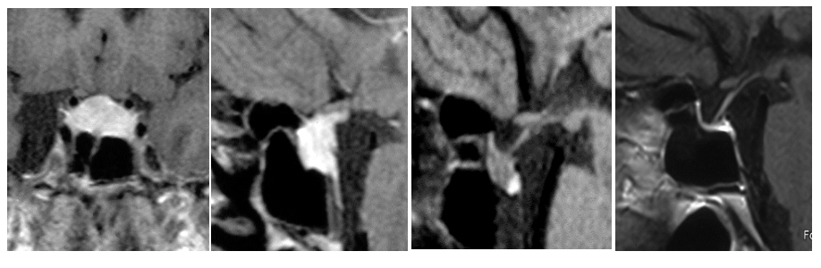
Figure 3. Contrast enhanced coronal and sagittal T1W images of lymphocytic hypophysitis spontaneous evolution from the presentation (panel A, B), after 4 (panel C) and 10 years (panel D) of follow-up resulting in secondary empty sella.
Figure 3. Contrast enhanced coronal and sagittal T1W images of lymphocytic hypophysitis spontaneous evolution from the presentation (panel A, B), after 4 (panel C) and 10 years (panel D) of follow-up resulting in secondary empty sella.