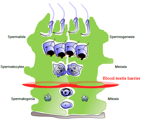
Figure 11. The position of the blood testis barrier in the seminiferous epithelium, which is formed by tight, occluding and adhesion junctions between adjacent Sertoli cells. This barrier restricts the diffusion of substances from the interstitum and blood vessels, and thus allows the Sertoli cell to determine the microenvironment above the junctions. This barrier effectively divides the seminiferous epithelium into two compartments, the basal compartment with free access to substances from outside the tubule, and the adluminal compartment, the environment of which is controlled by the Sertoli cell. Meiosis and the differentiation of spermatids occurs in the adluminal compartment. The inter-Sertoli cell junctions transiently remodel to allow germ cells to move from the basal to the adluminal compartments, whilst protecting the functionality of the barrier. Diagram provided by Jenna Haverfield.