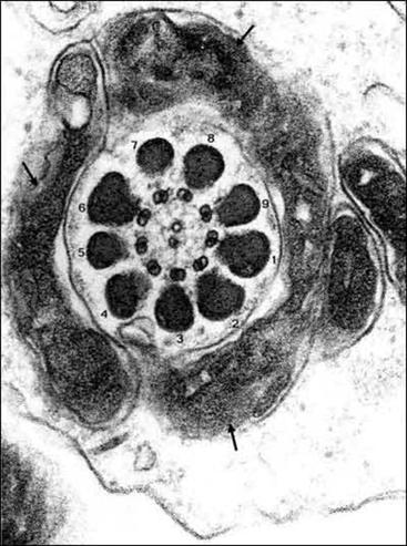
Figure 8. A cross-section through the developing mid-piece of the sperm tail shows the aggregation of mitochondria (arrows) surrounding the outer dense fibres (labelled 1-9) which in turn surround the axoneme composed of 9 doublet microtubules surrounding two central microtubules. Reproduced with permission from “Visual atlas of human sperm structure and function for assisted reproductive technology” Ed A.H. Sathanathan 1996.