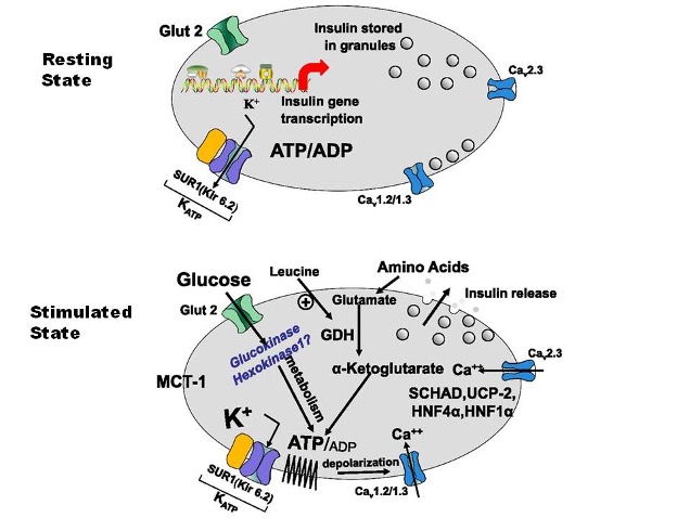
Figure 2. Insulin secretion by Pancreatic β cells. In the stimulated state, glucose is transported into the β cell by the GLUT2 transporter which undergoes phosphorylation by glucokinase and glucose is then metabolized. This results in an increase in the ATP/ADP ratio and initiation of a cascade of events that is characterized by closure of the K-ATP channel, decreased flux of potassium across the membrane, membrane depolarization, and calcium influx. This cascade ultimately results in insulin release from storage granules. The K-ATP channel shown is composed of four small subunits, Kir6.2, that surround a central pore and four larger regulatory subunits constituting SUR1. In the resting state, the potassium channel is open, modulated by the ratio of ATP to ADP. Leucine also stimulates insulin secretion by allosterically activating GDH and by increasing the oxidation of glutamate; this then increases the ATPADP ratio leading to the cascade of events beginning with closure of the KATP channel.
MCT-1: Monocarboxylate transporter-1, SCHAD: Short chain 3-hydroxyacyl-CoA dehydrogenase, SUR1: Sulfonylurea receptor 1, Kir 6.2: Potassium Inward Rectifying Channel 6.2, UCP-2: Uncoupling protein 2, HNF4α: Hepatocyte Nuclear Factor 4α, HNF1 α: Hepatocyte Nuclear Factor 4α, K+: Potassium, ATP: Adenosine Triphosphate, GDH: Glutamate Dehydrogenase, GLUT-2: Glucose Transporter 2