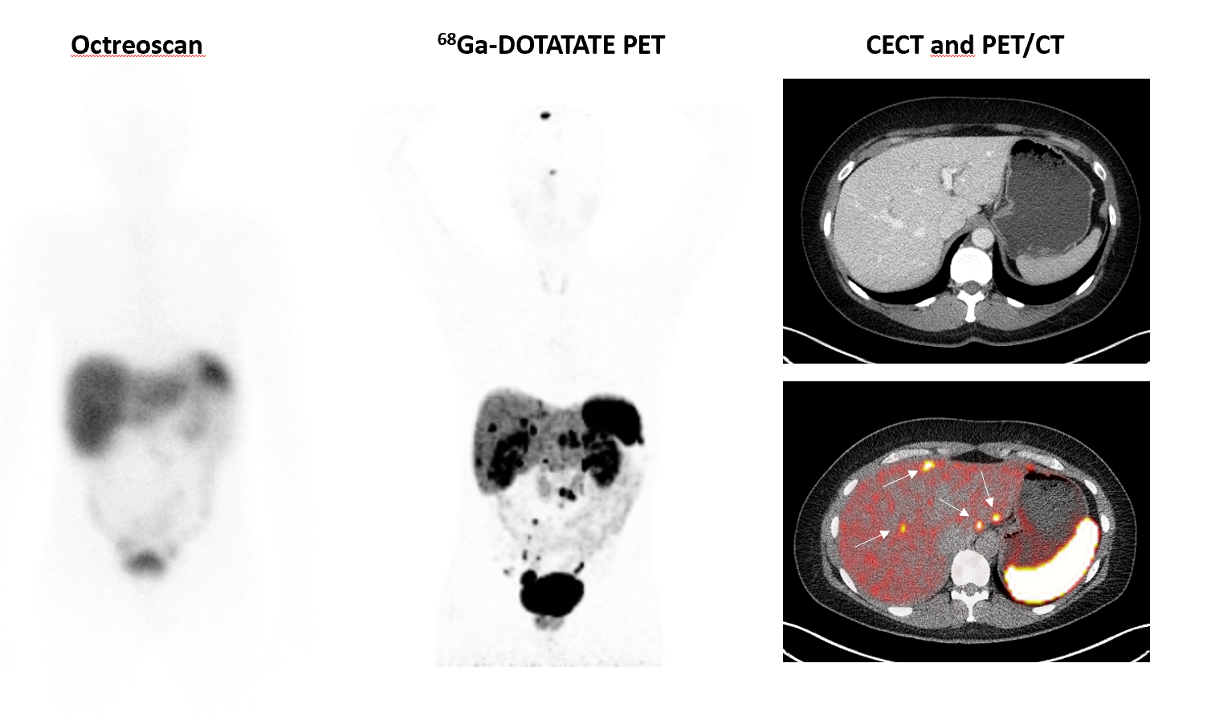
Figure 4. 68Ga-DOTA-SSA PET imaging. 68Ga-DOTA-SSA PET staging is superior to anatomical imaging and 111In-pentreotide SPECT (Octreoscan). In this case of a patient with stage IV small intestinal NET, PET imaging detected more lesions than Octreoscan, scanned within 3-month timeframe without anatomical progression. In the same patient, multiple liver metastases are detected on hybrid PET/CT imaging (arrow), which were not visible on contrast-enhanced CT (CECT).