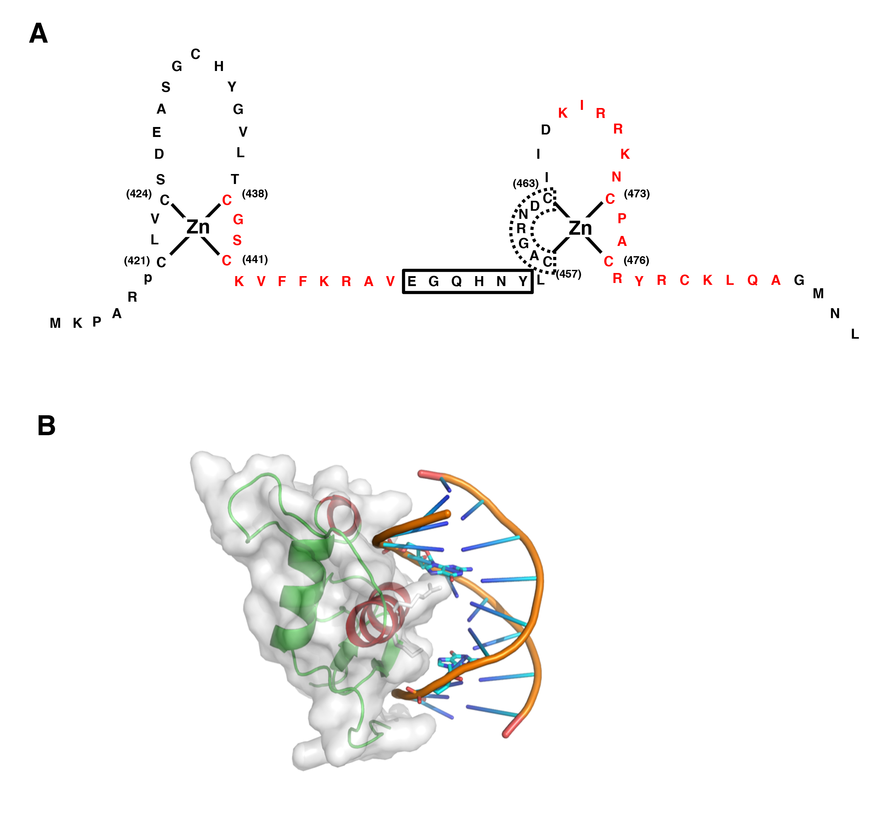
Figure 4: Structure of GR DBD and its interaction with DNA GRE
A: Zinc finger structures in DBD of hGR. Numbered eight cysteine (C) residues chelate Zn2+ to form two separate finger structures. Red-colored amino acid residues form -helical structures. Box with bold line indicates lever arm, while that with dashed line shows D-box. Modified from (30).
B: 3-Dimensional model of the physical interaction between the GR DBD and DNA GRE. The N-terminal’s first -helix of the GR DBD lies in the major groove of the double-stranded DNA, while the C-terminal part of the second -helix is positioned over the minor groove. The image was created and donated by Dr. D.E. Hurt (National Institute of Allergy and Infectious Diseases, NIH, Bethesda, MD)