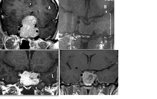
Figure 3. 26 yrs-old man complaining of visual disturbance was submitted to a sellar MRI (A): sellar mass with 5 cm in the maximal diameter with supra, infra and left parasellar invasion. Serum PRL levels were above 1000 ng/mL. After one year on cabergoline, 0.5 mg/day, there was no tumor reduction and PRL levels were around 800 ng/mL. He was then submitted to transsphenoidal surgery and radiotherapy in another medical service. Sellar MRI (B) after two months of the surgery showed a pituitary mass with 3.1 cm in the maximal diameter. Despite chiasmal decompression, visual dysfunction was not reversed and, during his follow-up, anterior pituitary function was lost. PRL levels on cabergoline, 1.5 mg/week, were 250 ng/ml. After two more years, sellar MRI depicted a lesion of 2.9 cm. in the maximal diameter (C). There was a progressive rise of PRL levels despite higher cabergoline doses (0.5 mg/d) and another sellar MRI (D), after two years, depicted a lesion of 3.1 cm in the maximal diameter. Another surgery was indicated.