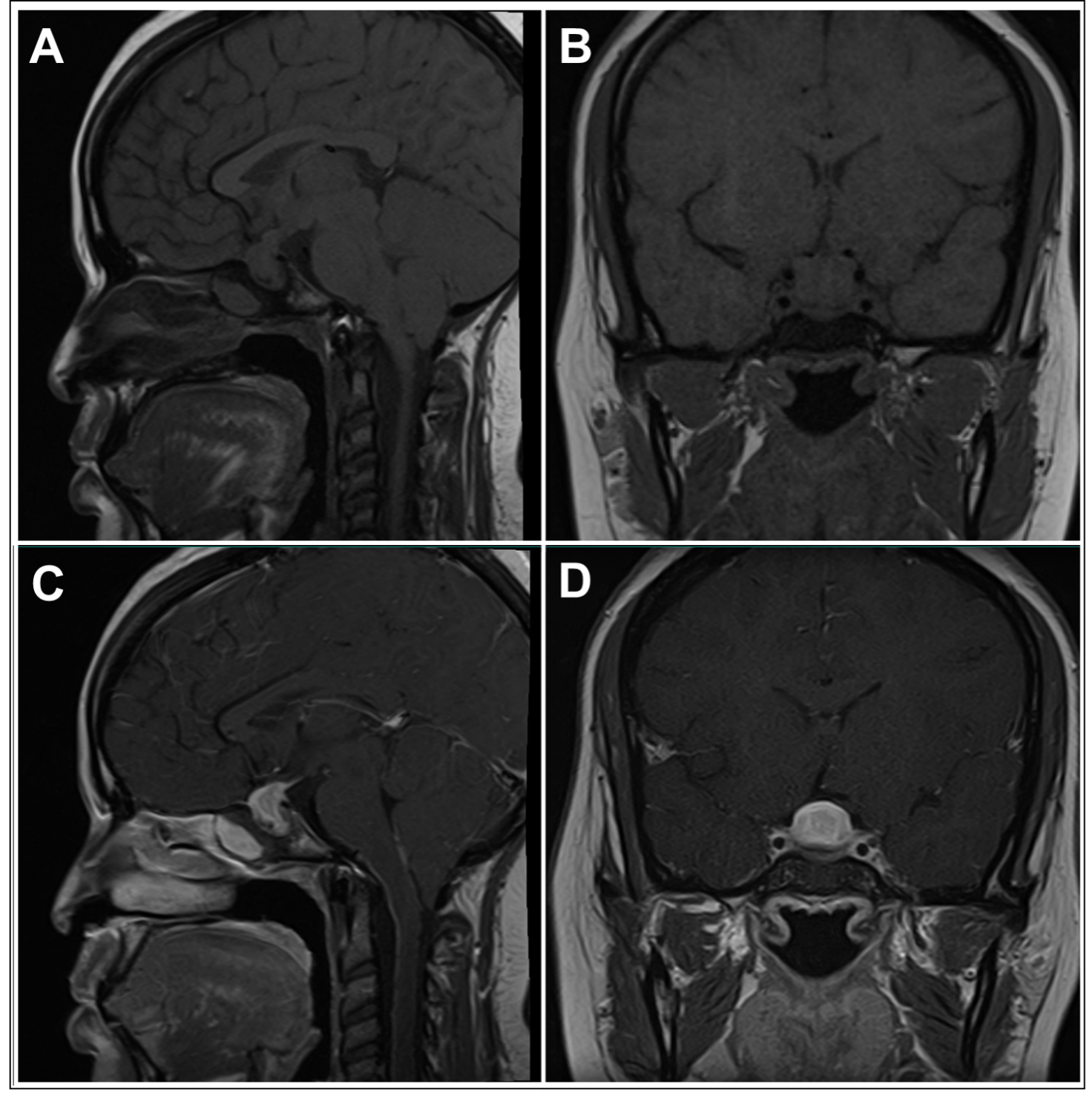
Figure 1. Magnetic resonance imaging findings in a case of primary hypophysitis. Panel A) T1-weighted image, sagittal section. Panel B) T1-weighted image, coronal section. Panel C) T1-weighted image post-gadolinium, sagittal section. Panel D) T1-weighted image post-gadolinium, coronal section. A homogeneous enlargement of the pituitary with thickening of the stalk can be seen. The mass shows intense and homogeneous enhancement post-gadolinium.