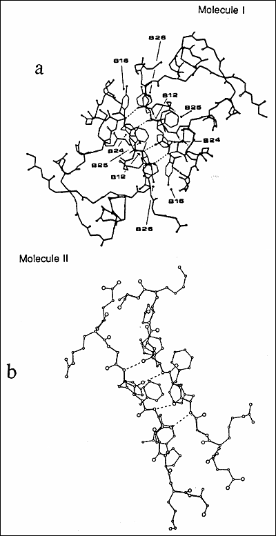
Figure 8.Structural illustration of the monomer-monomer interface in the insulin dimer. The dimer is viewed along the crystallographic 2-fold axis. The side chains of residues ValB12, TyrB16, PheB24, PheB25, and TyrB26 which form the core of the insulin dimer are illustrated in the figures. Four hydrogen bonds are formed from the main-chain atoms of PheB24 and TyrB26 are illustrated as dotted lines. In the figure (b) is a magnified view of the dimer interface in (a).