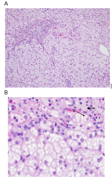
Figure 3. Liver Biopsies in Patients with LAL-D. A) Image of the portal tract and hepatocytes with mainly microvesicular steatosis. With microvesicular steatosis, the fat does not cause the nucleus to be pushed out to the side. B) Larger magnification of the portal tract. FM points to the foamy appearing cytoplasm, these are macrophages with something being stored in them. GC is pointing to a giant cell.