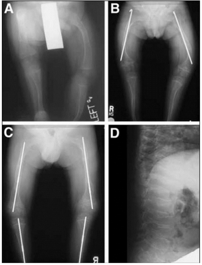
Figure 2. A, B: Radiographs of lower extremities of type III OI child. Shows osteoporosis, flared metaphyses, and placement of intramedullary Rush rod. C, D: Radiographs of child with type III OI. Shows lower long bones osteoporotic with cystic formation and “popcorn” metaphyses, and placement of telescoping intramedullary rods. Lateral view of spine shows anterior and central compression of multiple vertebrae.