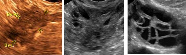
Figure 3: Ovarian sonographic imaging of women in their mid-30’s. Figure 3a is from a woman with premature ovarian failure and there are no visualized antral follicles (the sonographically anechoic regions measuring approximately two to nine millimeters within the ovary). Figure 3b is from a woman with tubal factor infertility, and for whom seeing a few follicles within a single plane of the ovary would be normal. Figure 3c is from a woman with polycystic ovarian syndrome. Though her ovary is arguably more multicystic than polycystic (which would typically have follicles concentrated on the periphery of the ovary), she met the criteria for PCOS and her ovary is clearly distinct from those shown in 3a and 3b. Of note, all three ultimately conceived with their own oocytes, so it should be remembered that the absence of visualized antral follicles makes conception far less probable, but not impossible.