Archives

Thyroid Cancer

Incidence and Distribution

The annual incidence of thyroid cancer varies considerably in different registries, ranging from 1.2-2.6 per 100,000 individuals in men and from 2.0-3.8 per 100,000 in women (106,107). It is particularly elevated in Iceland and Hawaii, being nearly two times higher than in North European countries, Canada and the USA. In Hawaii, the incidence rate of thyroid cancer in each ethnic group is higher than that registered in their country of origin (108), and it is particularly common among Chinese males and Filipino females. Most of the differences are probably due to ethnic or environ¬mental factors (such as spontaneous background radiation) or dietary habits (109), but different standards of medical expertise and health care may also play a role in the efficiency of cancer detection. The American Cancer Society indicated incidence in the USA of nearly 10/100,000 population in 2003. The reported incidence has been increasing at more than 5%/yr for a decade.

In sharp contrast with these data concerning the incidence of clinical thyroid cancer, is the prevalence found in autopsy series or screening programs. Autopsy studies indicate a surprising frequency ranging from 0.01 to over 2.0% (110,111).A survey of consecutive autopsies at Grace-New Haven Hospital found 2.7% of thyroids to harbour unsuspected thyroid cancer (111). Another 2.7% had discrete benign adenomas, and nearly half showed nodularity. The high prevalence may be attributed to careful examination of the gland, but probably also reflects a highly selected group of older patients dying in a hospital. Up to 6% of thyroid glands in autopsied adults in the United States, and over 20% in Japan, also harbour microscopically detectable foci of thyroid carcinoma, which are believed to be of no biologic significance. Altogether autopsy studies suggest that thyroid cancer is in many instances not diagnosed dur¬ing life or is not the immediate cause of death. Both suggestions are in agreement with the rather leisurely growth of the majority of thyroid tumors, especially the frequent small papillary types.
The annual mortality from thyroid cancer in 2003 was 5 per million for men and 6 per million for women (112). The discrepancy between incidence and mortality reflects the good prognosis for most thyroid cancers. Recent statistics suggest about 6 deaths /million in the USA.

A classification of thyroid tumors is given in Table 18-1.

Table 18-1. Neoplasms of the Thyroid(Adapted, and Revised, from WHO Classification) 12
  1. I.              I. Adenomas (fig.18-1, below)

A. Follicular

  1. Colloid variant
  2. Embryonal
  3. Fetal
  4. Hurthle cell variant

B. Papillary (probably malignant)

C. Teratoma

II. Malignant Tumors

A. Differentiated

  1. Papillary adenocarcinoma
    1. Pure papillary adenocarcinoma
    2. Follicular variant of papillary thyroid carcinoma
    3. Other variants: tall cell, columnar cell, oxyphyl, solid sclerosing
  2. Follicular adenocarcinomas (variants: Hurthle cell carcinoma or oxyphyl carcinoma, clear-cell carcinoma, insular carcinoma)
    1. Minimally invasive
    2. Extensively invasive

B. Medullary carcinoma

C. Undifferentiated

  1. Giant cell
  2. Carcinosarcoma

D. Miscellaneous

  1. Lymphoma, sarcoma
  2. Squamous cell epidermoid carcinoma
  3. Fibrosarcoma
  4. Mucoepithelial carcinoma
  5. Metastatic tumor
 

Thyroid tumors are rare in children and increase in frequency in each decade.  Carcinomas are two-three times as frequent in women as in men. In the past, it was generally believed that thyroid tumors were more frequent in areas of endemic goiter, and reports from Colombia and Austria support this association (113) (see Chapter 11). More recent studies suggest that in iodine deficient countries the number of nodules is increased and, as a consequence, also the number of thyroid cancers is increased (114). Surveys conducted in the United States found no relation between usual geographic residence and incidence of thyroid cancer.

ETIOLOGY

Most, if not all, thyroid adenomas are monoclonal, as, presumably, are most carcino¬mas (115). Colloid nodules may be either mono-or poly-clonal. Thus tumors represent the persistent growth of the progeny of one cell which has somehow escaped the mechanisms which maintain normal cell division at about once each 8.5 years (116).

The process of oncogenesis is conceived to be a series of events induced by genetic and environmental factors which alter growth control. At the phenomenologic level these factors may be considered as "initiators" and "promoters". Initiators include such agents as chemicals and irradiation which induce tumors, and promoters are agents such as phenobarbital, which in rats augments TSH secretion and radically increases tumor development. In man x-ray treatment is the sole known initiator, and other than elevated TSH, no promoters are known. Compounds such as phenobarbi¬tal, dilantin and PCBs, which are known thyroid tumor promoters in animals through liver microsomal hormone degrading enzyme induction leading to increased thyroid hormone metabolism, do not appear to have a detectable adverse effect in man in doses usually employed (117).

Oncogenes (Fig. 18-11)

We now begin to understand oncogenesis in more details. More than 30 "oncogenes" have been recognized in the human genome. The most likely genetic events in thyroid cancer are reported in Fig. 18-11. These genes, normally silent, can be¬come activated by chromosomal translocations, deletions, or mutations, and then can "transform" normal cells into a condition of uncontrolled growth. Most onco¬genes appear to be closely related to normal growth factors, genes that control cell division, or to hormone receptors. In general, these genes, when turned on, promote cell growth and cell division and depress differentiation. Typically activation of one such gene may not be enough to produce malignancy, but if accompanied by ex¬pression of another oncogene, or if gene mutation or reduplication occurs, the cell may progress toward a malignant potential. Information on expression of oncogenes in human thyroid tissue is rapidly accumulating. Expression of c-myc is stimulated in normal thyroid cells by TSH, and the proto-oncogene is expressed in adenomas and carcinomas. Activating mutations of h-ras at codons 12, 13, and 61, and over expression of h-ras, are found in adenomas and carcinomas, but h-ras mutations are also found in nodular goiter tissue (118), suggesting that h-ras mutations could be an early event in oncogenesis (119). Other studies, it should be noted, find ras mutations uncommon (120).

Figure18-11

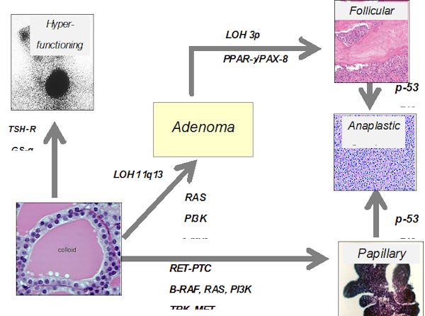
Figure 18-11. Possible role of oncogene activation, receptor or G-protein mutation, or tumor repressor gene alteration in the induction of thyroid carcinoma.

Santoro and co-workers (121) cloned an oncogene which is frequently and specifically expressed in papillary thyroid cancers. This oncogene is found on chromosome 10 and involves an intrachromosomal rearrangement of the tyrosine kinase domain of the ret oncogene so that it is attached to one of three different promoters, produc¬ing retPTC-1, retPTC-2, and retPTC-3. As a mean, one of these translocation products is found in about 20% of PTC, although in different series a large variation is observed (20-70%). This rearrangement leads to constitutive expression of the oncogene. It has been shown that intra-thyroidal expression of the ret/PTC1 oncogene can induce thyroid cancer (122). BRAF mutations, in the form of point mutations, are the most frequent alterations in papillary carcinoma, and undifferentiated cancers that have arisen from papillary tumors (123), approaching 40% of all PTCs (124).

Recently a mutational change has been associated with follicular cancers. In 5 of 8 follicular cancers, Kroll et al (125) found translocation of the DNA binding domain of PAX8 to domains A-F of the peroxisome proliferator-activater receptor (PPAR) gamma1 gene. The fusion oncogene is able to transform thyrocytes, so appears to be able to produce malignancies (126). Although initially thought to be exclusively present in follicular cancers, it is now known to be present in follicular adenomas as well (127).
Mutation or deletion of the p53 tumor suppressor gene is found in some differentiated thyroid cancers, and many undifferentiated cancers, suggesting that this genetic deletion may be one of the final steps leading to anaplastic cancer growth. A proliferation of studies in this field has provided many clues to thyroid tumorigenesis. Simian virus 40-like sequences are found in many thyroid cancers, as well as other cancers, and the Tag gene sequence found is known to be oncogenic in animal models (128). Mutated and non-functional thyroid hormone receptors are recognized in up to 90% of PTC by one author, suggesting a role in oncogenesis, but other workers find these mutations to be rare (129,130). The tumor suppressor gene TSG101 is over-expressed in most PTCs (131). Overexpression of many other genes -galectin-3, Thymosin beta-10,hTERT, CD97, CD26, VEGF-has been detected, but of course a question always is whether these changes represent the cause or the result of oncogenesis.

Mutations in the proteins involved in the normal TSH-receptor-G protein-adenyl¬cyclase-kinase signal transduction pathway also play a role in tumor formation. Activating TSH receptor mutations have been found by Parma and co-workers (132) to be the cause of most hyperfunctional nodules, and are now known to be common in "hot" nodules in patients with multi-nodular goiter.. These mutations involve the extracellular loops of the transmembrane domain and the transmembrane segments, and are proven to induce hyperfunction by transfection studies. However these mutations are not associated with cancer formation. Mutations of the stimulatory GTP binding protein subunit are also present in some patients with hyperfunctioning thyroid adenomas (133). TSH-R mutations are, however, unusual in thyroid cancer (134), (excepting hyperfunctional adenomas). TSH-R expression tends to be lost as cancers de-differentiate, and persistence of expression is associated with a better prognosis (135).

In addition to positive genetic factors, oncogenesis frequently involves loss of tumor suppressor genes. This has been proven in hereditary retinoblastoma. These genes are normally present on both sets (maternal and paternal) of chromosomes. In retinoblastoma the inherited lack of one suppressor (RB) gene does not cause disease, but if a genetic event (deletion, recombination, mutation, etc.) causes failure of expression of the second allele, cancer ensues. The presence of tumor-specific suppressor genes is often detected because of lack of heterozygosity of chromosomal markers associated with deletions of segments of genetic material. Evidence for characteristic chromosomal abnormalities within tumor cells may lead to recognition of a tumor suppressor gene. Deletions of the tumor suppressor genes, p53 and the RB gene, have been detected in differentiated and undifferentiated thyroid cancer (136). Many chromosomal rearrangements are found in Hurthle cell tumors, and correlate with tumor recurrence (137).

Ret oncogene and Medullary Thyroid Cancer

Studies on patients with MENI and MEN II indicated linkage to chromosomes 11 (138) and 10, respectively. Subsequent studies demonstrated that the ret oncogene is present at 10q11.2. Germline mutations have been detected in this oncogene in all patients with MEN II and MEN III (or IIB), and familial MTC (139). RET is a cell-membrane receptor of the growth factor family, with tyrosine kinase function. In up to 97% of patients with MenIIA, mutations are found in codons 609,611,618, 620, and 630 in exons 10 and 11. These all involve substitutions of other aminoacids for cysteine, and are thought to cause activation of the gene by aberrant disulphide bonding causing dimerization. Similar changes are seen in Familial MTC. In patients with the MENIIB syndrome, almost all,if not all, mutations involve an amino acid substitution of threonine for methionine at codon (918) in exon 16, and are thought to induce a change in substrate phosphorylation. Somatic mutations in ret are present in up to half of patients with sporadic MTC and are almost always in codon 918 (140,141). Gene mutations in this codon are thought to imply a poor prognosis.

Familial tumors

Apparent familial thyroid cancer development has been reported by several clinicians, including cases which seem to show a dominant pattern of inheritance (142,143). Thyroid carcinomas occur rarely as part of several familial syndromes, which may involve hereditable loss of tumor suppressor genes. Papillary cancer can occur as an independent familial syndrome in 5-10% of patienst in different series. Whether the recurrence of PTC represents a true familial aggregation or rather the fortuitous association of PTC in the same family, is still a matter of discussion. However, recent evidence seem to support the existence of a true familial PTC syndrome based on the demonstration that familial PTC display the features of “genetic anticipation” (the disease recurs at an earlier age and at an higher aggressiveness in the 2nd generation compared to the first one) typical of familial diseases (144). In addition, a germline alteration consisting in short telomeres has been demonstrated in familial cases of PTC (145,146), which may be responsible for genomic instability leading eventually to thyroid cancer.

Other, more rare forms of familial thyroid tumors are those associated with complex hereditable diseases. Cowden’s disease is a familial syndrome which includes a variety of hamartomas, multinodular goiter, and carcinomas of several tissues including breast, colon, lung, and thyroid, especially in women (147). Thyroid carcinoma also co-occurs in patients with familial adenomatous polyposis of the colon (148), and can occur in the absence of bi-allelic inactivation of the APC gene. Differentiated thyroid carcinoma is reported to co-occur with chemodactomas of the carotid body, which can be inherited in a familial autosomal dominant form (149). Thyroid carcinoma is also associated with Gardner’s syndrome (150) and Carney’s Syndrome (151). Papillary thyroid carcinoma has been associated with papillary renal neoplasia in a distinct hereditable tumor syndrome. Some patients in the families also have nodular thyroid disease. The predisposing gene has been chromosome 1q21 (152). These syndromes are listed in table 18.5.

Table 18.5. RARE SYNDROMES WITH HEREDITABLE THYROID TUMORS (NR9)
Syndrome Clinical Presentation Thyroid Pathology Gene and Location
Familial Papillary Carcinoma associated with papillary renal ca- Papillary cancer locus on 1q21
Familial non-medullary thyroid ca   PTC locus at 2q21
Thyroid tumors with oxyphilia   Benign nodules and PTC locus at 19p13.2
PTC without Oxyphilia   PTC Locus at 19p13
Familial Polyposis Large intestine polyps and other GI tumors Papillary cancer APC on 5q21
Gardner’s Syndrome Small and large intestine polyps, osteomas, fibromas, lipomas Papillary cancer APC on 5q21
Turcot’s Syndrome Large intestine polypsBrain tumors Papillary cancer APC on 5q21
Cowden’s Disease Multiple hamartomas and breast tumors Follicular adenoma and cancer Unknown
Carney Complex Pigmented adrenal nodules, pituitary adenomas, spotty skin pigmentation, myxomas Thyroid adenomas PRKAR1A located on 17q23-q24, while Carney complex type 2 has been mapped to chromosome 2p16.


Experimental Thyroid Tumor Formation

Thyroid tumors have been induced experimentally in rodents by several procedures having as their common denominator a prolonged increase in pituitary thyrotropin production and thyroid stimulation. Goitrogenic drugs, if administered to animals for a prolonged period, can induce tumors, as numerous investigators (153) have demonstrated. These tumors are typically papillary adeno-carcinomas, and are associated with a diffuse hyperplasia of the thyroid gland. Old rats of some strains appear to develop thyroid cancers spontaneously

Roentgen irradiation of the thyroid and administration of 131I have both induced carcinomas in the experimental animal (154). A combination of 131I injury to the thyroid cell and prolonged administration of a goitrogen is especially likely to produce carcinomas, as shown by Doniach (155). Cell metabolism is altered by 131I, even when small amounts are administered. In the rat, 5 µCi prevents subsequent response to a goitrogenic drug (156). With larger doses the colloid is sparse, the follicles are variable in size, and large eosinophilic acinar cells appear. Very large doses of 131I (producing several thousand rads) to the rat thyroid radically alter cell metabolism, liberate TG within 1 or 2 weeks, and subsequently reduce the efficiency of hormone synthesis. 131I iodine irradiation in rats in doses so low as not to alter hormone biosynthesis immediately inhibits DNA synthesis and cell replication, as shown by a failure to respond to subsequent goitrogenic challenge. The cells also have a shortened life span. Similar inhibition of hyperplasia follows x-irradiation to the thyroid.

Therapeutic doses of 131I to patients also induce atypical nuclei, which may remain for many years (157). The doses of RAI needed to produce neoplastic change in the thyroid glands of animals closely parallel those given in the treatment of thyrotoxicosis in humans. The morphologic changes are intensified by a goitrogenic stimulus and reduced by thyroid hormone treatment.

The effects of radiation may be twofold. The nuclear morphologic changes may derive from an abnormality in cell division or replication of nucleic acids, which may predispose to carcinomatous change. Also, the damaged cell produces less thyroid hormone, and thereby ultimately comes under intense TSH stimulation, as in experiments with goitrogens. Thus, it seems certain that chronic TSH stimulation in animals is associated with the evolution of a neoplasm, especially if it is combined with radiation damage to the cell nuclei. Experimental thyroid tumors induced by 131I are initially TSH dependent. At first, they can be transplanted successfully only into thyroidectomized animals that are producing much TSH. After serial passages through several generations, the tumors may become autonomous and will then grow in a normal host. Partial or complete dependence on TSH is also observed in some human papillary and follicular tumors.

External Radiation and Thyroid Cancer

Duffy and Fitzgerald (158) first made the important observation that a high proportion of children with thyroid carcinoma had received therapeutic x-irradiation to the upper mediastinum or neck during childhood for control of benign lesions such as enlarged thymus, tonsils, or adenoids. Their finding has been amply confirmed (159-166). Winship and Rosvoll (167) studied 562 children with thyroid carcinoma from all parts of the world. Among those for whom adequate historical data were available, 80% had a history of prior x-ray treatment. This relationship is not so obvious for carcinomas developing after age 35. Significant x-irradiation to the head, neck, and chest in childhood increases the frequency of thyroid cancer by 100-fold (168), and the incidence is proportional to the dose, reaching at least 1.7% at 500 rads, or 5.5 cases per million exposed persons per rad each year (Fig. 18-6). Our own data dislose a 7% incidence by 30 years after irradiation (163). The latent period averages 10 -20 years, but tumors occur even after 20-40 years (Fig. 18-7). There appears to be no true threshold, since even doses as low as 9 rads increase the incidence of cancer (160). It is in fact probable that “natural” background radiation may produce many of the spontaneous tumors (169). There is a direct dose-response relationship through 1,000 rads (168). Higher doses of irradiation also induce tumors, and the true dose-response curve in the range 1,000 -5,000 rads in humans is not known. Benign nodules occur with nearly 10 times the frequency of cancers. Interestingly, the type of tumor induced is not different from those occurring spontaneously, and there is no relation between dose and latent period. For some reason, women are more prone to develop radiation-induced tumors than men, and both ethnic and familial factors may influence tumor development (170).

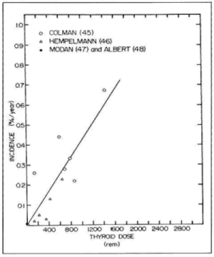
Figure 18-6. Estimated dose response for thyroid cancer in humans from external irradiation. The incidence of carcinomas each year is plotted against the original thyroid irradiation dose. (From Maxon H, Thomas SR, Saenger EL, Buncher ER, and Kereiakes JG. American J Med, 63:967, 1977)

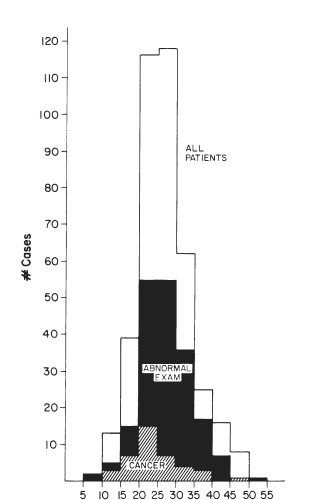
Figure 18-7. Distribution of patients with a history of irradiation to the head and neck, according to the time after irradiation at which they were examined. The majority of patients were seen 20 - 35 years after irradiation, but the incidence of tumors peaked 5 - 10 years earlier. Tumors continued to occur through 40 years after irradiation, and it is not clear that there is a finite latency period.

Probably any x-ray exposure of the thyroid has some carcinogenic potential, although the risk may decrease with age. Adults were extensively treated by x-irradiation for Graves’disease from 1930 to 1950. An increased incidence of carcinoma has been reported in these patients (171). A significant incidence of thyroid neoplasia was observed in patients who received x-ray therapy for thyroid disease (172). These patients were treated at ages up to 34, received 500-1,500 rads, and developed tumors 10-27 years after treatment. In a study of survivors of the atomic blasts at Nagasaki and Hiroshima, an increased incidence of thyroid cancer was found among persons who had received large amounts of radiation (173). Thus, the thyroid of the adult is sensitive to the carcinogenic action of x-rays, although not so sensitive as that of the child.

Radiation-associated tumors of the thyroid continue to occur, although x-ray treatment of thymic enlargement and tonsillar or adenoid hypertrophy has been discontinued since 1959. A recent analysis of 1787 patients treated with X-ray for Hodgkin’s disease found 1.7% to have thyroid cancer (174). The most dramatic and terrifying data emerged from the area around Chernobyl, where thousands of people of all ages received large doses of radiation from external fallout and ingested isotopes, especially short-lived isotopes of iodine. In this epidemic the risk of thyroid cancer is highest among children who were under 9 years and especially under 5 years old at the time of the Chernobyl explosion, and presumably ingested iodide via milk from cows grazing on contaminated forage. The latent period in these children is amazingly short (6 to 7 years), the tumors tend to be relatively aggressive, and are frequently associated with thyroid autoimmunity (175).

Radiation-associated tumors are generally found among younger patients. They are rarely undifferentiated, but some have been fatal. In a review of x-ray associated thyroid tumors at the University of Chicago Thyroid Clinic (163), the latent period among children treated predominantly in adolescence for tonsillar enlargement or acne averaged 20 years. It appears that the peak incidence of lesions is at 10 -25 years after exposure (Fig. 18-7, above), and it is probable that the occurrence of new cancers decreases over time. Among 100 consecutive patients seen in 1973 and 1974, only because they knew of prior radiation exposure, 15% had lesions suggestive of tumor and 7% had cancer proven at operation (162). Favus et al. (176) found a similar incidence of cancer (60/1056) in irradiated patients called back for evaluation. Although one case-controlled study suggests a lack of effect of radiation, the evidence, reviewed by Maxon et al (168), clearly confirms the importance of this problem.

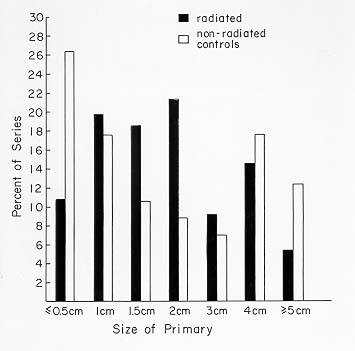
Fig 18-8. The size of radiation associated and non-radiation associated tumors was statistically non-different.

Based on these facts, it has been accepted by most physicians in the field that patients with a history of thyroid irradiation (over 20 rads, and certainly 50 rads) should be located and advised to have an assessment. This evaluation should consist at least of a physical examination and thyroid ultrasound. If one or more clear-cut nodules is found, then FNAC should be performed followed by surgical intervention in case of malignant result. Benign nodules are also found in these glands, with an incidence much higher than that of cancers. Serum TG levels tend to be elevated in irradiated patients, and antithyroid antibodies are more commonly present, but these tests are not of diagnostic value. When excised these glands often show multiple benign as well as malignant nodules as well as areas of fibrosis and hyperplasia (177).

M.P., 52-Year-Old-Woman: Thyroid Radiation and Multiple Gland Abnormalities

This patient was first seen with a history of irradiation for acne during her teens. She subsequently developed telangiectasia of the skin of her face. The month before the examination, she had observed a lump on the right side of the neck. Examination disclosed a 1-cm nodule in the right lobe of the thyroid and some irregularity of the left lobe. Thyroid scintiscan showed a cold nodule of the lower pole of the right lobe. Ultrasound examination of the right lobe identified a partially cystic nodule and a small cystic structure of the left. The FTI was slightly elevated at 10.9. RAIU was above normal. Thyroid antibodies were not present. Thyroid needle aspiration showed cells indicative of malignancy.
Routine blood tests disclosed alkaline phosphatase of 107 units (normal, 25-100 units), calcium 10.9 and 11.5 mg/dl (normal, 8.5-10.2 mg/dl), and phosphate 2.7 ng/dl. Repeated assay of FTI again demonstrated an elevated value of 15 (normal = 6-10.5). The level of parathyroid hormone was 0.65 ng/ml (with a coincident calcium level of 11.5 mg/dl), values indicative of primary hyperparathyroidism.

The patient was treated with potassium iodide for 1 week and admitted for ex¬ploratory surgery. A right upper para-thyroid adenoma weighing 908 mg was found. The adenoma showed areas of cystic degeneration and fibrosis. The thyroid gland was multinodular and was suspicious on frozen section for follicular carcinoma. There was extensive fibrosis around and adherent to the thyroid gland.A near-total thyroidectomy was performed. The gland weighed 17 g. Multiple nodules in the gland measured 1-18 mm in diameter. An 18-mm nodule in the right lobe was iden¬tified as follicular carcinoma. There were, in addition, multiple follicular adenomas and multiple Hurthle cell tumors, focal hyperplasia, and colloid nodules in the right and left lobe.

Postoperatively the patient received thyroid hormone. When seen 1 month after surgery, her calcium level was 9.4 mg/dl, phosphorus 3.5 mg/dl, and parathyroid hormone 0.28 ng/ml (normal). The FTI was 9.2 while taking 0.15 mg L-T4.

This patient developed a cystic parathyroid adenoma with hyperparathyroidism, multiple adenomas of the thyroid, follicular carcinoma, and multiple functioning adenomas that produced thyrotoxicosis. All of these tumors occurred concurrently in a gland showing changes typical of prior radiation exposure.

D.C., 19-Year-Old-Girl: Development of Thyroid Carcinoma, After X-Ray Therapy, While Receiving Thyroid Hormone

At age 10 the patient had respiratory distress and was found to have a superior-anterior mediastinal mass. There was left cervical lymphadenopathy and bilateral supraclavicular lymphadenopathy. Biopsy revealed Hodgkin’s disease of the nodular sclerosing variety. The result of staging laparotomy was negative. She was treated with 4,000 rads to a neck mantle field.

One year later the results of thyroid function tests were normal, but two years after x-ray treatment the FTI was 4.1 and the TSH level was 24 µU/ml. Thyroid hormone replacement therapy was begun, and the patient was carefully monitored over sub¬sequent years with periodic FTI and TSH determinations. Six years after irradiation a2-cm nodule was noted in the left lobe of the thyroid. This nodule was found to be cold on 123-I scan. Fine needle aspiration revealed cells suggestive of malignancy.

At surgery a papillary adenocarcinoma with capsular and vascular invasion was found, and a near-total thyroidectomy was performed. Postoperatively residual thy¬roid tissue was ablated by administration of 30mCi131I.The skeletal survey findings were negative; chest x-ray films and bone films were normal. She has remained free of evidence of thyroid carcinoma or Hodgkin’s disease in the subsequent three years.

This history demonstrates the occurrence of thyroid carcinoma, in a gland heavily irradiated during therapy for Hodgkin’s disease, while the patient was taking ade¬quate replacement doses of thyroid hormone. Possibly the period of X-ray induced hypothyroidism played a role in tumor induction. The tumor developed within six years of the radiation therapy. Fortunately, the tumor was not metastatic and has presumably been eradicated by surgery and RAI ablation of residual tissue.
In our series, postradiation carcinomas averaged 1.7 cm in size (Fig. 18-8), and 14% were below 0.5 cm.

The size distribution was similar to that of non-X-ray-associated tumors. They were more frequently multicentric than those in non irradiated glands and as aggressive, or more so, in behavior than tumors arising without known irradiation (178). The tumors are mainly papillary or follicular, but an occasional anaplastic cancer is also found.In examining patients, it should be remembered that benign and malignant salivary gland neoplasms, neuromas, parathyroid adenomas (179), laryngeal cancer, skin malignancies, and breast cancer also occur with undue frequency in this group of patients.
Thyroid nodularity and cancer also occur as a sequela of nuclear fallout. In the accident at Rongelap in the Marshall Islands (180), individuals received 200-1,400 rads. The incidence of nodularity was 40%, and nearly 6% proved to have cancer. Children in Utah exposed to small amounts of fallout from atomic bomb testing have been proven to develope nodules and possibly carcinomas.

Lack of Association of 131I Treatment and Thyroid Carcinoma

Iodine-131 treatment induces abnormalities in the thyroid gland that persist for many years (181). Giant nuclei, increased mitotic activity, hyperchromatic nuclei, and other abnormalities appear. It seems reasonable that these nuclear changes could lead to carcinomatous degeneration. Chromosomal damage in circulating lymphocytes has also been reported after 131I administration (182). Patients have developed thyroid nodules or tumors after 131I therapy for Graves’ disease; it has been suggested, but not proved, that the highest incidence has been among those treated during childhood. Some of the lesions found in 131I-treated children may actually have been carcinomas, but there has been debate (183) among the various pathologists who examined the specimens. Several reports of isolated instances of cancer after 131I treat¬ment of adults for Graves’ disease have appeared, but the large United States Public Health Service cooperative study failed to show an increased risk in this group (184-186). Studies by Holm et al (186) also failed to show an increase in cancer incidence among persons given 131-I either for diagnosis or for therapy for thyrotoxicosis. These patients were adults, and usually in the 40-60-year age group. Also, very large radiation doses may be less carcinogenic than small ones, and in half or more of these patients, the thyroid has been totally destroyed. Lastly, the follow-up time averages 8-13 years, which may be too soon to see radiation-induced neoplasia. Thus the evidence is reassuring but the question cannot be considered closed.

Thyroid Hyperplasia and Cancer

Chronic stimulation of the thyroid with TSH probably can lead to carcinogenesis in humans, as it can in animals. There are several reports of intensely hyperplastic congenital goiters, untreated for long periods, in which carcinomas have finally developed (187-191). Fortunately, most patients with congenital goiter are recognized and treated with replacement thyroid hormone at sometime during early childhood, so that chronic TSH stimulation does not occur. Interestingly, activating mutations of the TSH-R, which are metabolically like chronic TSH stimulation, lead to benign and not malignant change, as described above.
Relation of Cancer To Other Thyroid Disease

The relationship of thyroid tumors to other thyroid disease is still debated. In the preceding section we discussed whether carcinomas arise from adenomas, occurring either singly or as a component of a multinodular gland. While this must happen rarely, it is not the ordinary course of events. In support of this view one may note, for example, that, whereas adenomas are rarely if ever papillary, approximately 80% of all thyroid carcinomas are papillary. If carcinomas arise from adenomas, one might expect that the majority would be follicular rather than papillary, and this is not the case. Also, although carcinomas, largely of the papillary type, occur in nontoxic nodular goiters with a reported frequency of 4-17% of cases, the age of diagnosis of papillary carcinomas does not follow that for nontoxic goiter (192). Papillary carcinomas occur in children and adolescents, and reach their highest frequency during the middle decades of life. Multinodular goiter, by contrast, is infrequent in childhood, but increases with each decade. The high frequency of carcinomas detected in nodular goiter appears to reflect the efficiency of selection of patients for operation on the basis of suspicious clinical findings in the gland. Although it remains unproven, it is likely that in many or most thyroid adenomas and carcinomas, one specific mutational event leads directly to the development of the specific neoplasm.

Parathyroid adenomas occur in a small percentage of patients with thyroid cancer. The converse relationship may also exist; 2-11% of patients with parathyroid adenomas also have thyroid cancer (193-195). An important reason for this association is the induction of both tumors by X-ray exposure.

Neoplasia and Autoimmune thyroid diseases

An increased incidence of cancer in Hashimoto’s thyroiditis has been reported, Further, focal thyroiditis may occur as an immunologic response to thyroid cancer. In most series in the past the coexistence of Hashimoto’s thyroiditis among thyroid cancer patients was between 2-4%, but the real association, particularly with microcarcinomas, is difficult to be assessed because Hashimoto’s thyroiditis is rarely operated upon. Recently this issue has gained new attention in the era of FNAC, and several reports have found that when a thyroid nodule is associated with autoimmune thyroiditis, the chance of malignancy is significantly higher (9-40%) than in nodules not associated with thyroid autoimmunity (196,197). One possible explanation for this finding might be that patients with autoimmune thyroiditis tend to have higher levels of serum TSH (potential thyroid carcinogen) compared with non-autoimmune patients.

Many reports on Graves’ disease stress a normal or low coincidence of cancer, but several series have reported a significant association between Graves’ disease and thyroid cancer, ranging from 3 to 10% (198-200). However, most of these series were surgical, and the patients were selected for surgery on the basis of suspicious nodules or large goiters. In Graves’ patients treated by radioiodine, no subsequent increase in the discovery of thyroid cancer has been reported.In our review, 4 of 50 patients with thyroid cancer had coincident Graves’ disease (201). Belfiore et al found the risk of thyroid cancer in Graves’ disease to be increased 2-3 fold (198). TSAb can stimulate thyroid cancers when Graves disease coexists, so the idea that TSAb might induce malignant change is tenable, but not proven. It also is possible, but unproven, that continued stimulation of a tumor may make it behave in a more aggressive manner (198,202). Patients with Graves disease and thyroid cancer who underwent total thyroidectomy and 131I ablation fared as well in follow-up as did patients without Graves’ disease.

Micro-carcinomas

The term micro-carcinoma refers to tiny carcinomas (<1 cm), usually papillary and often sclerotic. Such tumors were frequently found in autopsy studies when the thyroids were sectioned completely at 1-2 mm intervals and every abnormality was studied, or during histology of glands operated for large multinodular goiters (203,204). Nowadays, with the extensive use of thyroid ultrasound, micronodules (<1 cm) are frequently discovered in the general population, and proven malignant in a significant percentage. These tumors are probably synonymous with the "occult" tumors described at autopsy, but since they are discovered during life can no longer be considered occult. The term microcarcinomas is now preferred to occult.

Tiny carcinomas, usually papillary and often sclerotic, have been found in 5.7% of thyroids of adults coming to autopsy in the United States (203). This prevalence is noted only when the thyroid is sectioned completely at 1-2 mm intervals and every abnormality is studied. The tumors have a mean diameter of 2 mm, and almost all are under 5 mm. The prevalence is best known in adults (204) and may be lower in young people. Since, in some glands, collections of psammoma bodies also exist in tiny scarred areas, it is hypothesized that such lesions may spontaneously regress. Because of their small size, they are detectable only at surgical or pathologic examination of the gland. These observations, now widely accepted, have provoked much discussion. Certainly most of these tiny tumors cannot be biologically significant, considering the low incidence of clinically recognized cancer. Most of the lesions are probably missed during routine surgical or autopsy pathologic studies. Their cause is unknown. They may be a variant of the occult sclerosing carcinomas described by Hazard, but the latter tumors are usually larger, have a predominant sclerotic component, and clearly do metastasize. It has been suggested that such lesions are in fact the cancers found after thyroid irradiation, but most likely they can explain only a small proportion of radiation-associated tumors. Most of these lesions would go undetected in a standard surgical pathologic examination, and the great majority of radiation-associated lesions are larger. In our own series, the radiation-associated le¬sions were on average 1.7 cm in diameter and only 14% were under 0.5 cm. Whether such "minimal" cancers are in fact the occasional precursors of clinically evident cancers is a moot point. It is clear that they are at present an important pathologic -but not clinical --entity.

PATHOLOGY (Figures 18-9a-h)

Pathologists are agreed that there are peculiar difficulties in the classification and diagnosis of malignant tumors of the thyroid. The histologic changes required for diagnosis of carcinoma include absence of a true capsule, invasion of surrounding normal tissue, invasion of blood and lymph channels, loss of normal follicular architectural arrangements, and cellular abnormalities such as an increase in the ratio of nucleus to cytoplasm, enlarged vesicular nuclei, nuclear folding, increased mitoses, and hyperchromasia of the nucleus. Recently, aneuploidy of nuclear DNA content has been added to this list. Obviously the presence of distant metastases is the most certain criterion. Most students of the disease agree that the ordinary criteria of malignancy have little prognostic value in thyroid tumors, except perhaps in the wildly growing anaplastic tumors. However, it may be noted that pathologists at the Mayo Clinic believe a histologic typing by their criteria provides significant information on prognosis.

Examples of the histologic patterns of several of these tumors are given in Figure 18-9. The papillary adenocarcinoma typically shows tumor cells around a fibrovascular core and, not infrequently, areas of follicular differentiation. Papillary lesions tend to be infiltrative, and encapsulation is rare. Lymphocytic "reactions" are prominent. The cell nuclei have a ground-glass or "cat’s eye" appearance and intracellular inclu¬sions are common. Vascular invasion is rare. Psammoma bodies are often abundant. Multiple intraglandular foci are frequent, especially in children. Areas of lymphocyte infiltration, and even extensive lymphocytic thyroiditis, are common, especially in papillary tumors. Many tumors look much like follicular cancers, but have the characteristic nuclei of papillary cancers. These constitute the "follicular variant" of papillary cancer, and behave more or less as do other papillary cancers.

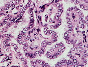
Figure 18-9. A) Papillary carcinoma of the thyroid. The structure is made up of complex (branches on branches) fibrovascular core structures covered by crowded, overlapping, vesicular nuclei (artifact of fixation). Little colloid is visible. Such histologic foci may be encapsulated, sclerosing, invasive, or multicentric.

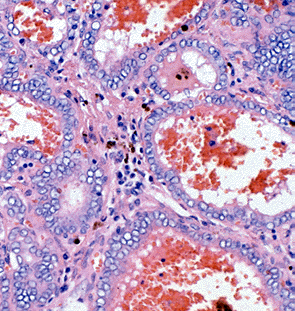
Figure 18-9 B) Follicular variant of papillary carcinoma with more typical vesicular nuclei, and hemorrhage in follicular lumens.

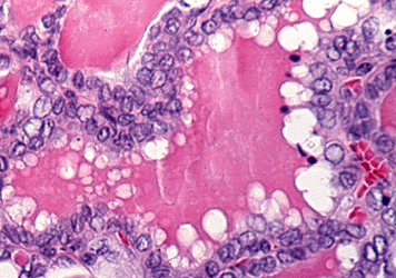
Figure 18-9. C) Follicular variant of papillary carcinoma with crowded nuclei showing nuclear folding and peripherally vacuolated colloid.

Follicular adenocarcinomas vary from those with a definite follicular pattern to those with solid sheets of cells. The lesions are more frequently encapsulated, but capsular and blood vessel invasions are typical. The nuclei are normo-or hyper-chromated, or may be quite vesicular. One variant, the so-called malignant adenoma, appears to be nearly benign and can be identified as malignant only by the demonstration of invasion of vessels or capsule, or because of the presence of distant metastases, which may also be composed of normal-appearing thyroid tissue. Hurthle cell carcinomas usually grow as solid sheets of large eosinophilic granular cells with much cytoplasm, and less often with a follicular pattern. An Hurthle cell appearance can also be observed in some papillary tumors. (Figure 18-9d)


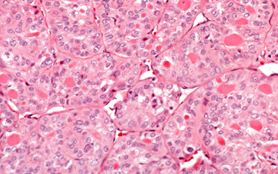
Figure 18-9. D) Poorly differentiated follicular carcinoma with oxyphillic features.


Medullary tumors (Fig. 18.9 f-g) have an ominous histologic pattern, with solid masses of cells with large vesicular nuclei (205). There may be considerable associated fibrosis, and deposits of amyloid are a helpful diagnostic point. At the time of initial histologic examination the pathologist should recognize these tumors as entities distinct from the undifferentiated cancers, for the medullary carcinomas have a much better prognosis. Medullary thyroid carcinoma (MTC) is associated with amyloid deposition in the surrounding tissues. Recent studies demonstrated that full-length calcitonin is the sole constituent of amyloid in MTC (206).

In the undifferentiated group of small-cell tumors, giant-cell tumors, and carcinosarcomas, or in the miscellaneous group, the histologic pattern has little resemblance to the original thyroid structure (Fig 18-9e-h).

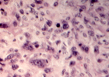
Figure 18-9. E) An anaplastic carcinoma of the thyroid with pleomorphic giant tumor cell nuclei.

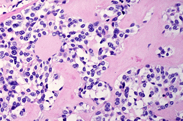
Figure 18-9. F) Medullary (C-cell) carcinoma of the thyroid with amyloid stroma.

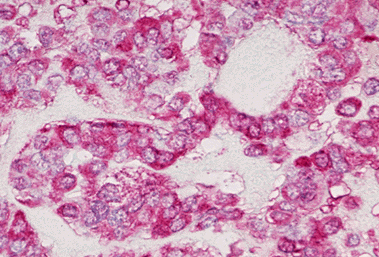
Figure 18-9. G) Immunohistochemical anti-calcitonin antibody stain of a medullary carcinoma showing strong red positivity.

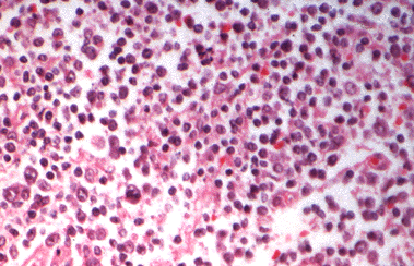
Figure 18-9. H) Large cell lymphoma of the thyroid.

The general experience of pathologists has been that, in the absence of irradiation, the substrate in which thyroid tumor forms is usually normal thyroid tissue or displays the changes of multinodular goiter or adenoma in approximately the proportion found in any sampling of the general population (207). There is a slightly increased frequency of association with benign adenomas and with Hashimoto’s thyroiditis (208). Lymphomas are associated with Hashimoto’s thyroiditis, and there is considerable evidence that lymphoma actually evolves from a gland with thyroiditis (209).

Multicentricity is a common feature of thyroid cancer, especially papillary cancer. Innumerable separate foci are sometimes found. Estimates of multicentricity range from 20 to 80% (210,211). Whether this phenomenon represents truly multicentric sites of origin or intrathyroidal dissemination is not clear. This multifocality is thought to be one cause of recurrences in patients treated by subtotal rather than total thyroidectomy.

Both papillary and follicular tumors may appear as small (less than 1.5-cm) tumors surrounded by a densely fibrotic reaction. Although it is frequently said that these "occult" (because they may be found incidentally at operation) tumors are benign, the original report by Hazard (212) and subsequent studies show that cervical lymph node metastases occur (213).
Occasionally pathologic examination suggests conversion of differentiated papillary or follicular cancers into anaplastic forms or conversion of a follicular adenoma into a follicular carcinoma.
An interesting aspect of thyroid tumor pathology is the frequency of metastatic tumors to the thyroid, 5% in the data of Silverberg and Vidone (214) for unselected autopsies and 24% for patients dying of metastatic malignant disease.

Course of the Disease

Clinical signs

Most frequently the tumor is discovered accidentally by the patient or physician as a lump in the neck or may be a fortuitous finding at ultrasound of the neck. Less and less frequently, nowadays, it may appear as a gradually enlarging, painful mass with associated symptoms of hoarseness, dysphagia or dysphonia, or there may be difficulty in breathing. Occasionally a arrives with metastatic nodules in the neck, pulmonary symptoms from metastases, or a pathologic fracture of the spine or hip. Usually there are no symptoms of hyper-or hypothyroidism, but in very rare instances the tumor, usually metastatic follicular carcinoma, can produce enough hormone to cause hyperthyroidism (215,216). (See also Chapter 13)

Upon examination of the neck, carcinoma of the thyroid characteristically appears as an asymmetrical lump in the gland. If it is still within the confines of the gland, it will move with the gland when the patient swallows and may be moveable within the gland. If it has invaded the trachea or neighboring structures, it may be fixed; this is a useful sign. Lymph nodes containing metastases may be found in the supr-aclavicular triangles, in the carotid chain, along the thyroid isthmus, and rarely in the axillary nodes. A sentinel or "Delphic" node above the isthmus may be present. Although carcinoma of the thyroid is typically firm or hard, rapidly growing lesions may sometimes be soft or even fluctuant. When the tumor is poorly differentiated or anaplastic, the lesions may undergo necrosis and discharge through sinuses that developed in the skin of the neck. Patients with thyroid cancer are also prone to develop other cancers, the risk being about double the average. Among these cancers is an excess incidence of leukemia, perhaps related to 131I therapy (217-219).

Age at diagnosis has an important bearing on the patient’s subsequent course. The adverse effect of age on the prognosis increases gradually with each decade (220). For practical assessment purposes, it is clear that patients diagnosed before age 45 have a much better prognosis than those detected later (221). Age is also directly related to the incidence of undifferentiated tumors and to overall mortality. Pregnancy does not seem to worsen the course of established or previously treated thyroid can¬cer (222). Overall, women have a better prognosis than men with cancer (223). Other characteristics of the tumor, including (as would be expected) extraglandular extension, gross invasion of the tumor capsule, and increasing size also carry a worsened prognosis (223).

Papillary Carcinoma-course of disease

Papillary carcinoma has a peak incidence in the third and fourth decades (224). It occurs three times more frequently in women than in men, and accounts for 60-70% of all thyroid cancers in adults and about 70% of those found in children. The disease tends to remain localized in the thyroid gland and in time metastasizes locally to the cervical or upper mediastinal nodes. The lesions are multicentric in 20% or more of patients, especially in children. Using rigid pathologic criteria, perhaps two-thirds of predominantly papillary thyroid cancers are found to have follicular elements. The natural history of these tumors is similar to that of pure papillary lesions (225). The metastases may conform to either histologic pattern. At present, the mixed tumors are lumped together with all other papillary cancers. This tumor tends to be indolent and may exist for decades without killing the host. In a Mayo Clinic series of papillary tumors that were detected because of lymph node metastasis or found incidentally during surgery of the thyroid gland, all the patients were unaffected by the tumors over several decades (224). However, in a recent series from one single institution in Italy, it is apparent that the way of presentation of papillary thyroid cancer has been changing in the last to decades compared to previous years. In particular the authors report increasing number of small tumors and less frequent lymph node metastases at presentation (226)

The frequent occurrence of occult or “minimal’ incidentally found thyroid cancers, usually papillary and under 0.5 cm in size, is described above. However, the term occult has been used in a variety of ways, including reference to tumors with malignant nodes but no obvious primary, or in reference to any tumor under 1.5 cm in diameter. Mayo Clinic reports of papillary tumors under 1.5 cm in diameter, treated with conservative subtotal thyroidectomy and node dissection, have stressed their non-lethal nature, but a 1980 follow-up report on 820 patients treated by this group notes that 6 patients eventually died after spread of tumor from such "occult" primaries (227). Patients with appropriately treated Clinical Class I or II lesions have 96-100% survival even after 15-30 years. Survival lowers to 87% for Class III and 35% for Class IV lesions at 15 years.

While the disease may be aggressive in children, it is distinctly less aggressive in young adults, as compared to patients over age 40 (223).Young patients tend to have small primary lesions and extensive adenopathy, but even with local invasion survival is good (228). When papillary cancer occurs in persons over the age of 45, it may show, on microscopic examination, areas of undifferentiation, and pursue a highly malignant clinical course. The lesions tend to be larger and more infiltrative, and to have fewer local metastases (229). It is possible that persons dying in older age actually have had their disease since youth, and that it has simply evolved into a more malignant phase (230,231).

Papillary carcinoma tends to metastasize locally to lymph nodes, and occasionally produces cystic structures near the thyroid that are difficult to diagnose because of the paucity of malignant tissue. In this case measurement of thyroglobulin in the fluid aspirate is the clue for the correct diagnosis. The presence of nodal metastasis correlates with recurrence (230-232) but has little effect on mortality in patients under age 45. In old studies, cervical adenopathy even seems to confer a protective effect on young people, but this assumption is not confirmed in present series. Indeed in patients over 45, the presence of nodes is associated with greater recurrence rates and more deaths (233,234). The tumors often metastasize elsewhere, especially to lung or bones.

Papillary tumors may metastasize to the lungs and produce a few nodules, or the lung fields may have a snowflake appearance throughout. These tumors are amazingly well tolerated and may allow relatively normal physical activity for 10-30 years. At times, particularly in the follicular variant of papillary thyroid cancer, the pulmonary metastases are active in forming thyroid hormone, and may even function as the sole source of hormone supply after thyroidectomy. The metastases may progress gradually to obstructive and restrictive pulmonary disease. They also may develop arteriovenous shunts, with hypoxia or cyanosis. Such shunts become more prominent during pregnancy, perhaps as an effect of the increased supply of estrogens.

The tall cell variant of papillary carcinoma comprises about 10% of total cases, and as noted by several authors appears to be more aggressive than other forms of the disease (235,236). .
The usual net extra mortality in papillary cancer is not great when compared to that of a control population, perhaps 10-20% over 20-30 years (231,232,234). Mortality is rare in patients diagnosed before age 40, and is much greater in the patients found to be in clinical stages III and IV at initial diagnosis. About one-half of patients ultimately dying from this lesion do so because of local invasion. Frazell and Duffy (237) have noted that papillary carcinoma is not always so benign; they reported 35 patients with "invasive papillary carcinoma," which had a very malignant course.

We found that risk of death from cancer was increased by extrathyroidal invasion (6 fold) or metastasis (47 fold), age over 45 years (32 fold) and size over 3 cm (6 fold). Thyroiditis, multifocality and the presence of neck nodes had no effect on disease-induced mortality.

P.P. 41-Year-Old Man: Long-Term Survival with Papillary Cancer

This man was first seen at age 41. At age 14, while living in Yugoslavia, he had developed a mass in his neck. Tracheostomy was required because of dyspnea, and a biopsy of the mass was performed. A diagnosis of papillary thyroid carcinoma was made, and he was treated with radiotherapy to the neck. Because of an abnormal chest x-ray film, presumed to be due to tuberculosis, he was given streptomycin and isoniazid for 2 months, but this therapy was discontinued when no improvement occurred. Extracts of calf thymus were injected. He was evaluated for hemoptysis at age 18, and chest x-ray films again showed the infiltrates without evidence of change. The patient was not given thyroid hormone replacement therapy at any time. At age 39 he developed fatigue, substernal chest pain, occasional cough without production of sputum, and occasional hemoptysis. A thyroid scintiscan at another hospital revealed poor visualization of the thyroid and uptake of 4%, and 131I uptake in the mediastinum.

Physical examination disclosed hyperpigmentation of the skin in the area of radiotherapy in the neck and supraclavicular areas. There was no evidence of a mass or lymph-adenopathy. The tracheostomy site was well healed. The lungs were clear, BP was 100/70, pulse 68, and respiration 16/min, and there was no cyanosis. Rou¬tine complete blood count, urinalysis, and blood chemistry test results were normal. Chest x-ray films showed multiple nodular densities throughout both lungs and a prominent left hilum. The results of a radiographic skeletal survey and a technetium pyrophosphate bone scan were normal. The FTI was 8.4 and TSH was 4.1 µU/ml. A whole body 131I scan showed multiple areas of abnormal uptake in the area of the thyroid, and satellite areas of focal uptake around the thyroid bed. There was also focal activity in the mediastinum, in both hila and in the lung fields. A 72-hour chest uptake was 8.9%. The serum TG level was 81 ng/ml.

The patient was treated multiple times with RAI and remained well while taking replacement T4 for ten years. At age 51, the tumor grew more rapidly, failed to accumulate 131I, and caused the patient’s death.

This patient developed thyroid carcinoma at age 14 and probably had lymph node and lung metastases at that time. He lived a normal life during the ensuing 27 years, without suppressive thyroxine treatment, and with only intermittent episodes of hemoptysis. The tumor responded at first to 131I but later was uncontrolled. The common benign course of metastatic papillary thyroid carcinoma over many years is clearly shown, as is the equally typical later exacerbation and death.

Follicular Carcinoma

Follicular carcinoma has a peak incidence in the fifth decade of life in the United States and accounts for about one-quarter of all thyroid carcinomas (223,238,239). In past decades, follicular carcinomas comprised up to 50% of thyroid malignancies in Europe. The high incidence may partly be explained by iodine deficiency, but, more likely, was due to histological miss-classification at a time when the follicular variant of papillary thyroid carcinoma was not recognized as papillary but rather was classified as follicular. It is a slowly growing tumor and frequently is recognized as a nodule in the thyroid gland before metastases appear. Variation in the cellular pattern ranges from an almost normal-appearing structure to anaplastic tissue that forms no follicles or colloid. The insular variant of follicular tumor tends to be more aggressive (240). The tumor is three times as common in women as in men. At operation one-half to two-thirds of these tumors are resectable. Tumors that are small and well circumscribed (not surprisingly) tend to be less lethal than those actively infiltrating local structures at the initial operation. Local adenopathy, which is uncommon, probably carries a greater risk, and extensive invasion of the tumor capsule and thyroid tissue increases mortality (241). Local direct invasion of strap muscles and trachea is characteristic of the more aggressive tumors (242). Resectability depends on this feature, and death may be caused by local invasion and airway obstruction. The "minimally invasive" variant has a far better prognosis than the highly invasive variant.
Follicular carcinomas tend to invade locally and metastasize distantly, rather than to local nodes, and are especially prone to metastasize to bone or lung. In a Massachusetts General Hospital series (231), one-half had metastasized at the time the diagnosis was originally established. Bony metastases are usually osteolytic, rarely osteoblastic, and the alkaline phosphatase level is rarely elevated. The tumor and metastases often retain an ability to accumulate and hold iodide, and are therefore sometimes susceptible to treatment with RAI. Indeed, some metastatic tumors synthesize thyroid hormone in normal or even excessive amounts. RAI therapy, as discussed below, improves survival in these patients (241).

Occasionally the primary lesion of a follicular tumor appears to be entirely benign, but distant metastases are found. Invasion of vessels or the capsule, apart from the metastasis, is the only reliable criterion of malignancy. This variant has been called the “benign metastasizing struma” or malignant adenoma. It has a more prolonged course than do other varieties of follicular tumor, and is the type that has offered the best opportunity for the therapeutic use of 131I.
The net extra mortality attributable to follicular cancer in the 10 -15 years after diagnosis is 30 -50% (231,233,235). Of the patients dying from the lesion, three-fourths do so from the effect of distant metastases and the remainder from locally invasive disease.

Hurthle CellTumors

Hurthle cell tumors are histologically distinct from other follicular tumors, but they pursue a similar course. They tend to invade and metastasize locally and have a strong propensity to recur after surgery. The course tends to be prolonged. These carcinomas often do not accumulate 131I. However, in a large survey, Caplan et al (243) found that 4.4% of Hurthle cell neoplasms were hot on scan and 8.9% were warm. Serum TG levels may be normal or elevated. Cheung et al recently studied the presence of ret/PTC gene rearrangements in Hurthle cell tumors and found that many expressed ret/PTC, and also had other evidence of a papillary cancer origin, including focal nuclear hypochromasia, grooves, and nuclear inclusions. Tumors with the ret/PTC gene rearrangement tended to have lymph node metastases, rather than hematogenous spread. Thus Hurthle cell tumors can be classified into Hurthle cell adenomas, Hurthle cell carcinomas, and Hurthle cell papillary thyroid carcinoma (244).

Insular Tumors

A subset of thyroid carcinomas which give a histologic picture of islands of cells -thus "insular" --has been identified (243). These tumors often look like anaplastic cancers, but sometimes are able to concentrate 131I and thus are amenable to this excellent treatment. Whether these are properly considered a variety of follicular cancer is uncertain. The important message is that the histology in this instance does not reliably predict the utility of 131I treatment, suggesting that all patients with thyroid cancer should at some point be studied to determine whether 131I treatment is possible.

Undifferentiated Tumors

Undifferentiated tumors occur predominantly in persons over 50 years of age and constitute an increasing proportion of lesions in each subsequent age decade. Of great interest is the pathologic evidence that such tumors arise, in perhaps half of the cases, in a long-standing benign lesion or in differentiated carcinoma (245). Although 131I therapy for differentiated cancers has been blamed for this dedifferentiation, current evidence is against this hypothesis. Spindle cell, and most giant-cell carcinomas of the thyroid grow rapidly and are very invasive. Local invasion may cause difficulty in breathing or swallowing, and tracheotomy is frequently required. These tumors metastasize to lymph nodes both locally and widely, but not characteristically to bone. Pulmonary metastases are frequent. Some patients present with a tender mass suggesting thyroiditis, and occasionally thyroid destruction induces hyperthyroidism (246). The outlook in this particular group of tumors is poor. By the time the diagnosis is made, the disease has spread in most patients beyond the area that can be attacked surgically, and they die within 6 months to 1 year. A few, perhaps 10%, of these tumors are entirely resectable when first discovered. There is nothing characteristic about the growth pattern of these tumors; their behavior is similar to that of any highly malignant tumor elsewhere in the body. The course of the epidermoid carcinoma and sarcoma of the thyroid is essentially the same. Small-cell anaplastic carcinomas are also found, but probably most tumors so classified in the past are actually lymphomas or lymphosarcomas.

Malignant Lymphomas

Lymphomas of the thyroid gland represent less than 5% of primary thyroid neo¬plasms (247-250). Unlike most other thyroid neoplasms, lymphomas usually appear as rapidly enlarging masses and local symptoms are common. Many patients note pain, hoarseness, dysphagia, and dyspnea or stridor. Hoarseness is often present in the absence of vocal cord paralysis. Rarely, patients may have the superior vena cava syndrome. The mean age at occurrence is 62 years. Primary lymphomas of the thy-roid are two to three times more common in women than in men.

The incidence of hypothyroidism at the time of appearance is variable, ranging from 0 to 60% (251,252). The co-occurrence of pathologic lymphocytic thyroiditis has ranged from 30 to 87%. These figures may underestimate the true incidence, as some patients have only had a biopsy examination and in others the entire gland has been replaced by the lymphoma. The frequent presence of thyroiditis has naturally led to the suggestion that the lymphoma might derive from preexisting thyroiditis.

The co-occurrence of thyroiditis may create difficulties in the proper interpretation of fine needle aspiration cytology. The clinical appearance must be carefully considered in accepting a diagnosis by fine needle aspiration of thyroiditis only or thyroiditis with lymphoma. An excisional or large needle biopsy may be necessary to make the correct diagnosis.

The majority of thyroid lymphomas are diffuse, large-cell lymphomas (formerly classified as diffuse histiocytic or reticulum cell lymphomas), diffuse mixed small and large cell lymphomas (formerly called diffuse mixed lymphocytic-histiocytic), or diffuse small cleaved-cell lymphomas (formerly classified as diffuse poorly differen¬tiated lymphocytic). Although older series include reports of Burkitt lymphoma, none were reported in larger, more recent series. Areas of diffuse large-cell lymphomas may have features similar to those characteristic of Burkitt’s lymphoma or the Reed-Sternberg cells of Hodgkin’s lymphoma.

Metastatic Carcinomas to the Thyroid

Melanomas, breast tumors, pulmonary tumors, gastric, pancreatic, and intestinal carcinomas, renal carcinomas, lymphomas, carcinomas of the cervix, and tumors of the head and neck may metastasize to the thyroid. Sometimes the first indication of one of these tumors may be the appearance of a lump in the neck. Unless there is evi¬dence for a primary site elsewhere, these tumors are easily mistaken for expanding tumors that have their origin in the thyroid gland. Usually by the time metastases appear in the thyroid, other metastases have occurred and the primary lesion may be discerned.

Cancer in Aberrant Thyroid Tissue

Thyroid tumors occasionally arise in lingual thyroids, along the thyroglossal duct (253), in substernal goiters, and even in struma ovarii. Whenever discovered, the therapeutic approach should the removal of the tumor assoiated with total thyroidectomy to allow subsequent treatment with radioiodine and follow-up based on serum Tg mreasurement.

MEDULLARY CARCINOMA

Hazard et al. (205) first described a unique tumor in the thyroid characterized by sheets of cells with large nuclei, amyloid deposits, fibrosis, multicentricity, and an unexpectedly benign course in view of the solid tumor appearance. Over 50% may have local or distant metastases at diagnosis. The tumors may metastasize locally, or to bones and soft tissues. The thyroid primary tumor and the metastases may show dense calcification on x-ray film. The course tends to be progressive, and 10-year survivorship varies from 50 to 70%. These tumors, which constitute 2-8% of all thyroid cancers, are derived from the "light," or "C," or "parafollicular" cells. These are calcitonin (CT)-secreting cells, distinct from thyroid acinar cells, and are of ultimo-branchial origin.

The tumors may occur sporadically (about 70% of the total) or as part of the MENII syndromes, which constitute about 10-20% of the cases, and are transmitted in families as dominant traits due to activating germline point mutations of the RET proto-oncogene (254,255). In contrast with thyroid epithelial cell tumors, the female to male ratio is near unity. MEN-II (or IIA) includes patients with medullary thyroid cancers, pheochromocytomas, and parathyroid hyperplasia or adenomas. MEN-III (or MEN-IIB) includes medullary thyroid carcinoma, mucosal neuromas, pheochromocytomas, which are usually bilateral and often malignant, occasionally cafe-au-lait spots, and possibly Gardner’s syndrome (mucocutaneous pigmented nevi and small intestinal polyps) (133,256-259). An occasional variant of medullary thyroid cancer appears to contain both CT and TG, suggesting the cells, surprisingly, have features of both medullary thyroid cancer and follicular cancer (260).

Gastrointestinal symptoms including diarrhea, constipation, and rarely megacolon occur in these patients and may occur before the thyroid tumor is detected. Hyperplasia of C cells often precedes the development of familial cancers (261). Medullary tumors derived from the C cells not only secrete CT, but in addition may produce serotonin (with a carcinoid syndrome), prostaglandins, corticotropin releasing factor, and adrenocorticotropic hormone (causing Cushing’s syndrome), histaminase, and somatostatin. Some of these substances, rather than calcitonin, may be the cause of the severe diarrea syndrome associated with metastatic medullary thyroid cancer. Interestingly, expression of somatostatin appears to correlate with improved prognosis (262). Medullary thyroid carcinoma cells in tissue culture have been found to produce Ghrelin, which is an endogenous ligand for the GH secretagogue receptor. MTC is known to produce several gastrointestinal hormones and neuroendocrine peptides, in addition to calcitonin, and including CGRP, ACTH, serotonin, chromogranin A, and vasoactive intestinal peptide, and to this we now add Ghrelin (263). Alcohol ingestion is reported to induce attacks of flushing and diarrhea and to stimulate CT secretion by the tumors (264). The CT secreted by these tumors rarely causes hypocalcemia (265).

Medullary tumors have an ominous histologic pattern, with solid masses of cells with large vesicular nuclei (212). There may be considerable associated fibrosis, and deposits of amyloid are a helpful diagnostic point. At the time of initial histologic examination the pathologist should recognize these tumors as entities distinct from the undifferentiated cancers, for the medullary carcinomas have a much better progno¬sis.

Ret oncogene and MedullaryThyroid Cancer

Studies on patients with MENI and MEN II indicated linkage to chromosomes 11 and 10, respectively (138). Subsequent studies demonstrated that the ret oncogene is present at 10q11.2. Germline mutations have been detected in this oncogene in all patients with MEN II A and MEN IIB, and familial MTC (139). RET is a cell-membrane receptor of the growth factor family, with tyrosine kinase function. In up to 97% of patients with MenII A, mutations are found mostly in codons 609, 611, 618, 620, and 630 in exons 10 and 11. These all involve substitutions of other aminoacids for cysteine, and are thought to cause activation of the gene by aberrant disulphide bonding causing dimerization. Similar changes are seen in Familial MTC. In patients with the MENIIB syndrome, almost all, if not all, mutations involve an amino acid substitution of threonine for methionine at codon 918 in exon 16, and are thought to induce a change in substrate phosphorylation. Somatic mutations in ret are present in up to half of patients with sporadic MTC and are almost always in codon 918 (140,141). Mutations in this codon are thought to imply a poor prognosis (266).

Calcitonin and CEA

The calcitonin assay provides a convenient screening procedure in families with this genetic trait (267). Family members at risk should firstly BE screened for ret oncogene mutation in their blood, and when when recognized as a carrier of the mutation, should be screened by neck ultrasound and calcitonin measurement to assess the presence of the disease. Every member of one of these families with either a thyroid mass or elevated calcitonin levels should have a thyroidectomy. If no thyroid nodule is detected and the serum calcitonin is normal according to the reference range for that specific age group, prophylactic thyroidectomy should be considered in order to take out the thyroid gland before the disease is initiated.

There is controversy regarding the more appropriate age for prophylactic thyroidectomy. Recent ATA guidelines for MTC have suggested different time for surgery according the the age and the mutation found (268) In MEN-IIA, the tumors follow a rather benign course somewhat akin to that of follicular cancer, and usually can be controlled by surgery. MEN-IIB tumors are much more aggressive and often cause death in the second or third decade. TSH suppressive treatment is not efficacious in MTC and has no logical basis.

Secretion of calcitonin by medullary cancer is remarkably increased by calcium or pentagastrin infusion (269). This procedure can be helpful in establishing a diagnosis if available. At present the infusion of pentagastrin (0.5 µg/kg over 5 seconds), with determination of calcitonin levels at 0, 1, 2, 5, 10, and 15 minutes, appears to be the best test. Alternatively stimulation with calcium gluconate, 20 mg/kg, can be done with infusion IV over 1 minute and testing as tor TRH. (This test can induce adverse cardiac effects, and close monitoring is reasonable.) Basal CT values are normally under (depending on the laboratory) 30 pg/ml (269). Values of 30-100 after pentagastrin indicate hyperplasia, and values over 100 typically indicate the presence of cancer. Calcitonin should drop to undetectable levels if the tumor is completely removed surgically. It should be noted that excess production of CT is not unique for medullary cancer, but can occur with granulomatous diseases and other cancers. An alternative is to monitor Calcitonin after Calcium infusion. Patients with the syndrome should also be studied with parathyroid hormone and catecholamine assays in order to determine the presence of other components of the syndrome.

The neoplastic C cells also produce carcinoembryonic antigen (CEA) in large amounts. Serum CEA levels are elevated in medullary cancer with the same frequency as are CT levels (270). Although CEA determination provides another parameter to follow, it does not offer any obvious advantage, and lacks the specificity of CT determinations. Tumor dedifferentiation is associated with a fall of CT and increasing CEA. This is an ominous sign.

Several specialized scanning procedures have been used in MTC. Total body imaging with Tl-201 chloride andTc-99m(V)DMSA have been successful in localizing metastases. 131I MIBG and 131I anti-CEA have been used both for localization and in attempts at therapy (271). Most recently, radiolabelled somatostatin has been used as a whole body scanning agent.
Since patients with MENIIA and IIB, and familial MTC, probably all have a germline mutation of the ret oncogene near the centromere of chromosome 10, and many patients with sporadic MTC have somatic ret mutations, molecular techniques can be used to detect at-risk subjects in recognized families (269). PCR amplification of exons 10, 11, and 16, followed by single strand conformational electrophoretic analysis, or use of specifically designed restriction enzyme sites, allows recognition of most mutations (272). This information is crucial in defining potential risk in young children and identifying need for operation or frequent pentagastrin testing (255), and presumably will also screen out members of families who will not need to be repetitively screened by pentagastrin stimulation tests. Unlike occasional false positives with CT assays, the results of genetic screening are unambiguous (273).
For readers with an interest in a somewhat broader view of oncogenesis, it may be noted that a syndrome entirely analogous to metastatic medullary thyroid carcinoma appears frequently in aged bulls (274). Histologically similar adenomas are also frequently found.The lesion may be due to excessive dietary calcium; whether a similar stimulus could operate in human disease is unknown.

Treatment of MedullaryThyroid Cancer (MTC)

MTC is unique since it can be detected in MEN-IIA or MEN-IIB families by genetic analysis and screening tests measuring pentagastrin-stimulated CT secretion before the disease reaches clinical detectability. In families with familial MTC, MENIIa, and IIB, repeatedly observed elevations to well above the normal range (e.g., peak values of 30-10pg/ml) constitute a basis for operation. In these patients focal tumors or C-cell hyperplasia may be found after near-total or total thyroidectomy is performed. Since C-cell hyperplasia precedes the development of malignancy, it is currently believed logical to operate at this stage of the disease even in young children with MENIIB. Patients with MENIIA or familial MTC are similarly followed, tend to develop tumor later, and 20-40% may never develop cancer (275). International Workshops on MTC have recommended prophylactic thyroidectomy before the age of 5 years in persons harboring mutations of codons 611, 618, 620, and 634 because of their high efficiency in transformation. Consensus on best treatment for patients with other mutations (codons 609,630,768,790,791,804,891) is not clear, with thyroidectomy recommended from ages 5 to 10 (268,276).

Patients with clinical tumor of any size, sporadic or familial, are also treated by near-total or total thyroidectomy. Invasive disease is resected if possible. Since cervical nodal metastasis occur in up to 90% of patients with palpable tumors, a careful mod¬ified radical neck dissection is performed with removal of nodes in the central and ipsilateral compartment. The exploration for nodes should include the upper mediastinum. Bilateral neck dissection may be appropriate in patients with hereditary tumors (277).

Management of Post-operative MTC patients who have positive CT assays

When post-surgical serum CT is undetectable in basal condition and after pentagastrin stimulation, the patient has an almost 100% chance of being in complete and stable remission. In this case follow up should be based on periodic measurement of serum CT, without any instrumental evaluation. After operation, patients with still-detectable serum CT levels should be screened for persistent disease. Imaging should consists of CT of the neck and chest, MRI of the liver and bone scintigraphy. PET scanning may also be informative in selected cases. Some centers used to ablate residual functioning thyroid tissue with 131I, as in papillary or follicular tumors, since this procedure may ablate tiny foci of residual cancer (278,279). However, the value of this procedure is not proven, and this procedure is almost universally abandoned. In patients with local invasive disease, who are above age 45-50, we believe that radiotherapy (5,500-6,000 rads) is useful, although there is disagreement on this point.(v.i.). Occasionally CT levels fall gradually over a year. It is also possible to follow the serum level of CEA as a tumor marker, or CT-related peptide (280). A discordant elevation of CEA in relation to CT may be an indicator of an aggressive tumor. However, in general, CT is the most informative marker.

What to do about the frequently-found elevated CT level without an obvious tumor source is less certain. Residual or recurrent resectable disease should be approached surgically. Neck dissection, if not done previously with a unilateral tumor, should be considered. In the absence of an identifiable source of the CT, careful, extensive, microdissection of the neck and upper mediastinum have been proposed in the hope to eliminate the source of CT (281), but the poor results of such extensive operation have dissuaded many from this approach. Catheterization of the superior vena cava and internal jugulars, with an attempt to localize the tumor by means of multiple venous sampling for CT (242) during pentagastrin stimulation, is possible. The technical success of this approach has been demonstrated, but often the CT level is not reduced to normal, even if operation is subsequently performed on the identified area (282). Radiotherapy may be given to identified non-resectable lesions.

Radiation therapy for MTC

In many cases, no source for the CT is found. Watchful waiting is the approach preferred by some experienced clinicians. Alternatively, mantle irradiation can be recommended. As shown by Simpson (283), medullary tumors are radiosensitive, although a full response may not be seen for many months. This irradiation has been shown to prolong local relapse-free time, but not to clearly improve ultimate prognosis (284). The effectiveness of radiotherapy was evaluated in 24 of 139 patients with medullary thyroid cancer who were given radiotherapy because of advanced local disease at presentation. Only one had normalization of calcitonin, but ten remained free of clinical recurrence. Local relapse was significantly reduced after radiotherapy, but there was no difference in ten year survival between those with and without treatment. These authors believe that radiotherapy does reduce the incidence of loco-regional relapse, but do not advise routine use of radiotherapy because of the relatively favourable long term survival of patients even with elevated calcitonin levels after operation (285). Other investigators question the value of X-ray therapy, contending that patients with MEN-II have multicentric foci from the start, and that radiotherapy may actually worsen prognosis (286). Systemic therapy is reserved for patients with proven symptomatic metastases, and the program is described below.

MTC is associated in MEN-II with parathyroid adenomas and hyperplasia, and in both MEN-IIA and MEN-IIB with pheochromocytomas that are often bilateral and malignant. Parathyroid hormone, VMA, and catecholamine assays should be done to evaluate these problems, and pheochromocytoma, if present, should be treated before the thyroid cancer. Occasionally neuromas cause problems requiring surgery of the intestine or other organs, including the larynx.

BIOLOGICAL FUNCTION OF THYROID TUMORS

Interesting abnormalities in iodide metabolism are typical of thyroid adenomas (described above) and in thyroid carcinomas. In general, the tumors are cold at 131I scintiscan, because they tend to lose the ability to accumulate iodide from the plasma, to bind iodide to TG, or to synthesize thyroid hormone (287). The pattern is one of dedifferentiation, the loss of those functions peculiar to the thyroid gland. Follicular carcinomas tend to retain iodide metabolism more completely than other tumors, and therefore may be susceptible to treatment with RAI. Localization of 131I in tumors, if present at all, is spotty rather than homogeneous.

Occasionally follicular thyroid cancers express high levels of Type2 iodothyronine deiodinase, and cause "consumptive hypothyroidism" (288). This surprising phenomenon has also been observed in patients who have highly vascular tumors which express the deiodinase (289).

In rare cases follicular tumors or their metastases produce significant amounts of thyroid hormone. In many instances the thyroid gland has been completely removed or destroyed, but the metastases have been sufficiently active to maintain the patient in a thyrotoxic state. More frequently, the metastases produce enough hormone to maintain the patient in a euthyroid condition, but not enough to produce thyrotoxicosis. This action indicates that the tumor may be responsive to thyrotrophic hormone, like the observation that thyrotropic hormone can induce growth of the tumors.
Carcinomas usually are associated with elevated serum TG levels. Growing differentiated tumors may cause levels of TG of > 2000 ng/ml. In Hurthle cell tumors and anaplastic cancers, TG levels are usually minimally elevated.

Differentiated thyroid cancers tend to retain a normal affinity and capacity for binding of TSH to their membrane receptors (290), and TSH stimulates cAMP production normally in tumor tissue. Perhaps in keeping with the clinically recognized unresponsiveness to TSH, undifferentiated tumors lack high-affinity TSH receptors. Genetic studies show that in these tumors the expression of the TSH receptor gene, and other thyroid differentiation genes is usually lost (291). In autonomous thyroid nodules, TSH stimulates cAMP accumulation normally; hypersensitivity to the hormone has sometimes been found (292,293).

Immunologic Studies on Tumors

Differentiated thyroid cancers contain TG and microsomal (TPO) antigens cross-reacting with antibody to normal thyroid antigens; this fact can be very useful in identifying metastatic deposits (294). Some tumors tend to lose the microsomal (TPO) antigen (295,296). There is strong evidence that the immune system mounts a defense against the tumors, although an imperfect one. Tumors commonly are infiltrated by lymphocytes, and there is less tendency for nodal metastasis, and a better prognosis, with tumors so involved (207). Antibodies to TG and TPO antigen are more common in cancer patients than in control subjects (254). We have shown, using in vitro assays, that 50% of patients develop immunity to TG, and some react to apparently specific thyroid tumor antigens (297). The tumor antigen is recognized in both autologous and heterologous tumors, and is not present in Graves’ disease tissue. Immunization with tumor preparation has been performed, and in a few cases has led to higher in vivo and in vitro reactivity and apparent partial tumor regression (298). Non-specific immune complexes are found in 10% of patients with differentiated cancers, and specific TG containing complexes are present at low levels in 30% of patients (299). With progress of thyroid cancer, the immune system gradually loses responsivity, as with other tumors (296,298,299).

In vivo, circulating antibodies to Tg and/or TPO are found in nearly one fourth of the patients, and the anti-TG antibodies cause interference in assay of this substance thus making follow-up of patients difficult. Their presence seems not to influence the tumor outcome (300,301). Interestingly, the disappearance of antithyroid autoantibod¬ies during follow-up, correlates with achievement of complete remission, suggesting that elimination of the antigen with successful therapy causes the disappearance of the corresponding autoantigen. Thus, the conversion from antibody positive to antibody negative may be interpreted as a marker of remission, while the persistence of the antigen may be regarded as indirect evidence of persistent disease (302).

The fibrosis and amyloid deposits in medullary cancers might suggest a host antitumor response, and indeed antitumor immunereactivity is detectable in vitro in most of these patients (303).
Sometimes autoimmunity may play an adverse role. Graves’ disease co-occurs with 4-8% of thyroid cancers, whether by chance or through a true association is uncertain. In some patients with the association of thyroid cancer and Graves disease, the thyroid-stimulatory autoantibodies seem clearly to stimulate the growth of the cancer (198,199,200), but overall, it is not clear that the co-occurrence of Graves’ disease causes thyroid tumors to behave more aggressively.

DIAGNOSIS

Most patients with thyroid carcinoma are recognized because of a positive or suspicious FNA done for the clinical or sonographical discovery of a thyroid nodule, either single or in "multinodular" goiter (304). Anaplastic cancers and lymphomas commonly present as a smooth goiter. Stony hardness, fixation to the trachea, and damage to recurrent laryngeal or cervical sympathetic nerves are other important clinical signs. The problems of differentiating carcinoma from other lesions of the thyroid have been reviewed in the preceding discussion of thyroid nodules. A few additional comments are given here. FNA and US studies, constitute the basic and most useful laboratory studies. TSH and fT4 are usually measured to verify metabolic status, and anti-TPO and TG antibodies may be useful in helping differentiate thyroiditis. Isotope scans have a limited role in initial diagnosis.

Thyroglobulin assay-

Although TG assay has been suggested as an important marker for thyroid cancer (305), practice shows that elevated TG levels can be caused by adenoma, multinodular goiter, and other diseases; thus the determination is of little value before operation. Chest xray may be informative but is often omitted. In lesions which extend outside the thyroid, or have metastasis, ultrasound of the neck, CAT scanning of the lungs, and MRI of the neck can provide useful information prior to surgery, and especially when following disease progress.

Fine Needle Aspiration cytology-

Currently, as described previously in the section on diagnosis of thyroid nodules, most reliance is placed on needle aspiration cytology. Fine needle aspiration cytology of cervical nodes under ultrasound guidance also must be remembered as a very useful technique (306).

Ultrasound

Typical features of ultrasonic scanning can suggest malignancy with rather good sensitivity. Microcalcifications, irregular margins, hypoechogeneicity, intranodular blood vessels and round shape are all in support of malignancy and may dictate the need for FNA. The US procedure has replaced other scanning procedures such as angiography, thermography, iodopaque "thyrography", and fluorescent scanning. US is described in the chapter on thyroid nodules and 6c. Very recently thyroid elastosonography has been proposed as a promising new tool to distinguish benign and malignant nodules.

Isotope scanning

Scintiscanning using 131-I is currently usually omitted in the initial evaluation of a possible malignancy. Most of the anatomic information needed is provided by US. Occasionally isotope scanning is useful in demonstration of hyperfunction in a nodule, lack of a lobe, extension below the sternum, or other factors. Demonstrate of failure of the involved area to concentrate RAI used to be important, since malignant lesion are invariably cold at scan. However it in no way proves the presence of cancer, nor does its absence rule out this possibility. Whole body scintiscanning is useful to determine whether lesions in lung or bone are thyroid tumor metastases. Significant accumulation of RAI by the metastasis definitely proves thyroid origin. Occasionally uptake of RAI can be demonstrated in metastases in the neck before surgery. This finding is almost certain evidence for cancer. Usually normal thyroid tissue must be destroyed and TSH elevated before a metastasis will accumulate 131I. 131I whole body scanning in management of previously diagnosed thyroid cancer is discussed later in this chapter. Some tumors accumulate iodide but do not organify it. They are delineated by radiotechnetium scans done during the early "iodide phase" of isotope distribution, but not by scans using131-I or 125-I done at 24 -72 hours, when the storage of organified isotope is primarily recorded. Conversely, some metastases accumulate small amounts of 131-I but are not shown on short-term technetium scans (307).
Recently, PET scan has been introduced and found informative for the imaging of metastatic disease devoid of 131-I uptake. 18-F Fluorodeoxyglucose Positron Emission tomography can localize tumor and determine tumor volume. Large deposits (>125ml) have a very adverse prognostic implication (308). It appears that stimulation of metastatic deposits by elevated TSH makes PET scanning more sensitive (309,310).

CHOICE OF OPERATIVE PROCEDURE

Which operative procedure is indicated when FNA is suspicious or indicative of cancer? (Table 18-6)

Suspicious FNA lesions have a nearly 70-80% chance to be malignant, while an FNA indicative of papillary thyroid cancer is almost always true positive at final histology. Thus, we recommend total (or near-total) thyroidectomy as the initial surgical procedure in these categories, regardless of the size of the nodule. "Near-total" thyroidectomy refers to a procedure which intentionally leaves small portions of thyroid tissue near parathyroid glands or at the entry of the recurrent nerve into the larynx, and is associated with a marked reduction in possibility of hypoparathyroidism and nerve damage. It is frequently used with intended 131I ablation of residual thyroid tissue.

Some authors prefer lobectomy with frozen section examination in case of suspicious FNA. .It must be noted that frozen section carries a high rate of false negative diagnosis, compared to final histology. For this reason, some authors prefer to do total (or near-total) thyroidectomy without performing frozen section. In addition, in many follicular lesions the diagnosis of malignancy can be made only from paraffin sections.

Table 18-6. Suggested Surgical Procedures in Thyroid Cancer

TYPE CLASS OPERATION
Papillary, Follicular 1, <1cm Lobectomy +/- contralateral STT* (if a < 1cm tumor is detected in a resected specimen, do not reoperate)
Papillary, Follicular 1, >1cm, or multicentric, or post-irradiation NTT**
Papillary, Follicular II NTT + MND***
Papillary, Follicular III Resection without mutilation
Papillary, Follicular IV Resection without mutilation
Medullary Any NTT , MND, see later discussion of extensive node dissection
Anaplastic Any TT or tumor resection if possible

* STT = Subtotal thyroidectomy ** NTT = Near-total thyroidectomy *** MND = Modified neck dissection

Among patients with papillary cancer within the gland, some will have cervical lymph node involvement and others will have no obvious spread. The utility of prophylactic neck dissection is controversial. Sone authoritative centers are in favour but other, including the authors of this chapter, prefer to perform central neck dissection only when there is a preoperative evidence of lymph node metastases at US or intraoperative evidence, The same attitude seems indicated for lymph node dissection of other node chains. Whenever a patient treated with lobectomy is found to have cancer at final histology, the question arise whether to perform completion thyroidectomy. The indication of several guidelines (39) are in favour of completion thyroidectomy, with the exception of patients with unifocal, small, intrathyroidal, papillary thyroid cancers without evidence of lymph node metastases and favourable histology.

The approach proposed here, is based on several observations. Multicentric involvement is reported to range from 25 to 90%. The wide variation of multicentricity (or intraglandular dissemination) can be explained in part by the finding that the incidence of multicentricity is doubled if one does whole gland histologic sections. There is little or no relationship between the size of a solitary nodule and the incidence of intraglandular dissemination, but an increasing degree of histologic malignancy is associated with the frequency of dissemination.

Mazzaferri et al., in their review of 576 cases of papillary carcinoma, found that total thyroidectomy significantly reduced the incidence of recurrences, and recurrences will presumably be correlated with deaths from disease (234). Samaan et al (311) also supported this procedure. Hay et al. evaluated the efficacy of different surgical approaches to treatment of patients with low risk papillary carcinoma at the Mayo Clinic and concluded that more extensive surgery was not associated with lower case specific mortality rates, but was associated with a lower risk of local regional recurrence. Their data supports the use of bilateral resection as the preferable initial surgical approach (312).
Total thyroidectomy carries an increased risk of hypoparathyroidism, recurrent nerve damage, and the necessity for tracheostomy (313). Accidental unilateral nerve damage may reach 5%, but fortunately bilateral injury is rare (314). All surgeons attempt to preserve those parathyroid glands that can be observed and spared, and an attempt is often made to transplant resected glands into the sternocleidomastoid muscles. Reports range from a 1 to a 25% incidence of hypoparathyroidism after total thyroidectomy (234,315).

TUMOR STAGING AFTER SURGERY

Tumor staging is intended to identify the risk of death or recurrence after initial treatment. The most used staging system is the TNM Staging system which combines simplicity with rather good predictive power.

Several other staging systems have been developed. One is the Clinical Class system developed at the University of Chicago and is based simply on the extent of disease (316). Other systems are designed to predict outcome. The EORTC classification pro¬posed by the European Thyroid Association is based on age, sex, histology, invasion, and metastases (317). The Dames classification includes data on age, extent and size of primary, distant metastases, and DNA ploidy (318). MACIS includes data on age, invasion, metastases, size, and completeness of surgery (319). All of the systems appear to be effective in categorizing patients into largely similar low and high risk groups.

Several groups have recently established new criteria for risk assessment based on pathological features combined with clinical features and with the response to initial therapy. The idea is to delay the risk assignement to a time when the response to initial therapy may be evaluated. The first proposal (called “Ongoing Risk Stratification”) came from the Memorial Center in New York (320), where patients were assigned to low or high risk category based on the results of follow-up after initial treatment. Patients in apparent complete remission at that time were defined as low risk, regardless of the initial risk stratification obtained soon after surgery. A second proposal came from an Italian study (321). These authors assigned patients to low or high risk group at the moment of the first evaluation done 8-12 months after surgery and radioiodine ablation (if performed). Patients free of disease (negative neck US, undetectable basal and stimulated serum Tg and no other evidence of disease) were classified at low risk. Patients with any evidence of ersistent disease (including detectable TG) were considered at high risk of recurrence. The authors demonstrated that nearly half of the patients could be shifted from the high risk category (at the time of surgery) to the low risk category. The system was named Delayed Risk Stratification (DRS). One advantage of these delayed risk stratification systems is that gives an idea of the risk of recurrence which is not considered in the TNM classification.

TSH SUPPRESSIVE/REPLACEMENT THERAPY

After operation all patients are kept on TSH-suppressive thyroid hormone therapy with l-thyroxine . At the time or the first post-surgical evaluation, individuals with current active cancer (other than medullary or lymphoma) should continue with TSH-suppressive therapy aimed to a TSH around 0.1 µU/ml. Pushing TSH below this level has not been associated with better outcome, while has been associated with more frequent side effects from clinical or subclinical hyperthyroidism. Patients who are considered free of disease, should have their replacement lowered to provide a TSH in the low-normal range, and ultimately as safety is assured, in the normal range.

CHILDHOOD THYROID CANCER

Some special features of thyroid cancer occurring in children deserve comment. It is, of course, an uncommon disease. The tumors are usually papillary or mixed histologically, and tend to grow slowly, with a high frequency (50 -80%) of neck metastases, but with a relatively favorable prognosis. Very young patients (under age 12) often have relatively aggressive disease. The association with x-ray exposure has already been discussed. As in adults, the incidence in girls is double that in boys. Multicen¬tricity of tumors is found in 30 -80%. Metastases to lungs, usually microscopic, are common (perhaps 20%), but tumor is rarely found in the bones. Lung metastases usually accumulate 131-I and can often be eradicated with this isotope, particularly those not visible with X-rays.

As with adult tumors there is no universally accepted surgical approach, but it is certain that sentiment has swung away from prophylactic and radical neck dissections to a more conservative position (322,323). The operations employed are as described above, and near-total thyroidectomy, done by an experienced surgeon, is favored. Thyroid remnants are destroyed with 131-I in patients with multicentric lesions and in all Clinical Stage II, III, and IV. Detection of metastases is attempted by131-I scanning, as described elsewhere in this chapter. Most childhood metastatic thyroid cancers are found to accumulate sufficient 131-I to allow useful and sometimes curative therapy, often with doses of 75-150 mCi. Presumably children are more likely to suffer side-effects of 131-I therapy, as described below.

Reproductive potential is diminished by large doses of 131-I (324), but an increased incidence of birth defects has to date not been encountered among the relatively few progeny studied (325-327). Thyroid hormone is given to suppress TSH to the 0.1uU/ml area in patients who have known residual or probable residual disease, even though this is known to cause some loss of bone mineral. Although the 10-year survival is from 90 to 95%, long term follow-up demonstrates an eight fold greater than normal mortality (230) and emphasizes the need for comprehensive therapy and long term follow-up.

RAI 131-I ABLATION

Most patients who have had a "total" thyroidectomy, and all patients who have had a subtotal resection, will have some functioning thyroid tissue remaining in the normal position after surgery, and will thus be candidates for 131I ablation. This is done to remove any possible residual tumor in the thyroid bed (thyroid ablation), to make subsequent scans and TG assays more interpretable, and (hopefully) to kill tumor cells elsewhere (adjuvant therapy). There is no unanimity regarding the use of postoperative 131I ablation in Stage I tumors, since absolutely convincing evidence of its value is lacking (234,328). But for all patients with papillary and follicular cancers as a group, 131I ablation correlates with improved survival (232). Our data demonstrate that postoperative 131I ablation correlated with decreased recurrences for all patients with papillary cancers over 1 cm in size. Samaan et al (311), in a review of 1599 patients, observed that 131I treat¬ment was the most powerful indicator for disease-free survival.

Ablation can be accomplished in most instances by one dose of 30 mCi 131I, giving the patients about 10 whole body rads (329). In our practice 80% of patients are ablated successfully with one dose of 30mCi, and the remainder require repeat therapy at the time of their second scan. Other clinicians find this dose insufficient, and give 50-150 mCi as an inpatient treatment. In part this difference may depend upon the surgeon, since small remnants of residual thyroid are more easily ablated than large amounts of residual tissue. Low dose (30 mCi) ablation of thyroid tissue after near-total thyroidectomy was recently reviewed by Roos et al. Surveying many studies, they concluded that 30 mCi was as effective as larger doses in inducing ablation, and since it could be administered without hospitalizing the patient, was an appropriate treatment (330). Doses of 100 mCi may provide more certain ablation with one dose (although at the expense of greater patient radiation) but there is little difference between ablation rates with does of 30-75 mCi. There is no data proving that one method or the other provides superior results in terms of survival. We do not routinely use ablation in patients under age 21 with tumors under 1 cm. Patients with tumors above this size, older patients, or those with multicentricity or a history of neck irradiation are advised to take 131I. This practice is followed in most clinics.

The indications for thyroid ablation, based on levels of evidence have been detailed in recent ATA guidelines (39). Three groups of patients are identified, one (at very low risk of recurrence) in which thyroid ablation is not indicated due to the lack of evidence of any benefit; a second group where the benefit, if any, are not evidence based. In this group, ablation should be offered in selected cases according to the judgement of the treating physician. Finally, a third group, including high risk patients, in which ablation has a strong indication based on good evidence that it may reduce cancer recurrence and possibly deaths.

Irrespective of the protocol and the dose used for ablation, there is always a subgroup of about 20% of patients that will not be successfully ablated with the first RAI course. The factors associated with ablation failure are not fully understood. Ablation failure does not correlate precisely with the dose, with the levels of TSH stimulation, the amount of thyroid residue or the level of urinary iodine excretion (331). In particular, it is not certain whether the use of doses higher than 3.70 GBq would result in any addi¬tional benefit, or whether there is a ’stunning’ effect of the diagnostic dose of 131I on the subsequent ablation rate, although likely to occur. A retrospective analysis was performed of all patients (n=389) with well-differentiated thyroid cancer treated at our institution between 1992 and 2001. The therapeutic dose was the only variable found to be associated with success (odds ratio, 1.96 per 1.85 GBq increment). Our results confirm the presence of a significant percentage of ablation failures (24.4%) despite the use of high ablative doses (3.70-7.40 GBq). Higher therapeutic doses are associated with higher rates of successful ablation, even when administered to patients with more advanced stages. Higher diagnostic doses were not associated with higher rates of ablation failure. (332).
The utility of radioactive iodide treatment of patients with papillary and follicular cancer was recently reviewed in a series of articles by Wartofsky, Sherman, and Schlumberger and their associates. Schlumberger concludes that routine radioactive iodide ablation is not indicated in patients with differentiated thyroid carcinomas of less than 1.5 cm in diameter, and advocates restricting RAI ablation to patients with poor prognostic indicators for relapse or death (333). Wartofsky points out a secondary benefit of postoperative low dose 131I ablation in that, for many patients, it provides a high degree of certainty and peace of mind when subsequent scans are negative and TG is undetectable. Another argument for radioactive iodide ablation and early detection of any recurrence is the data presented by several groups, including Schlumberger and colleagues, that there is a reciprocal relationship between the success of cancer therapy and the size and duration of the lesions.
In patients with Stage II to IV disease, we proceed to destroy all residual thyroid and to treat demonstrable metastases if they can be induced to take up enough 131I. Use of 131I therapy is investigated in these patients, regardless of the histologic characteristics of the resected lesion, although significant uptake rarely is found in Hurthle tumors (243,334) or in patients with anaplastic lesions.

Preparation for 131-I ablation

The "traditional" approach has been to induce hypothyroidism prior to the ablative dose in order to raise TSH and stimulate uptake of RAI in residual thyroid or tumor. This may be done by simply leaving the patient without T4 therapy for 3 weeks post op. Alternatively patients can be given thyroid hormone suppressive therapy for 6 weeks or so after operation, so that any malignant cells disseminated at the time of thyroidectomy will not be stimulated by TSH. The value of this measure is admittedly unknown. Patients then receive 25 µgL-T3 bid for 3 weeks, and therapy is then stopped for at 2-3 weeks to allow endogenous TSH (which may reach 20-60 µU/ml) to stimulate uptake of the 131I by the remaining fragments of thyroid tissue or metastatic lesions in the neck or elsewhere before proceeding with 131I therapy. Diagnostic scans are no longer indicated by several groups and ATA guidelines (39), based on the evidence that they do not offer additional information compared to the post-therapy scan and based on the possibility of stunning. However, if one wants to do it, the usual scanning dose should be no higher than 1 mCi 131-I, and scans are read at 48 or 72 hours, when body background has diminished. If TSH is sufficiently elevated the initial scan can reveal distant metastases as well as residual thyroid gland. If large thyroid tissue remnants are present, TSH may not become very elevated, but will do so after the first ablation dose. Patients with Class I and Class II disease under age 45 are given 30 mCi as an out-patient treatment. Older patients with Class II disease and patients with Class III or IV disease are given doses of 75-100 mCi as an inpatient treatment. A post-therapy whole body scans should be mandatory 3-5 days after the ablative dose of 131I (or after therapeutic doses), since occasionally unsuspected metastasis may be visualized on scans at this time. Serum Tg is always measured at the time of 131-I therapy. At 24 hours after initial ablation, we replace hormone therapy at suppressive doses

Some physicians proceed without prior scanning directly to 131-I ablation 2-4 weeks after surgery and perform a post-therapy scan 5-7 days later. Presumed benefits of this approach are patient convenience, less expense, and avoidance of possible thyroid "stunning" by the scan dose. In fact, stunning has not been demonstrated with the 2mCi 131-I dose, although it may occur. Arguments for doing a pre-ablation scan include finding out the actual percent uptake of the treatment dose in the neck and elsewhere, establishing if in fact there is uptake, and recognizing disease that may dictate a larger initial dose. The final word on these different approaches is not in. Variations on this approach were studied by Pacini et al (335), who compared induced hypothyroidism with rhTSH stimulation. The Pisa group found that either thyroid hormone withdrawal, or hormone withdrawal plus 2 doses of rhTSH, produced higher percentage uptakes and more frequent ablation with 30 mCi doses (in about 80% of cases), compared to rhTSH alone. These results have been confirmed in other series (336), including a randomized, international study (337) which brought the approval of Thyrogen in the preparation of thyroid ablation.

Half-Dose Protocol and Thyroid Hormone Withdrawal

An alternative to rhTSH stimulation for the initial follow-up is the "half-dose" protocol (338). Half the usual dose of thyroxine is given for six weeks. TSH is tested in the fifth week, and if over 20 uU/ml, scanning is done in the sixth week, or preparation is prolonged if needed. On this protocol patients usually feel quasi-normal and conduct normal activities, in contrast to their function during withdrawal. On the half-dose protocol, FT4 falls to just below normal, and TSH on average reaches about 60uU/ml in the sixth week. Patients who start with TSH below 0.1 U/ml may take longer to reach a satisfactory level for Tg testing, which is generally considered to be with TSH at least 30 U/ml.

Recombinate human TSH (Thyrogen)

During induced hypothyroidism, patients may experience a wide range of hypothyroid signs and symptoms which may be severe and may result in a substantial impairment of the patients’ lives and ability to work, and occasional tumor growth. Recombinant human TSH (Thyrogen) has been developed to meet the need for safe, adequate exogenous TSH stimulation in patients with papillary and follicular thyroid carcinoma. In vitro studies have shown that rhTSH can effectively stimulate cAMP production and the growth of human fetal thyroid cells. The in vivo biological efficacy of rhTSH was demonstrated in normal subjects, in whom it is able to increase serum T4 and T3 concentrations and stimulate thyroidal radioiodine uptake. A single dose of 0.1 mg rhTSH is a potent stimulator of thyroid function in normal subjects (339).

A first clinical trial of recombinant human TSH (rhTSH-THYROGEN) (phase I/II) was completed in 1994 in 19 patients after a recent thyroidectomy for differentiated thyroid cancer (340).The encouraging results of this limited study were confirmed in a larger multicenter phase III study conducted between 1992 and 1995 in the USA in 127 patients (341) and in a second phase III multicentric trial, which included USA and European centers (342). The results of this trial can be summarized as follows: scans were similar after rhTSH and thyroid hormone withdrawal in 92% of the patients, with no difference between the two dose regimens investigated. When the results of 131I WBS and post-rhTSH Tg levels were combined, the detection rate increased to 94%. Among the patients with persistent or recurrent disease, 80% had concordant scans, 4% had superior rhTSH scans and 16% had superior withdrawal scans. Interestingly, serum TG levels were detectable in 80% during thyroid hormone therapy and were detectable in all following either rhTSH stimulation or withdrawal of thyroid hormone treatment. However, the TG level reached after rhTSH stimulation was in general lower than that obtained after thyroid hormone withdrawal. Tissue RAI uptakes obtained in the patients undergoing hormone withdrawal were twice the values found after rhTSH, indicating that withdrawal provided a much greater stimulus to thyroid or tumor tissue. However, as noted, the diagnostic results were nearly equal. Quality of life was much better during rhTSH than during hypothyroidism induced by thyroid hormone withdrawal, and side effects were minimal, mainly consisting in mild and transient nausea or headache in less than 10% of patients. No patient has developed detectable anti-rhTSH antibodies, even after receiving repeated courses of rhTSH in successive clinical trials .All together these clinical trials have clearly shown that rhTSH is an effective and safe alternative to thyroid hormone withdrawal during the post-surgical follow-up of papillary and follicular thyroid cancer, although not as sensitive as scanning after hormone withdrawal in some patients. Another factor to consider is the cost, wehich is rougly $ 2000 per treatment, although for the majority of patients in USA this is covered by their insurance. A few patients have been reported with metastases demonstrated on withdrawal scans that were not evident on rhTSH scans (343). It has been found that Thyrogen administration induces a reduction of serum vascular endothelial growth factor, even in the absence of thyroid tissue (344). The clinical significance of this observation, if any, is unknown, but it does imply possible action of rhTSH on receptors other than in thryoid tissue. Use of rhTSH in managing thyroid cancer has recently been extensively reviewed (345). Thanks to many studies confirming the properties of rhtsh in stimulating iodine uptake and Tg production, rhTSH has now considered a standard method of preparation for both thyroid ablation and post-surgical follow-up in patients with any form of differentiated thyroid cancer.

Options in Follow-up scans and treatment-including recently described variations

After surgery and thyroid ablation, the next step is follow-up. The first important time for follow-up is between 8 and 12 months after initial treatment. At this time we want to understand whether the patients have evidence of complete remission or some evidence of persistent or recurrent disease. In the past, the conventional preparation for follow-up was to obtain a diagnostic total body scan with 131I after induction of hypothyroidism, with the same methodology as described for ablation, in order to stimulate uptake of 131-I by residual thyroid tissue or tumor cells and production of TG. In recent years it has become common to omit the diagnostic scans after initial ablation, at least in patients deemed to be at low risk, and relying entirely on measurement of stimulated (after rhTSH administration) serum TG when anti-Tg antibodies are negative (39). In patients known to have residual disease because of elevated baseline TG or ultrasound evidence of metastatic lymph nodes, is to give therapeutic 131-I without preliminary scanning. In several large series, it was demonstrated that at this time of the follow-up, more than 80% of the patients will have evidence of complete remission (negative neck US and undetectable stimulated serum Tg levels). These patients do not require additional tests or imaging and their suppressive hormone therapy should be shifted to replacement targeting serum TSH in the low-normal range. In subsequent years, the chance of these patients to have a recurrence is extremely low (<1%) and thus their follow uo should be solely based on basal Tg measurement and neck US once a year. On the contrary, when neck US is positive for local disease, or the basal or stimulated Tg is elevated the patient should be screened for the localization of the disease and treated accordingly.

FOLLOW -UP TREATMENT BASED ON TG ASSAYS

As assays for thyroglobulin (TG) have become more sensitive and reliable, measurement of TG assumes more and more importance in determining the management of patients followed after thyroidectomy and radioactive iodide ablation treatment for thyroid cancer. Serum TG levels, in the absence of antibodies interfering in the assay, correlate well with tumor burden, although detectable tumor may well be present even in the presence of negative TG assays in individuals who are on replacement thyroid hormone (346). Pacini et al (347) reported that in a retrospective study of 315 patients who had undetectable serum TG in the hypothyroid state at the time of the first control body scan after thyroid ablation, no useful information was provided by the body scan. Thus they propose that diagnostic 131I whole body scans can be avoided in patients with undetectable levels of stimulated TG after initial ablation, and that the patients can be monitored with clinical examination, ultrasound, and serial TG measurements on thyroxin treatment during the subsequent follow-up.
However, some concerns may be noted. The TG assay used in their study recognized a value of <3ng/ml as "undetectable". Also, of the 90 patients in the study with thyroid bed RAIU, 54 received a second course of treatment, and seven received two additional courses. This seems to question the recommended approach (347). In a second study (348) they found that an undetectable TG, when hypothyroid at the time of the first control scan after ablation, predicted complete and permanent remission. They propose that subsequent 131-I scanning is unneeded, and follow-up clinically and by TG assays while on thyroxin, would be sufficient. Mazzaferri and Kloos (349) studied retrospectively 107 patients who were “clinically free of disease” and had undetectable or very low serum TG levels during thyroid hormone therapy. The Tg levels on treatment were all 1 ng/ml or less, and 95% were under 0.5 ng/ml in their assay, which was a commercial (Nichols Institute) chemoluminescent antibody assay. In response to the administration of two doses of recombinant TSH and assay of TG on samples taken on the fifth day, 20% were found to have a value above 2, with values ranging from above 2 up to 18 ng/ml. Twenty percent of the patients who had low or undetectable Tg had elevation above 2 ng/ml after rhTSH stimulation, and many of these patients ended up with therapy. However, the authors found that radioactive iodide whole body scans often failed to localize the source of the elevated TG, which was found after post-therapy scans or by other imaging methods. This study suggests that even with a TG level below 1 while on replacement therapy, persistent disease may sometimes be present and be detected by stimulation using recombinant TSH or thyroid hormone withdrawal.

Wartofsky (350,351) comments on these studies and supports the idea that TG testing, both on suppression and after TSH stimulation, can help in determining therapy. He suggests that, in patients with a serum TG<0.5ng/ml on suppression, and in a low risk category, that stimulation by recombinant TSH and measurement of TG, rather than scanning, is satisfactory. If the TG remains <1, the patients can be evaluated annually with such a stimulation test. In patients with slightly higher TGs, up to 2, he suggests measuring a recombinant TSH stimulated TG, and scanning. In patients with higher TGs, he suggests that thyroid hormone withdrawal and radioactive iodide treatment, without initial scanning, may be appropriate. In a study done by the rhTSH-Stimulated Thyroglobulin Study Group (352) and published in 2002, a cut off level of 1 ng/ml for stimulated TG was taken as the safe level for patients with low risk. This group would presumably be monitored by repeat rTSH stimlated TG assays rather than scans. It is of interest that in this study 14 of the patients with stimulated TG <2ng/ml underwent isotope scanning and 9 were positive. These "could be interpreted as false negative" tests. Five had uptake outside the thyroid bed. This group suggests that patients with stimulated TG above 2 would have subsequent thyroid hormone withdrawal and possible 131-I therapy without scanning. A recent “consensus” statement by a group of thyroidologists also supports the categorization of patients into high and low risk groups, and use of TG as described above for following low risk patients (353).

Whether this approach, with omission of whole body scans, has any adverse effect on long term outcome is not yet known.

STUNNING FROM "TRACER" RAI-

The amount of 131I used for scanning varies from 2-10 mCi, in various clinics, or even larger amounts. Clearly the larger doses detect more lesions, but this rarely alters treatment plans. More importantly, doses of 5-10 mCi have been shown to decrease tumor uptake of the subsequent treatment dose due to "stuning" of the tumor (354). The exact importance of this phenomena is uncertain, but use of a 2mCi dose has seemed to be a reasonable compromise. A study by Lassmann et al (355) call this assumption into question, since they found that even a 2mCi scan dose reduced RAIU by up to 50% in a second procedure done 6 weeks later.This study was carefully performed on a few patients, but it is possible that prolonged stimulation of the thyroid by elevated TSH may have adversely altered iodide kinetics in the remnants during the second RAIU procedure.
Although a reduction of uptake after prior low dose scans has been reported, it is uncertain that this limits the effectiveness of treatment. It was found that ablation rates were equal in individuals who had, or had not, received scans prior to their treatment dose of 131-I (356).

Ingestion of large amounts of iodide, or exposure to contrast dye within 6 (or more) weeks, can prevent RAIU. Uptake can be enhanced by prescribing a low iodine diet for at least two weeks before scanning and therapy, although also this assumption has been questioned by a recent study (331). It is useful to give magnesium citrate to induce bowel emptying prior to the scan. As discussed above, the value of routine follow-up diagnostic scans has been challenged by two European study, who suggest that such scans rarely give information of value in patients who have TG <1 after thyroid ablation (357,358). They suggest testing TG after thyroid hormone withdrawal or rhTSH, and directly treating those with TG above 10 by administration of 100mCi 131-I. This concept is of interest and awaits further examination, but has obvious problems as noted.

HIGH DOSE RAI THERAPY FOR INVASIVE OR METASTATIC DISEASE

Efficacy and morbidity of high activity of 131-I therapy was assessed in 38 patients with locally advanced or metastatic differentiated thyroid cancer (16 follicular, 20 papillary, one Hurthle cell, one insular) who were treated with high activity radioiodine therapy (9 GBq) as the cancers had previously not responded to standard activities of 5.5 GBq.

After high activity treatment, 9.7% of patients suffered grade 3 and 3.2% suffered grade 4 WHO haematological toxicity. Significant salivary gland morbidity was observed (30% dry mouth, 27% salivary swelling). In this study repeated treatment with high activity (9 GBq) in patients with advanced differentiated thyroid carcinoma appeared to be of no apparent benefit and lead to late morbidity (359). However, other investigators have differing results. A retrospective analysis was conducted on 124 differentiated thyroid cancer patients who underwent dosimetric evaluation using MIRD methodology over a period of 15y. One hundred four RAI treatments were performed. A complete response at metastatic deposits was attained with absorbed doses of >100Gy. No permanent BM suppression was observed in patients who received absorbed doses of<3Gy to BM. The maximum administered dose was 38.5 GBq (1,040 mCi) with the BM dose limitation. Dosimetry-guided RAI treatment allowed administration of the maximum possible RAI dose to achieve the maximum therapeutic benefit. Estimation of tumor dose rates helped to determine the curative versus the palliative intent of the therapy (360).

131-I Treatment using empirically determined doses of 100-250mCi

Patients who have significant uptake of 131I in metastases (usually above 0.5% of the tracer) are given 150-250 mCi 131I. This dose can be tolerated without acute radiation sickness, and is below the level that would promote pulmonary fibrosis if diffuse pulmonary metastases are present, unless uptake in the lungs exceeds 50% (see below). Although use of these empirically derived doses is the most common practice, some centers do careful dosimetry with a tracer dose of 131-I prior to therapy, in order to judge the appropriate, or maximal safe, dose. This requires 2-5 days of observation. The methodology and results have been recently discussed (361). Whether administration of maximally large individual doses is more effective than use of somewhat smaller doses of 131-I has not been established. In perhaps four-fifths of patients accumulating 131I, it is possible to administer a dose of RAI that should be useful in destroying tumor. For normal thyroid tissue 10,000-15,000 rads is destructive, and a dose of 20,000 rads or more is probably needed for therapy of cancer. Assuming, for example, a standard 150 mCi 131I dose, and delivery to tumor of about 100 rads per micro Curie retained per gram, a 1% tumor uptake distributed through 10 g of metastatic tissue could provide an effective treatment.

Some groups have attempted to measure tumor volume by use of quantitative PET scanning (362). The effective half-life can be determined from serial counts of the tracer over the metastasis. If 10 g of tumor in the neck accumulated 1% of a 150 mCi dose, and isotope turnover in the tumor was extremely slow, the radiation dose might be as follows: Rads =74X0.19X 150,000X0.01X6 / 10= 12,654 rads.

The question of whether a sub-cancericidal dose should be delivered in patients with low levels of tumor isotope accumulation needs further investigation, since radiobiologic studies suggest that radiation could preferentially spare the more radioresistant cells, ultimately leaving a more lethal tumor. It may be possible to give conventional x-ray therapy after 131I in those instances in which 131I uptake is present but the total dose delivered to the metastasis is less than adequate. This procedure may provide another therapeutic approach to the thyroid cancer patient, but it has not yet been given adequate trial. Maxon et al (363) report that radiation doses of at least 30,000 rads for thyroid ablation, and 8,000 for therapy to metastasis, improve the rate of response.

Lithium was found to be a potent adjuvant in 131I therapy of metastatic well differentiated thyroid cancer. Koong et al administered 600 mg of lithium carbonate orally followed by 300 mg three times daily and adjusted the dose to maintain a lithium concentration of 0.6 – 1.2 mEq/L, which is effective in blocking 131I release from the thyroid. The estimated 131I radiation dose to metastatic tumor tissue was increased on average 2.29-fold. It is possible that lithium would be of value in managing a large fraction of patients treated with radioactive iodide. It must be noted that lithium has a narrow therapeutic index and that serum concentrations must be monitored. Care must be taken in patients with reduced renal function. It would be imperative in patients who are to receive "maximal 131I therapy" that the measurement of 131I retention in blood and body be performed while the patient is receiving lithium prior to therapy. The use of lithium might also reduce the required 131I dosage for ablation of thyroid remnants (364).

It is useful to do a scintiscan on patients who have received therapeutic doses of 131I at 5 -7days following the treatment, thus using the treatment dose as a more pow¬erful scanning dose. While often offering no new information, this may reveal unsuspected metastasis, especially in younger patients who have previously had 131I treatment. Fatourechi et al found that 13% of follow-up scans demonstrated abnormal foci of uptake not seen on diagnostic scans, and changed management in 9% of their patients (334,365).

The 131-I treatment cycle is repeated at 24-52 weeks, as long as there is no evidence of systemic radiation damage, and as long as the metastases continue to accumulate iodide. The total 131I dosage may vary from 150 to (rarely) 2,000 mCi. Both papillary and follicular cancers respond to 131-I therapy. Small metastases from papillary cancer, especially if functional in the lungs but not large enough to be visualized on X-ray, are typically cured. Follicular tumors often have relatively few metastases and high uptake, thus seem ideal targets for therapy. However portions of the metastases, especially in bone, appear to be resistant and finally continue growth despite 131-I treatment. Nevertheless 131-I therapy is beneficial even in advanced and aggressive tumors. Pelikan et al report their experience on the use of radioactive iodide in treating advanced differentiated thyroid carcinoma and report that up to 50% of patients who have distant metastases can be cured by 131I therapy (366). Aggressive high dose radioiodine therapy has been advocated for treatment of advanced differentiated thyroid cancer by Men-zel and colleagues. These physicians gave repeated doses of 300 mCi (11.1 GBq 131I) with mean accumulated total activities of, on average, 55 GBq per patient. Repetitive high dose therapy appeared beneficial in the majority of patients with papillary carcinoma, but the majority of follicular thyroid cancer patients had progressive disease despite treatment (367). The National Thyroid Cancer Treatment Cooperative Study Registry Group recently evaluated the therapy of high risk papillary and non-Hurthle cell follicular thyroid carcinoma. The study confirmed the utility and benefit of radioactive iodide therapy to reduce recurrence and cancer-specific mortality among patients in the high risk group (368). Pittas et al. (369) reviewed an extensive series of 146 patients with documented bone metastasis from thyroid carcinoma seen at Memorial Sloan Kettering in New York City. Bone metastases were most common in vertebrae, pelvis, ribs, and femur, and multiple lesions were present in more than half the cases. Overall ten year survival rate was 35%, and from diagnosis of initial bone metastasis, was 13%. Favorable prognostic signs for survival included radioiodine uptake by the metastases and absence of non osseous metastases. Hurthle cells had a favorable response to treatment, rather surprisingly, whereas un-differentiated thyroid tumors fared the worst.

Arterial embolization has been combined with radioactive iodide treatment for management of large bone metastasis from differentiated thyroid carcinoma with apparent improvement in effect over the use of radioactive iodide alone. In the study by VanTol et al, (370) embolization was not accompanied by any severe complications.

rhTSH is now available for routine use, and allows 131I therapy without induction of hypothyroidism (340). This will probably increase acceptance of scanning and therefor increase the frequency of the procedures. Iodide depletion by dietary control and diuresis, including furosemide or mannitol administration, can also double the fractional uptake of 131I in metastases (371,372). Finally, when the diagnostic scan shows no 131I uptake, even with TSH, the potential benefits from this mode of therapy have been probably exhausted. However, before giving up to 131-I therapy, some authors suggest using high doses of 131-I and obtaining a post-therapy scan, which in some cases may show areas of uptake not seen in the diagnostic scan (see below).

131-I Therapy with "Negative" scans

In some patients tracer studies fail to show uptake, and serum TG is elevated. Some investigators recommend treating these individuals with large doses of 131I (100¬-150 mCi) and report that tumor uptake can be visualized after treatment, and that serum TG may fall (373,374) (Fig. 18-18). The clinical efficacy of this approach is not known. In a few cases reported by Schlumberger et al. (375) and Pineda et al. (376). TG became undetectable, which clearly is a striking and hopeful result. As of this date, there is no data proving that this treatment improves prognosis (377).
The utility of radioactive iodide treatment of patients with papillary and follicular cancer was recently reviewed in a series of articles by Wartofsky, Sherman, and Schlumberger and their associates (333). Sherman and Gopal analyzed the use of 100 mCi doses of 131I for treatment of scan negative TG-positive patients and conclude that this must, at this point, be considered an experimental procedure of uncertain benefit. They argue against its use in young patients with elevated although apparently stable TG values and without radiographic evidence of disease. Fatourechi et al. (378) analyzed results of this treatment in a series of patients treated at the Mayo Clinic and concluded that it rarely produced significant effect, although it possibly helped stabilze disease in patients with micro metastases in the lung. It is clearly ineffective in patients who have metastases large enough to be detected on chest X-ray of CAT . Wartofsky et al. (333) sug¬gests that, rather than initial treatment with 131I of patients who are scan negative and TG-positive, thorough imaging studies are appropriate. These might include a CAT scan of the chest, an MRI of the neck, 99mTc-MIBI, or 18-fluorine fluorodeoxyglucose PET scanning, or even 99mTc-tetrafosmin, or 201TI thallium. Localization of malignant tissue by any of these means may allow surgical excision or external radiotherapy. This series of articles provides many very useful thoughts on management of diffi¬cult patients with recurrent thyroid carcinoma.

Figure 18-18. Changes in serum TG after therapeutic doses of radioiodine in patients with negative basal whole-body scans. The arrows indicate the administration of therapeutic doses of 131-I. The numbers represent individual patients. In most patients, thyroglobulin decreased, although only in a few to a level that would indicate absence of residual disease. (From Pacini et al, J Nuc Med 28:18888-1891, 1987)

Figure 18-18. Changes in serum TG after therapeutic doses of radioiodine in patients with negative basal whole-body scans. The arrows indicate the administration of therapeutic doses of 131-I. The numbers represent individual patients. In most patients, thyroglobulin decreased, although only in a few to a level that would indicate absence of residual disease. (From Pacini et al, J Nuc Med 28:18888-1891, 1987)

Maximal dose protocols

The therapeutic protocol used at Memorial Hospital in New York, by Maxon (363), and as well at some other centers, has for years been designed to give maximal-tolerable radiation doses to cancer patients (231). The dose is calculated on the basis of prior isotope tracer kinetics. The aim is to give a blood dose of under 200 rads, or less than 120 mCi retained at 48 hours, or 80 mCi retained at 48 hours if diffuse lung metastases are present. This method has theoretical advantages since it potentially provides the most cancericidal dose, but the difficulties of calculating the dose and the occasional adverse reactions have so far prevented this method from being generally employed. The dosimetric approach has been carefully reviewed by Van Nostrand et al (361).

Figure 18-19a

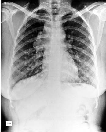
Figure 18-19a. Chest radiographs of a patient with extensive follicular thyroid carcinoma prior to (upper) and after 131-I therapy (lower). With 131-I therapy there was resolution of the large pulmonary metastases, but a tumor in the cervical vertebrae progressed and caused instability of the spine.

Figure 18-19b

Figure 18-19b. Chest radiographs of a patient with extensive follicular thyroid carcinoma prior to (upper) and after 131-I therapy (lower). With 131-I therapy there was resolution of the large pulmonary metastases, but a tumor in the cervical vertebrae progressed and caused instability of the spine.

RADIATION PRECAUTIONS

Before radiation therapy, female patients should be carefully screened for pregnancy and lactation. Confirmed or possible pregnancy constitutes a firm contraindication to therapy because of the risk of damage to the fetus.

A patient who has ingested many milliCuries of 131I can cause serious radiation contamination, and appropriate precautions must be followed. If less than 30 mCi 131I is given, it is permissible to have the patient dispose of urine and feces into general sewage. If amounts of 131I greater than 30 mCi are given, the patient should be kept in a private room in the hospital until less than 30 mCi is retained in the body. Urine can be directly disposed in sewage, or can be collected by the patient and stored in bottles behind protective lead shielding. After physical decay, usually after about 6 weeks, it may be discarded in the sewage. Contaminated bedding and utensils should be stored for 10 half-lives (80 days), thoroughly washed, and monitored for residual contamination before being used again. Alternatively, disposable bedding and utensils may be used.

Table 18-9. Radiation Exposure to Personnel During Care of a Patient Who Has Received 100 mCi 131I

Distance From Source – e.g The Patient Reason for Exposure Rate (mrad/hr) Allowable Duration of Exposure Permitted on Basis of 0.1Rad/Week
1/2 in. Direct handling of therapy dose or urine after therapy 136000 None
1 ft. Giving personal hygiene to treated patient 240 0.5 hr/week
3 ft. Making the bed, mopping the floor 27 5.0 hr/week
9 ft. In chair across the room 3 50.0 hr/week allowable exposure cannot be reached

Personnel caring for a patient who has received 131I therapy are often concerned about exposure to excessive radiation. This is almost never a real problem. Isotope can, at a practical level, only be passed from the patient to another person via saliva or urine. Monitoring by means of a portable counter is important in making certain that no person receives more than an allowable radiation dose from the isotope in the patient’s body. Table 18-9 gives a rough estimate of the amount of radiation received while performing ordinary hospital tasks at various distances from a patient who has received 131I. In general, all ordinary patient care can be performed without hazard. It is best to avoid close contact between hospital personnel and patient during the first 48 hours after therapy because of undue apprehension that may be induced. However, even after doses of up to 100 mCi, normal personal actvities such as eating at the same table, or driving in the same car, carry no risk to others.
The US Nuclear Regulatory Commission has published new guidelines which allow release of patients treated with isotopes from the hospital if the total effective radiation exposure from the treated person to any other individual is not likely to exceed 5mSv (0.5 rads). Grigsby et al (379) found that when using precautions such as those described above in a group of patients given on average about 100mCi 131-I, the exposure to other individuals in their household and to pets did not exceed this level. Recently, guidelines for the optimal radiation protection after treatment with 131-I have been proposed by the American Thyroid Association (380).

Radiation damage from 131-I Therapy

The use of RAI in large doses is not without hazard. The radiation dose delivered to the whole body, the gonads, or bone marrow is usually assumed to be the same as that of the blood. The blood dose depends on the amount of isotope administered; its distribution space and turnover; the degree of heterogeneity of distribution in the tumor; the uptake, synthesis, and secretion of labeled compound by the tumor; and perhaps other variables. The radiation is usually largely due to inorganic iodide, since little protein bound 131I ordinarily appears in the blood. Sometimes tumor destruction is such that much PB131I appears in the blood and can yield a major fraction of the total whole body radiation dose. As a rough estimate, the blood, gonadal, or bone marrow radiation may be assumed to be 0.3 -1.5 rads/mCi 131I administered (381), or 45-150 rads per treatment with 100 mCi. The genetic risks are discussed in Chapter 11 and are not reviewed here. Ordinarily, when 131I therapy is needed for carcinoma, the necessity of treating the patient outweighs the risks of genetic damage.

Various unwanted effects of radiation may occur in patients receiving large doses of 131I. Mild radiation sickness is seen. Metastatic deposits or surrounding tissues may become painful over 2-4 weeks from radiation-induced inflammation. Damage to the salivary glands can cause sialadenitis, and xerostomia, and can lead to loss of teeth (382). Increasing salivary flow following treatment is partially protective. Ovarian function is often temporarily suppressed (383), and if there are pelvic metastases that collect 131I, the gonads may receive a sterilizing dose. Sperm count may be reduced for months (384). Leukemia occurs with increased frequency in patients who have received large doses of 131I (usually>600 mCi) for cancer (217). Transient or permanent alterations in liver function and lymphoma of the parotid gland have been reported as possible sequelae (385). Pulmonary fibrosis has occurred in patients with functioning lung metastases who have received unusually large doses or who have very active metastases (386). Leukopenia, thrombocytopenia, and anemia are encountered with accumulating doses. A mild effect on the bone marrow is seen with each therapeutic dose, and after several hundred milliCuries, aplastic anemia may develop (387). The hemoglobin level, white cell count, differential count, and platelets should be monitored periodically in order to judge recovery of the marrow between treatments and to prevent excess total radiation damage to the marrow. Large radiation doses may cause transient swelling of metastasis in the brain or spinal canal.

Lin et al (388) recently reviewed pregnancies following 131I treatment of well differentiated thyroid carcinoma among a group of 58 pregnant women and found no evidence of demonstrable adverse effects, but suggest that it would be wise to avoid pregnancy during the first six months after the last administration of 131I. With the exception of possibly increased rate of miscarriages, no other adverse effect of radioiodine has been found on the outcome of 2113 pregnancies after radioiodine treatment and on their offspring (318).

Two special complications need be noted. Occasionally withdrawal of hormone suppression, in preparation for isotope therapy, leads to rapid growth of the tumor, and reinstitution may not seem to return the patient to the prior condition. Special care should be taken if metastases are present in areas such as brain or spinal column, where growth could cause serious sequelae. Glucocorticoids are occasionally given prophylactically in an effort to prevent tumor swelling in this situation .
RAI was introduced into the treatment of thyroid carcinoma with the hope that it would be a panacea for this disease. Unfortunately, the results have not been universally beneficial. Most tumors in children appear to be treatable, and among adults 80-90% of metastatic carcinomas accumulate sufficient 131I to warrant a serious therapeutic trial. Patients who harbor this form of the disease are fortunate, since 131I may totally eradicate the metastases. Even multiple pulmonary metastases occasionally disappear after 131Itherapy (Fig. 18-19a, above). The final value of 131I therapy has been difficult to define, largely because of a lack of controlled series and because of other treatment (especially thyroid hormone) given at the same time (387). Mazzaferri (232) found that 131I ablation and therapy significantly improved the prognosis in papillary cancer by decreasing recurrences. Varma et al. (389) found that RAI treatment had no effect on the survival of persons under age 40 but did lower the death rate of patients over age 40. Leeper (390) concluded that 131I treatment appears clearly to benefit patients under 40 years of age with papillary cancer, but the course of this cancer in older patients is rarely affected; follicular cancers in older patients are treatable, and survival is prolonged even if the disease is not eradicated. Soft tissue lesions, especially of the lung and mediastinum, respond best to 131I. Osseous lesions are often highly functional but are infrequently totally destroyed by 131I (Fig. 18-9b) (391). Lesions detected on whole body scans, with negative bone X-rays, are most likely to be cured. In another report, 59 of 400 patients were considered candidates for 131I therapy after using antithyroid drugs or TSH to stimulate uptake. Of these, 61% with metastatic disease were benefited (362). The follicular, papillary, and mixed cancers responded equally well. Numerous reports indicate that ablation of metastasis with 131I is associated with a better prognosis than failure to ablate, but obviously this outcome may relate to the histologic nature and function of the lesion rather than to the therapy per se.

Disseminated pulmonary metastasis can sometimes be eradicated by 131I, but radiation pneumonitis or fibrosis may be produced and may be fatal (321,386). On first observation of pulmonary metastases, this therapy should be considered, but no more than 75 mCi should ever be deposited in the lungs in one treatment. Progress of the lesion and pulmonary function should be carefully evaluated before and be-tween treatments (389,391,392-394). Occasionally patients present with locally advanced papillary thyroid cancer which is not surgically resectable. In some instances preoperative treatment with radioactive iodide sufficiently reduces the extent of the lesion to allow subsequent definitive surgery (395). One of the most informative study regarding the effects and limitation of 131-I therapy is given by the series of the Institut Goustave-Roussy periodically updated by Schlumberger et al (396). In their most recent publication the authors report on 444 patients treated with 131-I for distant metatsases and the results identified three groups of patients with different outcome after therapy: a group very likely to be cured after a few courses of RAI, represented by young patients with micronodular disease, usually in the lung; a second group whose metatsases can be stabilized but not cured after more than 600 mCi as cumulative doses and a third group including older patients with macronodular disease, partuclarly in the bones, who do not responde to RAI and progress rapidly to the exitus. It is apparent from this study that, continuing RAI therapy after 600 mCi usually has no benefit.

FOLLOW-UP OF CANCER PATIENTS: THE SERUM TG ASSAY

After initial ablation patients are given TSH-suppressive doses of l-thyroxine. This therapy as dual aims: to replace the thyroid hormone function and to inhibit the growth of potential residual disease..Two-three months later serum TSH, free thyroid hormones and Tg concentrations are measured during l-thyroxine treatment. The results of these tests will disclose whether the l-thyroxine dose is adequate in suppressing TSH levels without inducing hyperthyroidism, but will give little information on whether the patient is in remission.
To this purpose, the most informative follow-up period is at 6-12 minths after initial treatment, when the ablative dose of radioiodine should have exerted its effect. At this point, the patients have been already attributed a risk estimate based on the results of the post-ablative WBS and serum Tg measurement (397,398). This information is important at the moment of the new evaluation. If uptake was seen outside the thyroid bed on the post-therapy WBS, the patient must be considered in high risk of recurrence or persistent disease, while if no uptake was seen outside the thyroid bed on the 131-I post-therapy scan, the Patient is considered at low-risk. The 6-12 month control, consists of careful neck ultrasound to detect the lymph node status and serum Tg levels (with negative anti-Tg) after stimulation with exogenous (better) or endogenous TSH. These tests are considered by many authors as sufficient to confirm complete remission (negative US and undetectable stimulated Tg). Other advocate the usefulness of a diagnostic WBS with radioiodine, aimed to ensure that thyroid ablation has been successful and to search for foci of 131-I uptake outside the thyroid bed. However, two independent studies (357,399) have shown that this diagnostic WBS are almost always negative (and sometimes false negative) thus adding very little information to that given by neck US and serum Tg measurement. Indeed, the result of serum Tg measurement is the more sensitive predictor of complete remission or persistent disease (provided that anti-Tg autoantibodies are negative) (400-404). Nearly all patients with local or distant disease have detectable or elevated serum Tg levels, while patients in stable remission have undetectable serum Tg concentrations. Compared to serum Tg measurement, the yield of the diagnostic 131-I WBS is lower. A significant proportion of patients may have elevation of serum Tg in the presence of falsely negative diagnostic WBS. A retrospective study by Cailleaux et al. (357) has shown that when serum Tg off therapy is undetectable, routine diagnostic WBS does not add any further in¬formation on the clinical status of the patient. Similar results have been obtained at the Department of Endocrinology, University of Pisa, (399) in a retrospective series of 315 patients who had undetectable serum Tg off l-thyroxine at the time of the first control after thyroid ablation. None of these patients had evidence of disease activity at WBS, and 99.4% were in complete and stable remission after 12 years of follow-up. Only two patients (0.6%) had recurrence of lymph node metastases which were treated with radioiodine therapy. Based on these studies it is possible that in the future the need for 131-I scanning may be dictated by the results of serum TG during hypothyroidism or by rhTSH-stimulation (342).
After this follow up, low-risk patients (those with an undetectable stimulated serum Tg, negative neck US and negative WBS, when performed) are considered as cured and may be followed with periodic serum Tg measurement during l-thyroxine therapy. Thyroxine therapy may be decreased to maintain a low but not suppressed serum TSH concentration (0.1-0.4 µU/ml). The risk of recurrence is in fact so low in these patients (representing more than 80% of the total) that overdosage of l-thyroxine is unjustified.

As noted the problem of antibody interference in the TG assay makes this test unreliable in 10-15% of patients. However another aspect of the antibodies should be remembered. If patients are free of thyroid cancer and the thyroid has been ablated, it appears that the antigenic stimulation necessary to maintain an anti-TG titer is gradually lost, and these antibodies disappear with a 3-6 year half-life (302). Thus, the level of anti.Tg antibodies may be used as a surrogate marker of disease. Other approaches, including the search of Tg mRNA in the blood, have been proposed, but none has entered clinical practice. Fugazzola et al (405) point out that the combination of TG RIA and TG mRNA assay offer better positive and negative predictive value than TG alone. In some studies TGmRNA analysis has proven much less reliable than serum TG assay (406).

In high risk patients, even if considered cured, suppressive doses of l-thyroxine may be continued for some years, because the risk of relapse is greater. Pujol et al evaluated a series of patients over an average of 95 months and compared those who had TSH values constantly under 0.05 mU/l to those who had all TSH values greater than 1mU/l. A lesser degree of TSH suppression was associated with an increased incidence of relapse, with a shorter average relapse-free survival (407). This observation was not sustained in another study (408). The objective of suppressive therapy in these patients should be to attain a serum TSH level of 0.1 µU/ml or less with normal free T3. In this situation, side effects such as osteoporosis, are not observed (409). Clinical and biochemical evaluation is performed annually. If serum Tg becomes detectable during follow-up, the patient should be evaluated for the presence of disease by neck us, first, and by other imaging if neck ultrasound is negative. Some authors prefer to avoid this procedure and give directly a therapeutic dose of 131-I followed by a post-therapy scan. In the absence of uptake after therapeutic doses of 131-I, any further administration of 131-I is not justified, and the site of Tg production should be searched for by other imaging techniques. If 131-I thyroid ablation has not been performed or if the patient has undergone only partial thyroid surgery (subtotal or lobectomy), follow-up should consist of clinical and ultrasound examination and serum Tg measurement. However, in this case, the sensitivity and specificity of serum Tg assay is reduced. In such patients, ant suspicious of persistent or recurrence disease should prompt to complete the initial treatment with completion thyroidectomy and/or radioiodine ablation.

During follow-up, patients may develop isolated metastases that can be approached surgically. Osseous metastases, especially from follicular cancer, may require radiotherapy or operative procedures for stabilization. Progressive growth of soft tissue or osseous metastases that are not amenable to thyroid hormone, 131I therapy, or radiotherapy should lead to consideration of systemic therapies.

UNDIFFERENTIATED CARCINOMAS

Undifferentiated carcinomas, if operable, may be treated by thyroidectomy and resection of all involved tissue in the neck. 5-10% can apparently be completely resected, and the patient can have a discrete survival, although they are never cured. Prophylactic X-radiation (5,000 rads) should be given postoperatively in all cases.. If the lesion is obviously inoperable, X-ray therapy should be the initial treatment. In some instances, the rapidly growing tumors melt almost completely for a time after X-ray therapy, giving the pa¬tient a gratifying respite from the disease.

Some anaplastic tumors have been treated with alkylating agents, and rarely, a rapidly proliferating and highly undifferentiated tumor has responded with a temporary regression. Chemotherapy is discussed below. Kim and Leeper (410) reported partial results combining low dose adriamycin (10 mg/m2) and hyperfractionated radiotherapy (160 rads twice daily) to 5760 rads. Time of survival was prolonged and local recurrences largely prevented. There has been no confirmation of this study, unfortunately.

A prospective protocol combining surgery, chemotherapy (CT),and hyperfractionated accelerated radiotherapy (RT) was employed in anaplastic thyroid carcinoma. The main toxicity was hematologic. High long-term survival was obtained when RT-CT was given after complete surgery. This protocol avoided local tumor progression, and death was mainly caused by distant metastases (411). The clinical behaviour and therapy of anaplastic carcinoma have recently been reviewed by K. Ain (412).
Despite any combination of therapies, anaplastic thyroid cancer remains one of the most aggressive human cancer, with 80% of the patients dying within 2 6 months and the remaining within 2 years. Novel treatment regimens are badly needed for this tumor.

Lymphomas

Once diagnosed, patients should be staged by chest X-ray, chest and abdominal CAT scans, bone marrow biopsy, and gallium scan. In 20-30% of patients, the lymphoma will be confined within the thyroid gland. Lymph nodes are involved in approximately 60% of the patients, and perithyroidal invasion is present in about half. When patients have undergone appropriate staging procedures, 20-30% will have Stage IIIE (lymphoma present in nodes below the diaphragm) or Stage IVE (distant extranodal metastases) disease. The selection of appropriate therapy requires prior staging of the patient. As these tumors are radiosensitive, external radiation therapy is a satisfactory treatment for Stage I-E (local disease only, no nodal involvement) and has been used for Stage II-E (nodal involvement above the diaphragm only) disease. The dose to the neck is usually 4,000 rads (40 Gy) over 4-5 weeks (248). The radiation port should include the mediastinum even in the absence of clinical involvement. When evaluating the long-term survival of patients with Stage I and Stage II disease, Souhami et al. found that of seven patients treated with radiation to the neck only, five died within 5 years, whereas in five patients receiving radiation to the neck and mediastinum, there were no deaths (249). Patients with unfavorable prognostic fac¬tors such as age >60yr, large mass or tumor necrosis, may be given chemo-therapy after radiotherapy. In radiation-treated patients with local extension or with malignant nodes, the survival after surgery is unaffected by the extent of surgery even when surgery was limited to a biopsy. With obvious extension or nodal involvement, surgery should be limited to obtaining an adequate diagnostic specimen since an attempt at complete excision may damage surrounding structures without improving survival. There is a suggestion that total thyroidectomy may improve the prognosis in patients with intrathyroidal disease only (240).

There is a growing tendency to use combination chemotherapy as the initial definitive treatment for Stage IE and IIE thyroid lymphoma, rather than radiation.. Programs may combine cyclophosphamide, adriamycin, vincristine, and prednisolone ("CHOP"). This approach may increase cure above the 30-50% found with radiotherapy alone (413). Currently most patients with Stage II-E, and patients with Stage III-E or Stage IV-E disease, as well as those who relapse after radiation therapy, are treated with chemotherapy, given every 3-4 weeks for 3-6 cycles, prior to radiation therapy if combined with that treatment. Although previous series report overall 5-year survivals of about 50%, certain subgroups have a more favorable prognosis. With appro¬priate staging to exclude Stage III-E and Stage IV-E patients, the Stanford group has reported a 3-year survival of 83% and at 3 years 75% of their StageI-E and Stage II-E patients had no evidence of disease (414). Matsuzuka et al (415) used both radiation therapy and six courses of "CHOP" chemotherapy, and report 100% survival for 8 years in a group of patients who received this treatment.

RADIATION THERAPY

The general indications for radiation therapy are given in Table 18-11. Other sources should be reviewed for details of the port, dose, and methods of irradiation.
Studies by Tubiana et al (416), Simpson (283), and Riccabona (417) have clearly established the efficacy of radiotherapy in all types of thyroid cancer. Although its therapeutic use is now well accepted, its prophylactic use (e.g., in papillary or follicular lesions with possible residual disease, or in papillary or follicular Stage III lesions after RAI ablation) is controversial. Currently, we would first treat all papillary or follicular invasive or possibly metastatic tumors with RAI. In patients under age 55, it is not clear that x-ray therapy should be added. Over age 55, 131I therapy should be followed by x-ray treatment for Stage III or potentially Stage III lesions, and any recurrence or metastasis not responding to 131I may also be treated (418).

The efficacy of external radiation therapy in the management of thyroid malignancy has been reviewed by Brierley et al, Lin et al, Farahati et al, and Tsang et al. (419-422). Brierley, recommends X-ray therapy in addition to 131I therapy in patients with papillary or follicular cancer who have probable or definitive microscopic residual disease. They gave 30 to 50 Gy over four weeks with the spinal cord dose less than 80% of the total. Radiotherapy was also advised for treatment of anaplastic thyroid carcinoma, in an attempt to achieve local control, using hyperfractionation, possibly with the addition of doxorubicin. Radiotherapy was used for treatment of lymphomas, with a total dose of 35 – 40 Gy given over four weeks, and was typically preceded by the use of CHOP (cyclophosphamide, doxorubicin, vincristine, and prednisone) delivered every three to four weeks in three to six cycles prior to the radiotherapy. Cause-specific survival was 82% inpatients who received combined therapy and 63% in those who got X-ray alone. Combined therapies were given to all patients except those with small bulk disease under 3 cm in size confined to the thyroid, who received radiation alone. Lin et al. (420) evaluated 699 patients with differentiated thyroid cancer who received external radiotherapy after surgery, compared to 172 who did not, and found no evidence that radiotherapy improved survival rate of patients with well differentiated thyroid cancer. These were patients with either neck nodes or local invasion.There were 172 patients who received no radiotherapy versus 32 who did. The patients were in clinical stages 2 and 3 at the time of treatment. Farahati et al (421) looked at patients with parathyroidal tumor invasion and found that adjuvant external radiotherapy improved recurrence-free survival in patients over 40 years of age. Benefit was confined to patients with papillary thyroid carcinoma. Tsang et al (422) also confirmed the benefit of radiotherapy in patients with papillary tumors who have microscopic or macroscopic residual disease.

Table 18-11. Indications for Radiation Therapy
Tumor Stage Treatment(15-20 MV Electrons or Co-60)
Papillary or Follicular Invasive, under age 50-55 Treat if invasive disease is thought not to be destroyed or if neck recurrence is proven present after 131-I. Dose is 4500-500 rads.
Invasive or possible residual, over age 50-55   5000 rads* to thyroid bed after RAI
Recurrent, any age   5000* rads to thyroid bed if RAI treatment is thought not to be definitive
Isolated lesion in bone   5000-6000 rads, as required for symptoms after RAI treatment
Medullary Stage III 4000-5000* rads to thyroid bed
Abnormal or increasing CT   5000* rads to mantle
Recurrent tumor   5000-6000* rads to thyroid bed
Isolated metastasis   5000-6000* rads for symptoms
Lymphoma Stage I-E, possibly II-E Probably chemotherapy used first, 5000 rads ** to thyroid and mantle sometimes follows
Anaplast All 4500-5500 rads** to thyroid and mantle

Note- Spinal cord dose not to exceed *3000 or ** 3500 rads.

In a recent study (423), Hurthle cell carcinoma of the thyroid gland was found to be a radiosensitive tumor. Adjuvant radiation therapy was successful in preventing recurrence in 4 of 5 patients. Salvage radiation therapy was successful in 3 of 5 patients treated with external beam radiation therapy. Palliative radiation therapy provided sustained symptomatic relief at 67% of irradiated sites. Radiation therapy may provide palliative relief from symptomatic metastases, control recurrent tumors, and prevent recurrence of advanced resected tumors.

The indications for MTC have been described above. Stage I-E lymphomas may be treated by chemotherapy or radiotherapy. Anaplastic lesions are currently given radiotherapy after operation. It is possible that chemotherapy should be used instead of or in addition to radiotherapy in these lesions, but studies are needed to establish this point.

The exact dose must be individually determined, but usually the maximal dose is 5,000-6,000 rads, using ortho-or megavoltage, and a fractionated technique over several weeks. Dosage must be planned to assure that the spinal cord receives less than 3500 rads in order to avoid myelopathy.

SYSTEMIC THERAPY FOR METASTATIC DISEASE

Sporadic experience indicates that bleomycin (424,425), adriamycin (426), vinblastine (427), methotrexate (426), cisplatinum and other agents (428) may have value in treating disseminated thyroid tumors. However, none of these drug has given more than palliation. With the advent of new molecules targeting specific metabolic pathways involve in the generation and maintainance of thyroid cancer growth, the so call “tyrosine kinase inhibitor’, chemotherapy has been almost universally abandoned. Recent ATA guidelines (39) states that whenever systemic therapy is needed, the patient should be offered the possibility to be entered in clinical trials using these new molecules. Metastatic papillary or follicular tumors refractory to conventional therapy grow slowly and systemic therapy is not indicated until the tumor is clearly growing progressively despite hormone suppression. The same apply to the treatment of MTC (264).

RE-DIFFERENTIATION’ THERAPY

RETINOIC ACID- Experimental data from in vitro studies suggest that retinoic acid can inducere-differentiation of thyroid cancer cells with regain of iodide concentrating ability. Simon et al (429) studied 28 patients with differentiated thyroid cancer using 1.5 mg/kg retinoic acid per day for five weeks. Iodide uptake increased in eight of the patients and thyroglobulin increased in 63%. Thus retinoid appeared to reinduce iodide uptake in half of the treated patients by redifferentiation. They gave 13-cis-retinoic acid. Side effects occurred in half of the patients but were generally well tolerated. A common side effect was dryness of the skin and mucosal surfaces. The reinduction of 131I uptake allowed radioiodine therapy in several of the patients. The effect on tumor size was uncertain. Retinoic acid redifferentiation therapy has recently been reviewed by Schmutzler and Kohrle (430). They note that, of twenty documented retinoic acid treated patients, at least eight had exhibited a decrease or stabilization in tumor size, and in serum TG levels, in addition to enhanced radioiodide transport. However, this relatively positive review seems enthusiastic, considering the small evidence of improvement observed in most cases. Clearly the ben¬efits of this experimental treatment are as yet uncertain, but it is nevertheless an exciting observation that retinoic acid can have an apparent beneficial redifferentiating effect on some thyroid tumors. Histone deactylase inhibitors also have been found to restore in part the function of NIS, TPO and TG in thyroid tumor cells in vitro (431), and valproic acid also has an action to restore NIS function, probably via the same mechanism (432).
PPARgamma agonists-In vitro studies show that PPAR gamma agonists can slow thyroid tumor cell growth and induce apoptosis (433). Results of clinical trials are awaited.

ONYX-015is an E1B deleted adenovirus that replicates in cells with impaired p53 function. p53 is commonly inactivated in anaplastic thyroid cancers. In vitro studies demonstrate that this virus induced cell death in in vitro trials in anaplastic cancer cell lines, and synergized with treatment with doxorubicin and pacitaxel (434).

Adenoviral vectors producing tk, IL-2, IL-12, and GM-CSF in a cell specific manner are currently under study in animals with encouraging results. Studies in humans are so far very limited, but the methods appear to be safe and effective, especially with the immunomodulator IL-12 (434,435).
A variety of ideas on possible treatments for thyroid cancer have been reviewed by Braga-Basaria and Ringel (436).

TYROSINE KINASE INHIBITORS

Several oncogenes involved in thyroid cancer progression are tyrosine kinase receptor. Molecules that block kinase activity at distal steps in the MAP kinase pathway are logical candidate drugs for thyroid cancer. TKI being tested against differentiated thyroid cancer in clinical trials include motesanib diphosphate, axitinib, gefitinib, sorafenib and sunitinib. None of these is specific for one oncogene protein but they target several TK receptors and pro-angiogenic growth factor receptors. The results of phase II-III clinical trials conducted so far are promising with partial responses ranging 14- 32% and stable disease 50-67% (437). In addition, very recently one of this drug, vandetanib, have been approved for the treatment of progressive, metastatic MTC, based in clear evidence of beneficial effects in clinical trials (438,439). All together, the preliminary results of these trials are promising and indicate that target therapy might become the first line treatment of metastatic refractory thyroid cancer patients in the near future.
Sunitinib is a multitargeted tyrosine kinase inhibitor (TKI) that is is currently approved for the therapy of renal cell carcinoma and gastrointestinal stromal tumor. Some eficacy was found in both differentiated thyoid cancer and medullary cancer in Phase II studies, with partial response rates of 13-30%.

Sorafenib is a similar agent, also achieving significant levels of partial responses and stabilized disease. Several Phase II stuies have been reported, and a Phase III trial has been completed.

Vandetanib has been studies in Phase II trials and in a randomized, placebo-controlled phase III study in patients with both hereditary and sporadic MTC, using vandetanib 300 mg or placebo. A 54% reduction in the risk of progression was observed and also objective tumor responses in 45% of patients. The FDA approved it for the treatment of symptomatic or progressive medullary thyroid cancer in patients with unresectable locally advanced or metastatic disease. Motesanib, Axitinib, Pazopanib, Lenvatinib and several other related TKIs that more specifically block VEGF receptors are in Phase II or III trials.

These agents appear to block progression and in some studies decrease tumor bulk, but are not specifically cytotoxic, and tend to loose efficacy over months, presumably by mutational events in the tumor. Future studies will surely investigate serial or combined treatments, and use with other possibly cytotoxic agents.

Progress in the last few years has reinvigerated therapy for these difficult tumors, but comes with a price. Since TRK Iinhibitors affect many sites in the body, side effects are very common and often serious, but generally tolerable The major side-effects include hypertension in 20-50% of cases, diarrhea in 37-77%, fatigue in 40-50%, weight loss in 20-60%, frequent nausea, skin sensitivity, and very frequent rashes, depending on the study, dosage,and drug (439). Some of the agents induce hypothyroidism during therapy.

Most reports available are the result of clinical investigations, often by consortia made up of physicians specializing in therapy of thyroid cancer, and patients are generally advised to enter such a trial if a newer agent is used.. However these agents gradually spread into more generaized usage, by therapists who may not have extensive experience. The MD Anderson group has published suggested institutional guidelines to help provide safeguards for non-investigative use of TKIs(440).

Presentation of this chapter is supported in part by Genzyme, the makers of rhTSH (THYROGEN)


References for cancer chapter

1. Hedinger, C, Dillwyn Williams, E, Sobin, L 1989. The WHO histological classification of thyroid tumors. A commentary on the second edition. Cancer 63:908-911
2.Namba, H, Matsuo, K, Fagin, J 1990 Clonal composition of benign and malignant human thyroid tumors. J Clin In 86:.
3.. Parma, J, Duprez, L, Van Sande, J, et al. 1993. Somatic mutations in the thyrotropin receptor gene cause hyperfunctioning thyroid adenomas. Nature 365:649-651
4.. Tonacchera, M, Chiovato, L, Pinchera, A, et al. 1998 Hyperfunctioning thyroid nodules in toxic multinodular goiter share activating thyrotropin receptor mutations with solitary toxic adenoma. J Clin Endocrinol Metab 83:492-8
5. Coclet, J, Foureau, F, Ketelbant, P, Galand, P, Dumont, J 1989. Cell population kinetics in dog and human adult thyroid. Clinical Endocrinol 31:655-665
6. Viglietto, G, Chiappetta, G, Martinez-Tello, F J, et al. 1995 RET/PTC oncogene activation is an early event in thyroid carcinogenesis. Oncogene 11:1207-10
7. Hamburger, J I 1980; Evolution of toxicity in solitary nontoxic autonomously functioning thyroid nodules. J Clin Endocrinol Metab 50:1089-1093
8. Silverstein, G, Burke, G, Cogan, R 1967. The natural history of the auto-nomous hyperfunctioning thyroid nodule. Ann Intern Med 67:539
9. Evered, D, Clark, F, Peterson, V 1974. Thyroid function in euthyroid subjects with autonomous thyroid nodules. Clin Endocrinol 3:149
10. Horst, W, Rosler, H, Schneider, C, Labhart, A 1967. 306 cases of toxic adenoma. Clinical aspects; findings in radioiodine diagnostics; radiochromatography and histology; results of 131I and surgical treatment. J Nucl Med 8:515
11.Parma, J, Duprez, L, Van Sande, J, et al. 1993. Somatic mutations in the thyrotropin receptor gene cause hyperfunctioning thyroid adenomas. Nature 365:649-651
12.Suarez, H, du Villard, J, Caillou, B, Schlumberger, M, Parmentier, C, Monier, M 1991. Gsp mutations in human thyroid tumors. Oncogene 6:677-679
13. see 10
13a. DeGroot, L 1970. Lack of iodide trapping in "cold" thyroid nodules. Acta Endocrinol Panam 1:27
14. Field, J, Larsen, P, Yamashita, K, Mashiter, K, Dekke, A 1973. Demonstration of iodide transport defect but normal iodide organification in nonfunctioning nodules of human thyroid glands. J Clin Invest 52:2404
15. Fragu, P, Nataf, B 1977. Human thyroid peroxidase activity in benign and malign thyroid disorders. J Clin Endocrinol Metab 45:1089
16. Demeester-Mirkine, N, Van Sande, J, Corvilain, H, Dumont, J 1975. Benign thyroid nodule with normal iodide trap and defective organification. J Clin Endocrinol Metab 41:1169
17. Burke, G, Szabo, M 1972. Dissociation of in vivo and in vitro "autonomy" in hyperfunctioning thyroid nodules. J Clin Endocrinol Metab 35:199
18. Sande Van, J, Mockel, J, Boeynaems, J, Dor, P, Andry, G, Dumont, J 1980. Regulation of cyclic nucleotide and prostaglandin formation in normal human thyroid tissue and in autonomous nodules. J Clin Endocrinol Metab 50:776
19. Smanik, P, Ryu, K-Y, Thel, K, Mazzaferri, E, Jhiang, S 1997. Expression; exon-intron organization; and chromosome mapping of the human sodium iodide symporter. Endocrinology 138:3555-8
20.Arturi, F, Russo, D, Schlumberger, M, et al. 1998. Iodine symporter gene expression in human thyroid tumors. J Clin Endocrinol Metab 83:2493-96
21. Thomas, C J, Buckwalter, J, Staab, E, Kerr, C 1976. Evaluation of dominant thyroid masses. Ann Surg 183:464
22. Sipple, J H 1984 Multiple endocrine neoplasia type 2 syndromes: historical perspectives. Henry Ford Hosp Med J 32:219-21
23.Schimke, R, Hartmann, W, Prout, T, Rimoin, D 1968. Syndrome of bilateral pheochromocytoma; medullary thyroid carcinoma; and multiple neuromas. N Engl J Med 279:1
24.Sapira, J, Altman, M, Vandyk, K, Shapiro, A 1965. Bilateral adrenal pheo-chromocytoma and medullary thyroid carcinoma. N Engl J Med 273:140
25. Duffy, B J, Fitzgerald, P 1950. Cancer of the thyroid in children. A report of twenty-eight cases. J Clin Endocrinol 10:1296
26. Clark, D 1955. Association of irradiation with cancer of the thyroid in children and adolescents. JAMA 159:1007
27. Modan, B, Ron, E, Werner, A 1977. Thyroid cancer following scalp irradiation. Therapeutic Radiology 123:741
28 DeGroot, L, Frohman, L, Kaplan, E, Refetoff, S e 1977. Radiation-Associated Thyroid Carcinoma. New York Grune & Stratton:539 pages
29. Refetoff, S, Harrison, J, Karanfilski, B, Kaplan, E, DeGroot, L, Bekerman, C 1975. Continuing occurrence of thyroid carcinoma after irradiation to the neck in infancy and childhood. N Engl J Med 292:171
30 DeGroot, L, Paloyan, E 1973. Thyroid carcinoma and radiation. A Chicago endemic. JAMA 225:487
31. Sokal, J 1959. The problem of malignancy in nodular goiter -- recapitulation and a challenge. JAMA 170:61
32. Veith, F, Brooks, J, Grigsby, W, Selenkow, H 1964. The nodular thyroid gland and cancer. N Engl J Med 270:431
32.1 Frates MC, Benson CB, Doubilet PM, Kunreuther E, Contreras M, Cibas ES, Orcutt J, Moore FD Jr, Larsen PR, Marqusee E, Alexander EK. Prevalence and distribution of carcinoma in patients with solitary and multiple thyroid nodules on sonography. J Clin Endocrinol Metab. 2006 Sep;91(9):3411-7.
33. Hoffman, G, Thompson, N, Heffron, C 1972. The solitary thyroid nodule. Arch Surg 105:379
33.1 Haymart MR, Repplinger DJ, Leverson GE, Elson DF, Sippel RS, Jaume JC, Chen H.Higher serum thyroid stimulating hormone level in thyroid nodule patients is associated with greater risks of differentiated thyroid cancer and advanced tumor stage.J Clin Endocrinol Metab. 2008 Mar;93(3):809-14.
34. Pacini, F, Fontanelli, M, Fugazzola, L, et al. 1994. Routine measurement of serum calcitonin in nodular thyroid diseases allows the preoperative diagnosisof unsuspected sporadic medullary thyroid carcinoma. J Clin Endocrinol Mertab 78:826-9
35. Rieu, M, Lame, M, Richard, A, et al. 1995. Prevalence of sporadic medullary thyroid carcinoma. the importance of routine measurement of serum calcitonin in the diagnostic evaluation of thyroid nodules. Clin Endocrinol (Oxf) 42:453-7
36. Niccoli, P, Wion-Barbot, N, Caron, P, et al. 1997. Interest of routine measurement of serum calcitonin. Study in a large series of thyroidectomized patients. J Clin Endocrinol Metab 82:338-341
37. Gagel, R 1997. The goitrous patient with an elevated serum calcitonin -- what to do? J Clin Endocrinol Metab 82:335
38. Hamlin, E, Vickery, A 1956. Needle biopsy of the thyroid gland. N Engl J Med 254:742
39. Walfish, P, Hazani, E, Strawbridge, H, Miskin, M, Rosen, B 1977. Combined ultrasound and needle aspiration cytology in the assessment and management of hypofunctioning thyroid nodule. Ann Intern Med 87:270
40. Gershengorn, M, McClung, M, Chu, W, Hanson, T, Weintraub, B, Robbins, J 1977. Fine-needle aspiration cytology in the preoperative diagnosis of thyroid nodules. Ann Intern Med 87:265
41.Baloch, Z, Sack, M, Livolsi, V, Gupta, P 1998. Fine-needle aspiration of thyroid. an institutional experience. Thyroid 8:565-9
42. Benvenga, S, Bartolone, L, Squadrito, S, Trimarchi, F 1997. Thyroid hormone autoantibodies elicited by diagnostic fine needle biopsy. J Clin Endocrinol Metab 82:4217-4223
43 Wilems, J-S, Lowhagen, T 1981. Fine needle aspiration cytology in thyroid disease. . Clin Endocrinol Metab 2:247
44 Gharib, H, Goellner, J, Johnson, D 1993. Fine needle aspiration cytology of the thyroid. A 12-year experience with 11;000 biopsies. Clin Lab Med 13:699-709
44.1 Tee YY, Lowe AJ, Brand CA, Judson RT.Fine-needle aspiration may miss a third of all malignancy in palpable thyroid nodules: a comprehensive literature review.Ann Surg. 2007 Nov;246(5):714-20.
45. Pacini, F, Fugazzola, L, Lippi, F, et al. 1992 Detection of thyroglobulin in fine needle aspirates of nonthyroidal neck masses. a clue to the diagnosis of metastatic thyroid cancer. J Clin Endocrinol Metab 74:1401-4
46. Chow, L S, Gharib, H, Goellner, J R, van Heerden, J A 2001 Nondiagnostic thyroid fine-needle aspiration cytology: management dilemmas. Thyroid 11:1147-51
47. Khurana, K, Richards, V, Chopra, P, Izquierdo, R, Rubens, D, Mesonero, C 1998. The role of ultrasonography-guided fine-needle aspiration biopsy in the management of nonpalpable and palpable thyroid nodules. Thyroid 8:511
48. Erdogan, M, Kamel, N, Aras, D, Akdogan, A, Baskal, N, Erdogan, G 1998. Value of reaspirations in benign nodular thyroid disease. Thyroid 8:1087
49. Bieche, I, Ruffet, E, Zweibaum, A, Vilde, F, Lidereau, R, Franc, B 1997. MUC1 Mucin gene; transcripts; and protein in adenomas and papillary carcinomas of the thyroid. Thyroid 7:725
50 Brousset, P, Chaouche, N, Leprat, F, et al. 1997. Telomerase activity in human thyroid carcinomas originating from the follicular cells. J Clin Endocrinol Metab 82:4214-4216
51 Khoo, M L, Beasley, N J, Ezzat, S, Freeman, J L, Asa, S L 2002 Overexpression of cyclin D1 and underexpression of p27 predict lymph node metastases in papillary thyroid carcinoma. J Clin Endocrinol Metab 87:1814-8
51a. Maruta, J; Hashimoto, H; Yamashita, H; Yamashita, H; Noguchi, S. Diagnostic applicability of dipeptidyl aminopeptidase IV activity in cytological samples for differentiating follicular thyroid carcinoma from follicular adenoma. Arch Surg 139 83-88 2004.
51b. Durand S, Ferraro-Peyret C, Selmi-Ruby S, Paulin C, El Atifi M, Berger F, Berger-Dutrieux N, Decaussin M, Peix JL, Bournaud C, Orgiazzi J, Borson-Chazot F, Rousset B Evaluation of gene expression profiles in thyroid nodule biopsy material to diagnose thyroid cancer. J Clin Endocrinol Metab. 2008 Apr;93(4):1195-202.
51c Shibru D, Hwang J, Khanafshar E, Duh QY, Clark OH, Kebebew E.Does the 3-gene diagnostic assay accurately distinguish benign from malignant thyroid neoplasms?Cancer. 2008 Sep 1;113(5):930-5.
52 Clark, O, Okerlund, M, Cavalieri, R, Greenspan, F 1979. Diagnosis and treatment of thyroid; parathyroid; and thyroglossal duct cysts. J Clin Endocrinol Metab 48:983
52.1 Leboulleux S, Girard E, Rose M, Travagli JP, Sabbah N, Caillou B, Hartl DM, Lassau N, Baudin E, Schlumberger M Ultrasound criteria of malignancy for cervical lymph nodes in patients followed up for differentiated thyroid cancer. J Clin Endocrinol Metab. 2007 Sep;92(9):3590-4
53. Marcocci, C, Vitti, P, Cetani, F, Catalano, F, Concetti, R, Pinchera, A 1991. Thyroid ultrasonography helps to identify patients with diffuse lymphocytic thyroiditis who are prone to develop hypothyroidism. J Clin Endocrinol Metab 72:209-13
54. Kendall, L, Condon, R 1969. Prediction of malignancy in solitary thyroid nodules. Lancet 1:1071
55. Miller, J, Hamburger, J 1965.. The thyroid scintigram. 1. The hot nodule. Radiology 84:66
56. Attie, J 1960. The use of radioactive iodine in the evaluation of thyroid nodules. Surgery 47:611
57. Dische, S 1964. The radioisotope scan applied to the detection of carcinoma in thyroid swellings. Cancer 17:473
58. Fujimoto, Y, Oka, A, Nagataki, S 1972. Occurrence of papillary carcinoma in hyperfunctioning thyroid nodule. Report of a case. Endocrinol Jpn 19:371
59. Scott, M, Crawford, J 1976. Solitary thyroid nodules in childhood. Is the incidence of thyroid carcinoma declining? Pediatrics 58:521
60.Messaris, G, Evangelou, G, Tountas, C 1973. Incidence of carcinoma in cold nodules of the thyroid gland. Surgery 74:447
61. Van den Bruel, A, Maes, A, De Potter, T, et al. 2002 Clinical relevance of thyroid fluorodeoxyglucose-whole body positron emission tomography incidentaloma. J Clin Endocrinol Metab 87:1517-20
62. Hakama, M Berlin; Springer-Verlag Different world thyroid cancer rates; in Hedinger CE (ed). Thyroid Cancer. International Union Against Cancer Monograph Series:Vol 12.
63. Young, J J, Percy, C, Asire, A e 1082 pages Surveillance; Epidemiology; and End Results. Incidence and Mortality Data; 1973-77. National Cancer Institute Monograph 57 NIH Publication No. 81:2330
64. Goldstein, R, Hart, I 1983. Follow-up of solitary autonomous thyroid nodules treated with 131I. New Engl J Med 309:1473
65. Gorman, C, Robertson, J 1978. Radiation dose in the selection of 131I or surgical treatment for toxic thyroid adenoma. Ann Intern Med 89:85
66. Paracchi, A, Ferrari, C, Livraghi, T, et al. 1992. Percutaneous intranodular ethanol injection. A new treatment for autonomous thyroid adenoma. J Endocrinol Invest 15:353-362
67. Lippi, F, Ferrari, C, Manetti, L, et al. 1996. Treatment of solitary autonomous thyroid nodules by percutaneous ethanol injection. results of an Italian multicenter study. The Multicenter Study Group. J Clin Endocrinol Metab 81:3261-3264
68. Monzani, F, Caraccio, N, Goletti, O, et al. S54-8 Treatment of hyperfunctioning thyroid nodules with percutaneous ethanol injection. Eight years' experience. Exp Clin Endocrinol Diabetes 106: Suppl 4
69. Hamburger, J I 1980; Evolution of toxicity in solitary nontoxic autonomously functioning thyroid nodules. J Clin Endocrinol Metab 50:1089-1093
70. McCowen, K, Reed, J, BL., F 1980. The role of thyroid therapy in patients with thyroid cysts. Amer J Med 68:853
71. Treece, G, Georgitis, W, Hofeldt, F 1983 Resolution of recurrent thyroid cysts with tetracycline instillation. Arch Int Med 143: 2285.
72. Monzani, F, Lippi, F, Goletti, O, et al. 1994. Percutaneous aspiration and ethanol sclero-therapy for thyroid cysts. J Clin Endocrinol Metab 78:800-802
73. Zingrillo, M, Torlontano, M, Ghiggi, M, et al. 1996. Percutaneous ethanol injection of large thyroid cystic nodules. Thyroid 6:403-8
74.Del Prete, S, Caraglia, M, Russo, D, et al. 2002 Percutaneous ethanol injection efficacy in the treatment of large symptomatic thyroid cystic nodules: ten-year follow-up of a large series. Thyroid 12:815-21
75. see 70
76. Papini, E, Bacci, V, Panunzi, C, et al. 1993. A prospective randomized trial of levothyroxine suppressive therapy for solitary thyroid nodules. Clin Endocrinol 38:507-513
77. Burch, H 1995. Evaluation and management of the solid thyroid nodule. Endocrinol Metab Clin N Am 24:663-710
78. Mainini, E, Martinelli, I, Morandi, G, Villa, S, Stefani, I, Mazzi, C 1995. Levothyroxine suppressive therapy for solitary thyroid nodule. J Endocrinol Invest 18:796-799
79. Cooper, D 1995. Clinical review 66. Thyroxine suppression therapy for benign nodular disease. J Clin Endocrinol Metab 80:331-334
80. Castro, M R, Caraballo, P J, Morris, J C 2002 Effectiveness of thyroid hormone suppressive therapy in benign solitary thyroid nodules: a meta-analysis. J Clin Endocrinol Metab 87:4154-9
81 Zelmanovits, F, Genro, S, Gross, J 1998. Suppressive therapy with levothyroxine for solitary thyroid nodules. A double-blind controlled clinical study and cumulative meta-analyses. J Clin Endocrinol Metab 83:3881-3885
82. Herms, A, Huysmans, D 1998. Treatment of benign nodular thyroid disease. N Engl J Med 312:601-4
83. Larosa, G, Ippolito, A, Luppo, L, et al. 1996. Cold thyroid nodule reduction with l-thyroxine can be predicted by initial nodule volume and cytological characteristics. J Clin Endocrinol Metab; 81:4385-7
83.a. Sdano MT, Falciglia M, Welge JA, Steward DL. Efficacy of thyroid hormone suppression for benign thyroid nodules: meta-analysis of randomized trials Otolaryngol Head Neck Surg. 2005 Sep;133(3):391-6
84. Wemeau, J L, Caron, P, Schvartz, C, et al. 2002 Effects of thyroid-stimulating hormone suppression with levothyroxine in reducing the volume of solitary thyroid nodules and improving extranodular nonpalpable changes: a randomized, double-blind, placebo-controlled trial by the French Thyroid Research Group. J Clin Endocrinol Metab 87:4928-34
85 see 77
86 Morita, T, Tamai, H, Ohshima, A, et al. 1989. Changes in serum thyroid hormone; thyrotropin and thyroglobulin concentrations during thyroxine therapy in patients with solitary thyroid nodules. J Clin Endocrinol Metab 69:227
87 Woolner, L, Beahrs, O, Black, B, McConahey, W, Keating, F J vol 12 Long term survival rates. in Hedinger Chr E (ed). Thyroid Cancer. International Union Against Cancer: Monograph Series
88. Fogelfeld, L, Wiviott, M, Shore-Freedman, E, et al. 1989. Recurrence of thyroid nodules after surgical removal in patients irradiated in childhood for benign conditions. N Engl J Med 320:835-840
89. Bennedbaek, F, Hegedus, L 1999. Percutaneous ethanol injection therapy in benign solitary solid cold thyroid nodules. A randomized trial comparing one injection with three injections. Thyroid 9:225-233
90. Khurana, K, Labrador, E, Izquierdo, R, Mesonero, C, Pisharodi, L 1999. The role of fine-needle aspiration biopsy in the management of thyroid nodules in children; adolescents; and young adults. A multi-institutional study. Thyroid 9:383
91.Tan, G, Gharib, H 1997. Thyroid incidentalomas. management approaches to nonpalpable nodules discovered incidentally on thyroid imaging. Ann Intern Med 126:226-31
92. Franceschi, S, Boyle, P, Maissonneuve, P, et al. 1993. The epidemiology of thyroid carcinoma. Critical Reviews in Oncogenesis 4:25
93. Parkin, D, Muir, C, Whelan, S, Gao, Y, Fenlay, J, Powell, J Vol. 6 Cancer incidence in five continents. IARC Scientific Publication 120 International Agency for Research on Cancer: Lyon
94 Goodman, M, Yoshizawa, C, Kolonel, L 1988. Descriptive epidemiology of thyroid cancer in Hawaii. Cancer 61:1272
95. Spitz, M, Sider, J, Katz, R, Pollack, E, Newell, G 1988. Ethnic patterns of thyroid cancer incidence in the United States; 1973-1981. Int J Cancer 42:549
95.1. .Davies L, Welch HG Increasing incidence of thyroid cancer in the United States, 1973-2002. JAMA. 2006 May 10;295(18):2164-7.
95.2. Enewold L, Zhu K, Ron E, Marrogi AJ, Stojadinovic A, Peoples GE, Devesa SS.Rising thyroid cancer incidence in the United States by demographic and tumor characteristics, 1980-2005.Cancer Epidemiol Biomarkers Prev. 2009 Mar;18(3):784-91
96. Mortensen, J, Woolner, L, Bennett, W 1955. Gross and microscopic findings in clinically normal thyroid glands. J Clin Endocrinol Metab 15:1270
97. Silverberg, S, Vidone, R 1966. Metastatic tumors in the thyroid. Pac Med & Surg 74:175
98. Hundahl, S A, Fleming, I D, Fremgen, A M, Menck, H R 1998 A National Cancer Data Base report on 53,856 cases of thyroid carcinoma treated in the U.S., 1985-1995 [see commetns]. Cancer 83:2638-48
99. Wahner, H, Cuello, C, Correa, P, Uribe, L, Gaitan, E 1966. Thyroid carcinoma in an endemic goiter area; Cali; Colombia. Am J Med 40:58
100. Belfiore, La Rosa, G, La Porta, G, et al. 1992. Cancer risk in patients with cold thyroid nodules. relevance of iodine intake; sex; age; and multinodularity. Am J Med 93:363
100.1. Sandeep TC, Strachan MW, Reynolds RM, Brewster DH, Scelo G, Pukkala E, Hemminki K, Anderson A, Tracey E, Friis S, McBride ML, Kee-Seng C, Pompe-Kirn V, Kliewer EV, Tonita JM, Jonasson JG, Martos C, Boffetta P, Brennan P. Second primary cancers in thyroid cancer patients: a multinational record linkage study.J Clin Endocrinol Metab. 2006 May;91(5):1819-25
101. Namba, H, Matsuo, K, Fagin, J 1990 Clonal composition of benign and malignant human thyroid tumors. J Clin In 86:.
102. Coclet, J, Foureau, F, Ketelbant, P, Galand, P, Dumont, J 1989. Cell population kinetics in dog and human adult thyroid. Clinical Endocrinol 31:655-665
103. Curran, P, DeGroot, L 1991. The effect of hepatic enzyme-inducing drugs on thyroid hormones and the thyroid gland. Endocrine Rev 12:135-150
104. Namba, H, Gutman, R, Matsuo, K, Alvarez, A, Fagin, J 1990 H-Ras protooncogene mutations in human thyroid neoplasms. J Clin Endocrinol Metab 71: .
105. Namba, H, Rubin, S, Fagin, J 1990. Point mutations of Ras oncogenes are an early event in thyroid tumorigenesis. Molecul Endocrinol 4:1474

106. Franceschi, S, Boyle, P, Maissonneuve, P, et al. 1993. The epidemiology of thyroid carcinoma. Critical Reviews in Oncogenesis 4:25.
107. Parkin, D, Muir, C, Whelan, S, Gao, Y, Fenlay, J, Powell, J Vol. 6 Cancer incidence in five continents. IARC Scientific Publication 120 International Agency for Research on Cancer: Lyon.
108. Goodman, M, Yoshizawa, C, Kolonel, L 1988. Descriptive epidemiology of thyroid cancer in Hawaii. Cancer 61:1272.
109. Spitz, M, Sider, J, Katz, R, Pollack, E, Newell, G 1988. Ethnic patterns of thyroid cancer incidence in the United States; 1973-1981. Int J Cancer 42:549.
110. Mortensen, J, Woolner, L, Bennett, W 1955. Gross and microscopic findings in clinically normal thyroid glands. J Clin Endocrinol Metab 15:1270.
111. Silverberg, S, Vidone, R 1966. Carcinoma of the thyroid in surgical and post-mortem material. Analysis of 300 cases at autopsy and literature review. Ann Surgery 164:291.
112. Hundahl, S A, Fleming, I D, Fremgen, A M, Menck, H R 1998 A National Cancer Data Base report on 53,856 cases of thyroid carcinoma treated in the U.S., 1985-1995 [see commetns]. Cancer 83:2638-48.
113. Wahner, H, Cuello, C, Correa, P, Uribe, L, Gaitan, E 1966. Thyroid carcinoma in an endemic goiter area; Cali; Colombia. Am J Med 40:58.
114. Belfiore, La Rosa, G, La Porta, G, et al. 1992. Cancer risk in patients with cold thyroid nodules. relevance of iodine intake; sex; age; and multinodularity. Am J Med 93:363.
115. Namba, H, Matsuo, K, Fagin, J 1990 Clonal composition of benign and malignant human thyroid tumors. J Clin In 86.
116. Coclet, J, Foureau, F, Ketelbant, P, Galand, P, Dumont, J 1989. Cell population kinetics in dog and human adult thyroid. Clinical Endocrinol 31:655-665.
117. Curran, P, DeGroot, L 1991. The effect of hepatic enzyme-inducing drugs on thyroid hormones and the thyroid gland. Endocrine Rev 12:135-150.
118. Namba, H, Gutman, R, Matsuo, K, Alvarez, A, Fagin, J 1990 H-Ras protooncogene mutations in human thyroid neoplasms. J Clin Endocrinol Metab 71.
119. Namba, H, Rubin, S, Fagin, J 1990. Point mutations of Ras oncogenes are an early event in thyroid tumorigenesis. Molecul Endocrinol 4:1474.
120. Karga, H, Lee, J-K, Vickery, A, Thor, A, Gaz, R, Jameson, J 1991. Ras oncogene mutations in benign and malignant thyroid neoplasms. J Clin Endocrinol Metab 73:832.
121. Santoro, M, Carlomagno, F, Hay, I, et al. 1992. Ret oncogene activation in human thyroid neoplasms is restricted to the papillary cancer subtype. J Clin Invest 89:1517-1522
122. Jhiang, S, Sagartz, J, Tong, Q, et al. 1996. Targeted expression of the ret/PTC1 oncogene induces papillary thyroid carcinomas. Endocrinology 137:375-378.
123. Nikiforova, MN; Kimura, ET; Gandhi, M; Biddinger, PW; Knauf, JA; Basolo, F; Zhu, Z; Giannini, R; Salvatore, G; Fusco, A; Santoro, M; Fagin, JA; Nikiforov, YE. BRAF mutations in thyroid tumors are restricted to papillary carcinomas and anaplastic or poorly differentiated carcinomas arising from papillary carcinomas. J Clin Endocrinol Metab 88 5399-5404 2003.
124. Giannini R, Ugolini C, Lupi C, Proietti A, Elisei R, Salvatore G, Berti P, Materazzi G, Miccoli P, Santoro M, Basolo F. The Heterogeneous Distribution of Distinct Tumor BRAF mutations in Multifocal Papillary J Clin Endocrinol Metab. 2007 Sep;92(9):3511-6
125. Kroff, T, Sarraf, P, Pecciarini, L, et al. 2000. PAX8-PPAR(1 fusion oncogene in human thyroid carcinoma. Science 289:1357-1360
126. Powell, JG; Allard, BL; Sahin, M; Wang, X-L; Wang, X; Hay, ID; Hiddinga, HJ; Deshpande, SS; Kroll, TG; Grebe, SKG; Eberhardt, NL; McIver, B. The PAX8/PPAR( fusion oncoprotein transforms immortalized human thyrocytes through a mechanism probably involving wild-type PPAR inhibition. Oncogene 2003.
127. Marques, A R, Espadinha, C, Catarino, A L, et al. 2002 Expression of PAX8-PPAR gamma 1 rearrangements in both follicular thyroid carcinomas and adenomas. J Clin Endocrinol Metab 87:3947-52
128. Vivaldi, A, Pacini, F, Martini, F, et al. 2003 Simian virus 40-like sequences from early and late regions in human thyroid tumors of different histotypes. J Clin Endocrinol Metab 88:892-9
129. Puzianowska-Kuznicka, M, Krystyniak, A, Madej, A, Cheng, S Y, Nauman, J 2002 Functionally impaired TR mutants are present in thyroid papillary cancer. J Clin Endocrinol Metab 87:1120-8
130. Takano, T; Miyauchi, A; Yoshida, H; Nakata, Y; Kuma, K; Amino, N. Expression of TR(1 mRNAs with functionally impaired mutations is rare in thyroid papillary carcinoma. J Clin Endocrinol Metab. 88:3447-3449, 2003.
131. Liu, R T, Huang, C C, You, H L, et al. 2002 Overexpression of tumor susceptibility gene TSG101 in human papillary thyroid carcinomas. Oncogene 21:4830-7.
132. Parma, J, Duprez, L, Van Sande, J, et al. 1993. Somatic mutations in the thyrotropin receptor gene cause hyperfunctioning thyroid adenomas. Nature 365:649-651
133. Suarez, H, du Villard, J, Caillou, B, Schlumberger, M, Parmentier, C, Monier, M 1991. Gsp mutations in human thyroid tumors. Oncogene 6:677-679
134. Matsuo, K, Friedman, E, Gejman, P, Fagin, J 1993. The thyrotropin receptor (TSH-R) is not an oncogene for thyroid tumors. Structural studies of the TSH-R and the -subunit of GS in human thyroid neoplasms. J Clin Endocrinol Metab 76:1446-1451.
135. Shi, Y, Zou, M, Farid, N 1993. Expression of thyrotrophin receptor gene in thyroid carcinoma is associated with a good prognosis. Clinical Endocrinol 39:269-274.
136. Fagin, J, Matsuo, K, Karmakar, A, Chen, D, Tang, S-H, Koeffler, H 1993. High prevalence of mutations of the p53 gene in poorly differentiated human thyroid carcinomas. J Clin Invest 91:179-184.
137. Wada, N, Duh, Q Y, Miura, D, Brunaud, L, Wong, M G, Clark, O H 2002 Chromosomal aberrations by comparative genomic hybridization in hurthle cell thyroid carcinomas are associated with tumor recurrence. J Clin Endocrinol Metab 87:4595-601.
138. Larsson, C, Skogseid, B, Oberg, K, Nakamura, Y, Nordenskjold, M 1988. Multiple endocrine neoplasia type I gene maps to chromosome 11 and is lost in insulinoma. Nature 332:85-87.
139. Quadro, L, Panariello, L, Salvatore, D, et al. 1994. Frequent RET proto-oncogene mutations in multiple endocrine neoplasia type 2A. J Clin Endocrinol Metab 79:590-594.
140. Romei C, Elisei R, Pinchera A, Ceccherini I, Molinaro E, Mancusi F, Martino E, Romeo G, Pacini F. 1996. Somatic mutations of the ret protooncogene in sporadic medullary thyroid carcinoma are not restricted to exon 16 and are associated with tumor recurrence. J Clin Endocrinol Metab.81:1619-22.
141. Elisei R, Cosci B, Romei C, Bottici V, Renzini G, Molinaro E, Agate L, Vivaldi A, Faviana P, Basolo F, Miccoli P, Berti P, Pacini F, Pinchera A. 2008. Prognostic significance of somatic RET oncogene mutations in sporadic medullary thyroid cancer: a 10-year follow-up study. J Clin Endocrinol Metab. 93:682-7.
142. Loh, K C 1997 Familial nonmedullary thyroid carcinoma: a meta-review of case series. Thyroid 7:107-13..
143. Fagin, J 1997. Familial nonmedullary thyroid carcinoma - The case for genetic susceptibility. J Clin Endocrinol Metab 82:342-344.
144. Capezzone M, Marchisotta S, Cantara S, Busonero G, Brilli L, Pazaitou-Panayiotou K, Carli A, Caruso G, Toti P, Capitani S, Pammolli A, Pacini F. Familial non-medullary thyroid carcinoma displays the features of clinical anticipation suggestive of a distinct biological entity. Endocr Relat Cancer 15:1075-81, 2008.
145. Marco Capezzone, Silvia Cantara, Stefania Marchisotta, Sebastiano Filetti, Maria Margherita De Santi, Benedetta Rossi, Giuseppe Ronga, Cosimo Durante, Furio Pacini. Short telomeres, hTERT gene amplification and increased telomerase activity in the blood of familial papillary thyroid cancer patients. J Clin Endocrinol Metab 93:3950-7, 2008.
146. Capezzone M, Cantara S, Marchisotta S, Busonero G, Formichi C, Benigni M, Capuano S, Toti P, Pazaitou-Panayiotou K, Caruso G, Carli AF, Palummo N, Pacini F. Telomere Length in Neoplastic and Nonneoplastic Tissues of Patients with Familial and Sporadic Papillary Thyroid Cancer. J Clin Endocrinol Metab. 2011 Aug 24. [Epub ahead of print]
147. Lloyd, K, Dennis, M 1963. Cowden's disease. A possible new symptom complex with multiple system involvement. Annals Int Med 58:136
148. Malchoff, C D, Malchoff, D M 2002 The genetics of hereditary nonmedullary thyroid carcinoma. J Clin Endocrinol Metab 87:2455-9
149. Parkin, J 1981. Familial multiple glomus tumors and pheochromocytomas. Ann Otol 90:.60
150. Camiel, M, Mule, J, Alexander, L, Benninghoff, D 1968. Association of thyroid carcinoma with Gardner's syndrome in siblings. N Engl J Med 278:1056
151. Stratakis, C, Courcoutsakis, N, Abati, A, et al. 1997. Thyroid gland abnormalities in patients with the syndrome of spotty skin pigmentation; myxomas; endocrine overactivity; and schwannomas (Carney Complex). J Clin Endocrinol Metab 82:2037-2043.
152. Malchoff, C, Sarfarazi, M, Tendler, B, et al. 2000. Papillary thyroid carcinoma associated with papillary renal neoplasia. genetic linkage analysis of a distinct heritable tumor syndrome. J Clin Endocrinol Metab 85:1758-1764.
153. Wollman, S 1963. Production and properties of transplantable tumors of the thyroid gland in the Fischer rat. Recent Prog Hormone Research 19:579.
154. Frantz, V, Kligerman, M, Harland, W, Phillips, M, Quimby, E 1957. A comparison of the carcinogenic effect of internal and external irradiation on the thyroid gland of the male Long- Evans rat. Endocrinology 61:574.
155. Doniach, I 1953. The effect of radioactive iodine alone and in combination with nethylthiouracil upon tumor production in the rat's thyroid gland. Br J Cancer 7:181.
156. Maloof, F, Dobyns, B, Vickery, A 1952. The effects of various doses of radioactive iodine on the function and structure of the thyroid of the rat. Endocrinology 50:612.
157. Dobyns, B, Didtschenko, I 1961. Nuclear changes in thyroidal epithelium following radiation from radioiodine. J Clin Endocrinol Metab 21:699
158. Duffy, B J, Fitzgerald, P 1950. Cancer of the thyroid in children. A report of twenty-eight cases. J Clin Endocrinol 10:1296.
159. Clark, D 1955. Association of irradiation with cancer of the thyroid in children and adolescents. JAMA 159:1007.
160. Modan, B, Ron, E, Werner, A 1977. Thyroid cancer following scalp irradiation. Therapeutic Radiology 123:741.
161. DeGroot, L, Frohman, L, Kaplan, E, Refetoff, S e 1977. Radiation-Associated Thyroid Carcinoma. New York Grune & Stratton:539 pages.
162. Refetoff, S, Harrison, J, Karanfilski, B, Kaplan, E, DeGroot, L, Bekerman, C 1975. Continuing occurrence of thyroid carcinoma after irradiation to the neck in infancy and childhood. N Engl J Med 292:171.
163. DeGroot, L, Paloyan, E 1973. Thyroid carcinoma and radiation. A Chicago endemic. JAMA 225:487.
164. Pifer, J, Hempelmann, L, Dodge, H, Hodges, F 1968. Neoplasms in the Ann Arbor Series of thymus-irradiated children; a second survey. Am J Roent; Rad Ther & Nucl Med 103:13.
165. Saenger, E, Silverman, F, Sterling, T, Turner, M 1960. Neoplasia following therapeutic irradiation for benign conditions in childhood. Radiology 74:.889.
166. Beach, S, Dolphin, G 1962. A study of the relationship between X-ray dose delivered to the thyroids of children and the subsequent development of malignant tumors. Phys Med Biol 6:583
167. Winship, T, Rosvoll, R 1961. Thyroid carcinoma in childhood. Cancer 14:734.
168. Maxon, H, Thomas, S, Saenger, E, Buncher, C, Kereiakes, J 1977. Ionizing irradiation and the induction of clinically significant disease in the human thyroid gland. Am J Med 63:967.
169. Hollowell, JG; Staehling, NW; Flanders, WD; Hannon, WH; Gunter, EW; Spencer, CA; Braverman, LE. Serum TSH, T4, and thyroid antibodies in the United States population (1988 to 1994): National Health and Nutrition Examination Survey (NHANES III). J Clin Endocrinol Metab 87:489-499, 2002.
170. Perkel, V, Gail, M, Lubin, J, et al. 1988. Radiation-induced thyroid neoplasms. Evidence for familial susceptibility factors. J Clin Endocrinol Metab 66:1316.
171. Hanford, J, Quinby, E, Frantz, V 1962. Cancer arising many years after radiation therapy. JAMA 181:404.
172. DeLawter, D, Winship, T 1963. Follow-up study of adults treated with Roentgen rays for thyroid disease. Cancer 16:1028.
173. Parker, L, Belsky, J, Yamamoto, T, Kawamoto, S, Keehn, R 1974. Thyroid carcinoma after exposure to atomic radiation. Ann Intern Med 80:600.
174. Hancock, S, Cox, R, McDougall, I 1991. Thyroid diseases after treatment of Hodgkin's disease. N Engl J Med 325:599.
175. Pacini, F, Vorontsova, T, Demidchik, E, et al. 1997. Post-chernobyl thyroid carcinoma in Belarus children and adolescents. Comparison with naturally occurring thyroid carcinoma in Italy and France. J Clin Endocrinol Metab 82:3563-3569.
176. Favus, J, Schneider, A, Stachura, M, et al. 1976. Thyroid cancer occurring as a late consequence of head-and-neck irradiation. N Engl J Med 294:1019.
177. Spitalnik, P, Straus, F 1978. Patterns of human thyroid parenchymal reaction following low-dose childhood irradiation. Cancer 41:1098.
178. Roudebush, C, Asteris, G, DeGroot, L 1978. Natural history of radiation-associated thyroid cancer. Arch Intern Med 138:1631.
179. Rao, D, Frame, B, Miller, M, Kleerekoper, M, Block, M, Parfitt, A 1980. Hyperparathyroidism following head and neck irradiation. Arch Intern Med 140:205.
180. Conard, R 1975. A twenty-year review of medical findings in a Marshallese population accidentally exposed to radioactive fallout. Published by Brookhaven National Laboratory (BNL No. 50424) Upton: New York.
181. Vickery, A The Williams & Wilkins Co Thyroid alterations due to irradiation. The Thyroid International Academy of Pathology Monograph No 5: Baltimore.
182. Cantolino, S, Schmickel, R, Ball, M, Cisar, C 1966. Persistent chromosomal aberrations following radioiodine therapy for thyrotoxicosis. N Engl J Med 275:739.
183. Sheline, G, Lindsay, S, McCormack, K, Galante, M 1962. Thyroid nodules occurring late after treatment of thyrotoxicosis with radioiodine. J Clin Endocrinol Metab 2: 8.
184. Dobyns, B, Sheline, G, Workman, J, Tompkins, E, McConahey, W, Becker, D 1974. Malignant and benign neoplasms of the thyroid in patients treated for hyperthyroidism. A report of the cooperative thyrotoxicosis therapy follow-up study. J Clin Endocrinol Metab 38:976.
185. Holm, LE, Dahlqvist, I, Israelsson, A, Lundell, G 1980. Malignant thyroid tumors after iodine 131-therapy. N Engl J Med 303:188.
186. Holm, L-E, Eklund, G, Lundell, G 1980. Incidence of malignant thyroid tumors in humans after exposure to diagnostic doses of iodine-131. II. Estimation of thyroid gland size; thyroid radiation; and predicted versus observed number of malignant thyroid tumors. J Natl Cancer Inst 65:1221.
187. Stanbury, J F g p 273 in Stanbury, JB; Wyngaarden, JB; Fredrickson, DS (eds). The Metabolic Basis of Inhterited Disease. New York: McGraw-Hill Book Co Inc.
188. McGirr, E, Clement, W, Currie, A, Kennedy, J 1959. Impaired dehalogenase activity as a cause of goiter with malignant changes. Scott Med J 4:232.
189. Elman, D 1958. Familial association of nerve deafness with nodular goiter and thyroid carcinoma. N Engl J Med 259:219.
190. Medeiros-Neto, G, Oliveira, N 1970. Follicular adenocarcinoma of thyroid associated with congenital hyperplastic goiter. Acta Endocrinol Panam 1:73.
191. Cooper, D, Axelrod, L, DeGroot, L, Vickery, A, Maloof, F 1981. Congenital goiter and the development of metastatic follicular carcinoma with evidence for a leak of nonhormonal iodide. Clinical; pathological; kinetic; and biochemical studies and a review of the literature. J Clin Endocrinol Metab 52:294.
192. Sloan, L 1954. Of the origin; characteristics and behaviour of thyroid cancer. J Clin Endocrinol Metab 14:1309.
193. Ellenbert, A, Goldman, L, Gordan, G, Lindsay, S 1962. Thyroid carcinoma in patients with hyperparathyroidism. Surgery 51:708.
194. LiVolsi, V, Feind, C 1976. Parathyroid adenoma and nonmedullary thyroid carcinoma. Cancer 38:1391.
195. Shapiro, S, Friedman, N, Perzik, S, Catz, B 1970. Incidence of thyroid carcinoma in Graves' disease. Cancer 26:1261
196. Fiore E, Rago T, Latrofa F, Provenzale MA, Piaggi P, Delitala A, Scutari M, Basolo F, Di Coscio G, Grasso L, Pinchera A, Vitti P.Hashimoto's thyroiditis is associated with papillary thyroid carcinoma: role of TSH and of treatment with L-thyroxine.Endocr Relat Cancer. 2011, 18:429-37.
197. Boi F, Lai ML, Marziani B, Minerba L, Faa G, Mariotti S. High prevalence of suspicious cytology in thyroid nodules associated with positive thyroid autoantibodies.Eur J Endocrinol. 2005;153:637-42.
198. Belfiore, A, Garofalo, M, Giuffrida, D, et al. 1990. Increased aggressiveness of thyroid cancer in patients with Graves' disease. J Clin Endocrinol Metab 70:830.
199. Pellegriti, G, Belfiore, A, Giuffrida, D, Lupo, L, Vigneri, R 1998. Outcome of differentiated thyroid cancer in Graves' patients. J Clin Endocrinol Metab 83:2805.
200. Filetti, S, Belfiore, A, Amir, S, et al. 1988. The role of thyroid-stimulating antibodies of Graves' disease in differentiated thyroid cancer. N Engl J Med 318:753.
201. Pacini F, Elisei R, Di Coscio GC, Anelli S, Macchia E, Concetti R, Miccoli P, Arganini M, Pinchera A. Thyroid carcinoma in thyrotoxic patients treated by surgery. J Endocrinol Invest 11:107, 1988.
202. Mazzaferri, E 1990. Thyroid cancer and Graves' disease. J Clin Endocrinol Metab 70:826.
203. Sampson, R, Woolner , L, Bahn, R, Kurland, L 1974. Occult thyroid carcinoma in Olmsted County; Minnesota. Prevalence at autopsy compared with that in Hiroshima and Nagasaki; Japan. Cancer 34:2072.
204. Fukunaga, F, Yatani, R 1975. Geographic pathology of occult thyroid carcinomas. Cancer 36:1095.
205. Hazard, J, Hawk, W, Crile, G J 1959. Medullary (solid) carcinoma of the thyroid. A clinicopathologic entity. J Clin Endocrinol Metab 19:152.
206. Khurana R,. Agarwal A, Bajpai VK, Verma N, Sharma AK, Gupta RP, Madhusudan KP.Unraveling the amyloid associated with human medullary thyroid carcinoma., Endocrinology. 2004 Dec;145(12):5465-70.
207. Meier, D, Woolner, L, Beahrs, O, McConahey, W 1959. Parenchymal findings in thyroid carcinoma. Pathologic study of 256 cases. J Clin Endocrinol Metab 19:162.
208. Lindsay, S, Dailey, M 1955. Malignant lymphoma of the thyroid gland and its relation to Hashimoto's disease. A clinical and pathologic study of 8 patients. J Clin Endocrinol Metab 15:1332.
209. Rayfield, E, Nishiyama, R, Sisson, J 1971. Small cell tumors of the thyroid. A clinicopathologic study. Cancer 28:1023.
210. Black, B, Kirk, T J, Woolner, L 1960. Multicentricity of papillary adenocarcinoma of the thyroid. Influence on treatment. J Clin Endocrinol Metab 20:130.
211. Iida, F, Yonekura, M, Miyakawa, M 1969. Study of intraglandular dissemination of thyroid cancer. Cancer 24:764.
212. Reed, R, Russin, D, Krementz, ET. 1966. Latent metastases from occult sclerosing carcinoma of the thyroid. JAMA 196:233.
213. Hazard, J 1960. Small papillary carcinoma of the thyroid. Lab Invest 9:86.
214. Chung AY, Tran TB, Brumund KT, Weisman RA, Bouvet M. 2012 Metastases to the thyroid: a review of the literature from the last decade. Thyroid. 22:258-68.
215. Studer, H, Veraguth, P, Wyss, F 1961. Thyrotoxicosis due to a solitary hepatic metastasis of thyroid carcinoma. J Clin Endocrinol Metab 21:1334.
216. Hunt, W, Crispell, K, McKee, J 1960. Functioning metastatic carcinoma of the thyroid producing clinical hyperthyroidism. Am J Med 28:995.
217. Pochin, E 1960. Leukemia following radioiodine treatment of thyrotoxicosis. Br Med J 2:1545.
218. Wyse, E, Hill, C, Ibanez, M, Clark, R 1969. Other malignant neoplasms associated with carcinoma of the thyroid. Thyroid carcinoma multiplex. Cancer 24:701.
219. Shimaoka, K, Takeuchi, S, Pickren, J 1967. Carcinoma of thyroid associated with other primary malignant tumors. Cancer 20:1000.
220. Halnan, K 1966. Influence of age and sex on incidence and prognosis of thyroid cancer. Cancer 19:1534.
221. Russel, M, Gilbert, E, Jaeschke, W 1975. Prognostic features of thyroid cancer. A long term follow-up of 68 cases. Cancer 36:553.
222. Rosvoll, R, Winship, T 1965. Thyroid carcinoma and pregnancy. Surg Gynecol Obstet 121:1039.
223. Franssila, K 1975. Prognosis in thyroid carcinoma. Cancer 36:1138.
224. Woolner, L, Lemmon, M, Beahrs, O, Black, B, Keating, F J 1960. Occult papillary carcinoma of the thyroid. Study of 140 cases observed in a 30-year period. J Clin Endocrinol Metab 20:89-113.
225. Passler, C; Prager, G; Scheuba, C; Niederle, BE; Kaserer, K; Zettinig, G; Niederle, B. Follicular variant of papillary thyroid carcinoma: a long-term follow-up. Arch Surg 138:1362-1366,2003.
226. Shattuck TM, Westra WH, Ladenson PW, Arnold A. Independent clonal origins of distinct tumor foci in multifocal papillary thyroid carcinoma. N Engl J Med. 2005, 352:2406-12.
227. McConahey, W, Taylor, W, Gorman, C, Woolner, L Field. Educational Italian Retrospective study of 820 patients treated for papillary carcinoma of the thyroid at the Mayo Clinic between 1946 and 1971. In Andreoli, M; Monaco, F; Robbins, J (eds). Advances in Thyroid Neoplasia: Rome.
228. C. Ceccarelli, F. Pacini, F. Lippi, R. Elisei, M. Arganini, P. Miccoli, A. Pinchera. Thyroid cancer in children and adolescent. Surgery 104:1143, 1988.
229. McDermott, W J, Morgan, W, Hamlin, E J, Cope, O 1954. Cancer of the thyroid. J Clin Endocrinol Metab 14:1336.
230. Schlumberger, M, De Vathaire, F, Travagli, J, et al. 1987. Differentiated thyroid carcinoma in childhood. Long term follow-up of 72 patients. J Clin Endocrinol Metab 65:1088.
231. Cady, B, Sedgwick, C, Meissner, W, Bookwalter, J, Romagosa, V, Werber, J 1976. Changing clinical; pathologic; therapeutic; and survival patterns of differentiated thyroid carcinoma. Ann Surg 184:541.
232. Mazzaferri, E, Young, R 1981. Papillary thyroid carcinoma. A ten year follow-up report of the impact of therapy in 576 patients. Am J Med 70:511.
233. Harwood, J, Clark, O, Dunphy, J 1978. Significance of lymph node metastasis in differentiated thyroid cancer. Am J Surg 136:107.
234. Mazzaferri, E, Young, R, Oertel, J, Kemmerer, W, Page, C 1977. Papillary thyroid carcinoma. The impact of therapy in 576 patients. Medicine 56:171.
235. Terry, J, St John, S, Karkowski, F, al., e 1994. Tall cell papillary thyroid cancer. Incidence and prognosis. Am J Surg 168:459.
236. Burman, K, Ringel, M, Wartofsky, L 1996. Unusual types of thyroid neoplasms. Endocrinol Metab Clinics North America 25:49-68.
237. Frazell, E, Duffy, B 1954. Invasive papillary cancer of the thyroid. J Clin Endocrinol Metab 14:1362.
238. Grebe SK, Hay ID. Follicular thyroid cancer. Endocrinol Metab Clin North Am. 1995, 24:761-801.
239. Elisei R, Molinaro E, Agate L, Bottici V, Masserini L, Ceccarelli C, Lippi F, Grasso L, Basolo F, Bevilacqua G, Miccoli P, Di Coscio G, Vitti P, Pacini F, Pinchera A. Are the clinical and pathological features of differentiated thyroid carcinoma really changed over the last 35 years? Study on 4187 patients from a single Italian institution to answer this question. J Clin Endocrinol Metab. 2010, 95:1516-27.
240. Chao, TC; Lin, JD; Chen, MF. Insular carcinoma: Infrequent subtype of thyroid cancer with aggressive clinical course. World J Surg, 2004, 28:393-6.
241. Young, R, Mazzaferri, E, Rahe, A, Dorfman, S 1980. Pure follicular thyroid carcinoma. Impact of therapy in 214 patients. J Nucl Med 21:733.
242. Justin, E, Seabold, J, Robinson, R, Walker, W, Gurll, N, Hawe, D 1991. Insular carcinoma. A distinct thyroid carcinoma with associated Iodine-131 localization. J Nucl Med 32:1358-1.
243. Caplan, R, Abellera, R, Kisken, W 1994. Hurthle cell neoplasms of the thyroid gland. Reassessment of functional capacity. Thyroid 4:243.
244. Cheung, C, Ezzat, S, Ramyar, L, Freeman, J, Asa, S 2000. Molecular basis of Hurthle cell papillary thyroid carcinoma. J Clin Endocrinol Metab 85:878-882.
245. Harada, T, Ito, K, Shimaoka, K, Hosoda, Y, Yakumara, K 1977. Fatal thyroid carcinoma. Anaplastic transformation of adenocarcinoma. Cancer 39:2588.
246. Oppenheim, al., e 1983. Analplastic thyroid cancer presenting with hyperthyroidism. Amer J Med 75:702.
247. Leedman, P, Sheridan, W, Downey, W, Fox, R, Martin, F 1990. Combination chemotherapy as single modality therapy for stage IE and IIE thyroid lymphoma. Med J Australia 152:40.
248. Butler, J, Brady, L, Amendola, B 1990. Lymphoma of the thyroid. Report of five cases and review. Amer J Clin Oncol (CCT) 13:64.
249. Souhami, L, Simpson, W, Carrothers, J 1980. Malignant lymphoma of the thyroid gland. Int J Radiat Oncol Biol Phys 6:1143.
250. Kini, S, Miller, J, Hamburger, J 1981. Problems in the cytologic diagnosis of the "cold" thyroid nodule in patients with lymphocytic thyroiditis. Acta Cytol 25:506.
251. Grimley, R, Oates, G 1980. The natural history of malignant thyroid lymphomas. Br J Surg 67:475.
252. Siroto, D, Segal, R 1979. Primary lymphomas of the thyroid. JAMA 242:1743.
253. Cignarelli, M, Ambrosi, A, Marino, A, Lamacchia, O, Cincione, R, Neri, V 2002 Three cases of papillary carcinoma and three of adenoma in thyroglossal duct cysts: clinical-diagnostic comparison with benign thyroglossal duct cysts. J Endocrinol Invest 25:947-54.
254. Moline J, Eng C. Multiple endocrine neoplasia type 2: an overview. 2011. Genet Med. 2011 13:755-64.
255. Sobol, H, Narod, S N, Y, Boneu, A, et al. 1989. Screening for multiple endocrine neoplasia type 2a with DNA-polymorphism analysis. N Engl J Med 321:996-1001.
256. Manning, P, Molnar, G, Black, M, Priestley, J, Woolner, L 1963. Pheochromocytoma; hyperparathyroidism; and thyroid carcinoma occurring coincidentally. N Engl J Med 268:68.
257. Gorlin, R, Sedano, H, Vickers, R, Cervenka, J 1968. Multiple mucosal neuromas; pheochromocytoma; and medullary carcinoma of the thyroid -- a syndrome. Cancer 22:293
258. Gagel, R, Robinson, M, Donovan, D, Alford, B 1993. Medullary thyroid carcinoma. Recent progress. J Clin Endocrinol Metab 76:.809-814.
259. Carney, J, Go, V, Sizemore, G, Hayles, A 1976. Alimentary-tract ganglioneuromatosis. N Engl J Med 295:1287.
260. Noel, M, Delehaye, M-C, Segond, N, et al. 1991. Study of calcitonin and thyroglobulin gene expression in human mixed follicular and medullary thyroid carcinoma. Thyroid 1:249.
261. Wolfe, H, Melvin, K, Cervi-Skinner, S, et al. 1973. C-cell hyperplasia preceding medullary thyroid carcinoma. N Engl J Med 289:437.
262. Pacini, F, Basolo, F, Elise, R, Fugazzola, L, Cola, A, Pinchera, A 1991. Medullary thyroid cancer. An immunohistochemical and humoral study using six separate antigens. Amer J Clin Pathol. 95:300.
263. Kanamoto N, Akamizu T, Hosoda H, Hataya Y, Ariyasu H, Takaya K, Hosoda K, Saijo M, Moriyama K, Shimatsu A, Kojima M, Kangawa K, and Nakao K. Substantial production of Ghrelin by a human medullary thyroid carcinoma cell line. J Clin Endocrinol Metab 86:4984-4990, 2001.
264. Cohen, S, Graham-Smith, D, MacIntyre, I, Walker, J 1973. Alcohol- stimulated calcitonin release in medullary carcinoma of the thyroid. Lancet 2:1172.
265. Melvin, K, Tashjian, A 1968. The syndrome of excessive thyrocalcitonin produced by medullary carcinoma of the thyroid. Proc Natl Acad Sci USA 59:1216.
266. Elisei R, Cosci B, Romei C, Bottici V, Renzini G, Molinaro E, Agate L, Vivaldi A, Faviana P, Basolo F, Miccoli P, Berti P, Pacini F, Pinchera A. Prognostic significance of somatic RET oncogene mutations in sporadic medullary thyroid cancer: a 10-year follow-up study. J Clin Endocrinol Metab. 2008, 93:682-7.
267. Melvin, K, Miller, H, Tashjian, A 1971. Early diagnosis of medullary carcinoma of the thyroid gland by means of calcitonin assay. N Engl J Med 285:1115.
268. Richard T. Kloos, Charis Eng, Douglas B. Evans, Gary L. Francis, Robert F. Gagel, Hossein Gharib, Jeffrey F. Moley, Furio Pacini, Matthew D. Ringel, Martin Schlumberger, and Samuel A. Wells Jr. 2009. Medullary Thyroid Cancer: Management Guidelines of the American Thyroid Association. Thyroid 19:565.
269. Barbot, N, Calmettes, C, Schuffenecker, I, et al. 1994. Pentagastrin stimulation test and early diagnosis of medullary thyroid carcinoma using an immunoradiometric assay of calcitonin. Comparison with genetic screening in hereditary medullary thyroid carcinoma. J Clin Endocrinol Metab 78:114-120.
270. Calmettes, C, Moukhtar, M, Milhaud, G 1977. Correlation between calcitonin and carcinoembryonic antigen levels in medullary carcinoma of the thyroid. Biomedicine 27:52.
271. Hoefnagel, C, Delprat, C, Zanin, D, Van Der Schoot, J 1988. New radionuclide tracers for the diagnosis and therapy of medullary thyroid carcinoma. Clin Nucl Med 13:159.
272. Pacini, F, Ceccherini, I, Martino, E, et al. Abstract No. 182. Screening for ret gene mutations in multiple endocrine neoplasia (MEN) type 2 and in sporadic medullary thyroid carcinoma (MTC). clinical applications. Sixty-eighth annual meeting of the American Thyroid Association Chicago; IL: September 28-October 1; 1994.
273. Lips, C, Landsvater, R, Hoppener, J, et al. 1994. Clinical screening as compared with DNA analysis in families with multiple endocrine neoplasia type 2A. N Engl J Med 331:828-835.
274. Black, H, Capen, C, Young, D 1973. Ultimobranchial thyroid neoplasms in bulls. Cancer 32:865.
275. Graze, K, Spiler, I, Tashijan, A J, et al. 1978. Natural history of familial medullary thyroid carcinoma. N Engl J Med 299:980.
276. Machens, A; Niccoli-Sire, P; Hoegel, J; Frank-Raue, K; van Vroonhoven, TJ; Roeher, H-D; Wahl, RA; Lamesch, P; Raue, F; Conte-Devolx, B; Dralle, H. Early malignant progression of hereditary medullary thyroid cancer. N Engl J Med 349 1517-1525 2003.
277. Scollo C, Baudin E, Travagli J-P, Caillou B, Bellon N, Leboulleux S, Schlumberger M. Rationale for central and bilateral lymph node dissection in sporadic and hereditary medullary thyroid cancer. J Clin Endocrinol Metab 88:2070-2075, 2003.
278. Deftos, L, Stein, M 1980. Radioiodine as an adjunct to the surgical treatment of medullary thyroid carcinoma. J Clin Endocrinol Metab 50:967.
279. Hellman, D, Kartchner, M, Van Antwerp, J, Salmon, S, Patton, D, O'Mara, R 1979. Radioiodine in the treatment of medullary carcinoma of the thyroid. J Clin Endocrinol Metab 48:451.
280. Kim, S, Morimoto, S, Kawa, i Y, Koh, E, Onishi, T, Ogihara, T 1989. Circulating levels of calcitonin gene-related peptide in patients with medullary thyroid carcinoma. J Clin Chem Clin Biochem 27:423
281. Moley, J, Wells, S, Dilley, W, Tisell, L 1993. Reoperation for recurrent or persistent medullary thyroid cancer. Surgery 114:1090-1095.
282. Frank-Raue, K, Raue, F, Buhr, H, Baldauf, G, Lorenz, D, Ziegler, R 1992. Localization of occult persisting medullary thyroid carcinoma before microsurgical reoperation. High sensitivity of selective venous catheterization. Thyroid 2:113.
283. Simpson, W 1975. Radiotherapy in thyroid cancer. Can Med Assoc J 113:115.
284. Brierley, J, Tsang, R, Simpson, W, Gospodarowicz, M, Sutcliffe, S, Panzarella, T 1996. Medullary thyroid cancer. Analyses of survival and prognostic factors and the role of radiation therapy in local control. Thyroid 6:305-310.
285. Fersht, N, Vini, L, A'Hern, R, Harmer, C 2001 The role of radiotherapy in the management of elevated calcitonin after surgery for medullary thyroid cancer. Thyroid 11:1161-8.
286. Samaan, N, Schultz, P, Hickey, R Medullary thyroid carcinoma. Prognosis of familial versus nonfamilial disease and the role of radiotherapy.
287. DeGroot, L 1970. Lack of iodide trapping in "cold" thyroid nodules. Acta Endocrinol Panam 1:27.
288. Kim, B W, Daniels, G H, Harrison, B J, et al. 2003 Overexpression of type 2 iodothyronine deiodinase in follicular carcinoma as a cause of low circulating free thyroxine levels. J Clin Endocrinol Metab 88:594-8.
289. Huang, S A, Fish, S A, Dorfman, D M, et al. 2002 A 21-year-old woman with consumptive hypothyroidism due to a vascular tumor expressing type 3 iodothyronine deiodinase. J Clin Endocrinol Metab 87:4457-61
290. Abe, Y, Ichikawa, Y, Muraki, T, Ito, K, Momma, M 1981. Thyrotropin (TSH) receptor and adenylate cyclase activity in human thyroid tumors. Absence of high affinity receptor and loss of TSH responsiveness in undifferentiated thyroid carcinoma. J Clin Endocrinol Metab 52:23.
291. Carayon, P, Thomas-Morvan, C, Castanas, E, Tubiana, M 1980. Human thyroid cancer. Membrane thyrotropin binding and adenylate cyclase activity. J Clin Endocrinol Metab 51:915.
292. Burke, G, Szabo, M 1972. Dissociation of in vivo and in vitro "autonomy" in hyperfunctioning thyroid nodules. J Clin Endocrinol Metab 35:199.
293. Van Sande, J, Mockel, J, Boeynaems, J, Dor, P, Andry, G, Dumont, J 1980. Regulation of cyclic nucleotide and prostaglandin formation in normal human thyroid tissue and in autonomous nodules. J Clin Endocrinol Metab 50:776.
294. Franklin, W, Mariotti, S, Kaplan, D, DeGroot, L 1982. Immunofluorescence localization of thyroglobulin in metastatic thyroid cancer. Cancer 50:939.
295. Goudie, R, McCallum, H 1963. Loss of tissue-specific autoantigens in thyroid tumors. Lancet 1:348.
296. Pontius, K, Hawk, W 1980. Loss of microsomal antigen in follicular and papillary carcinoma of the thyroid. An immunofluorescence and electron-microscope study. Am J Pathol 74:620.
297. Aoki, N, DeGroot, L 1979. Lymphocyte blastogenic response to human thyroglobulin in Graves' disease; Hashimoto;s thyroiditis; and metastatic thyroid cancer. Clin Exp Immunol 38:523.
298. Amino, N, Pysher, T, Cohen, E, DeGroot, L 1975. Immunologic aspects of human thyroid cancer. Cancer 36:963.
299. Mariotti, S, DeGroot, L, Scarborough, D, Medof, M 1979. Study of circulating immune complexes in thyroid diseases. Comparison of Raji cell radioimmunoassay and specific thyroglobulin-antithyroglobulin radioassay. J Clin Endocrinol Metab 49:679.
300. Pacini, F, Mariotti, S, Formica, N, et al. 1988 Thyroid autoantibodies in thyroid cancer. Incidence and relationship with tumour outcome. Acta Endocrinol (Copenh) 119:373.
301. Rubello, D, Casara, D, Girelli, M, Piccolo, M, Busnardo, B 1992. Clinical meaning of circulating antithyroglobulin antibodies in differentiated thyroid cancer. a prospective study. J Nucl Med 33:1478
302. Chiovato, L; Latrofa, F; Braverman, LE; Pacini, F; Capezzone, M; Masserini, L; Grasso, L; Pinchera, A. Disappearance of humoral thyroid autoimmunity after complete removal of thyroid antigens. Ann Intern Med 139:346-351, 2003.
303. Rocklin, E, Gagel, R, Feldman, Z, Tashijan, A J 1977. Cellular immune responses in familial medullary thyroid carcinoma. N Engl J Med 296:835.
304. Wu, P-C, Leslie, P, McLaren, K, Toft, A 1989. Diffuse sclerosing papillary carcinoma of thyroid. A wolf in sheep's clothing. Clin Endocrinol 31:535.
305. Van Herle, A, Uller, R 1975. Elevated serum thyroglobulin. A marker of metastases in differentiated thyroid carcinomas. J Clin Invest 56:272.
306. Cignarelli, M, Triggiani, V, Ciampolillo, A, et al. 2001. High frequency of incidental diagnosis of extrathyroidal neoplastic diseases at the fine-needle aspiration biopsy of laterocervical lymph nodes in patients with thyroid nodules. Thyroid 11:65.
307. Meighan, J, Dworkin, H 1969. Failure to detect 131I positive thyroid metastases with 99mTc. J Nucl Med 11:173.
308. Wang, W, Larson, S, Fazzari, M, et al. 2000. Prognostic value of [18F]-Fluorodeoxy-glucose positron emission tomographic scanning in patients with thyroid cancer. J Clin Endocrinol Metab 85:1107-1113.
309. Van Tol KM, Jager PL, Piers A, Pruim J, de Vries EGE, Dullaart RPF, Links TP. Better yield of 18-Fluorodeoxyglucose-positron emission tomography in patients with metastatic differentiated thyroid carcinoma during thyrotropin stimulation. Thyroid 12:381, 2002. .
310. Chin, BB; Patel, P; Cohade, C; Ewertz, M; Wahl, R; Ladenson, P. Recombinant human thyrotropin stimulation of fluoro-D-glucose positron emission tomography uptake in well-differentiated thyroid carcinoma. J Clin Endocrinol Metab 89 91-95 2004.
311. Samaan, N, Schultz, P, Hickey, R, et al. 1992. The results of various modalities of treatment of well differentiated thyroid carcinoma. A retrospective review of 1599 patients. J Clin Endocrinol Metab 75:714-720
312. Hay, I, Grant, C, Bergstralh, E, Thompson, G, van Heerden, J, Goellner, J 1998. Unilateral total lobectomy. Is it sufficient surgical treatment for patients with AMES low-risk papillary thyroid carcinoma? Surgery 124:958-966.
313. Rustad, W, Lindsay, S, Dailey, M 1963. Comparison of the incidence of complications following total and subtotal thyroidectomy for thyroid carcinoma. Surg Gynecol Obstet 116:109.
314. Thompson, N, Harness, J; 1970. Complications of total thyroidectomy for carcinoma. Surg Gynecol Obstet 131:861.
315. Tollefson, H, DeCosse, J 1964. Papillary carcinoma of the thyroid. The case for radical neck dissection. Am J Surg 108:547.
316. DeGroot LJ, Kaplan EL, McCormick M, Straus FH. Natural history, treatment, and course of papillary thyroid carcinoma. J Clin Endocrinol Metab. 1990, 71:414-24.
317. Tennvall J, Biorklund A, Moller T et al. Is the EORTC prognostic index of thyroid cancer valid in differentiated thyroid Carcinoma? Cancer 57:1405, 1986.
318. Pasieka JL, Zedenius J, Azuer G et al; Addition of nuclear content to the AMES risk-group classification for papillary thyroid cancer: Surgery 112:1154, 1992.
319. Hay ID, Bergstralh EJ, Goellner JR, et al. Predicting outcome in papillary thyroid carcinoma. Surgery 114: 1050, 1993.
320. Tuttle RM, Tala H, Shah J, Leboeuf R, Ghossein R, Gonen M, Brokhin M, Omry G, Fagin JA, Shaha A.2010 Estimating risk of recurrence in differentiated thyroid cancer after total thyroidectomy and radioactive iodine remnant ablation: using response to therapy variables to modify the initial risk estimates predicted by the new American Thyroid Association staging system. Thyroid. 20:1341-9
321. Castagna MG, Maino F, Cipri C, Belardini V, Theodoropoulou A, Cevenini G, Pacini F. 2011. Delayed risk stratification, to include the response to initial treatment (surgery and radioiodine ablation), has better outcome predictivity in differentiated thyroid cancer patients. Eur J Endocrinol. 165:441-6.
322. Klapp, C, Rosvoli, R, Winship, T 1967. Is destructive surgery ever necessary for treatment of thyroid cancer in children? Ann Surg 165:745
323. Liechty, R, Safaie-Shirazi, S, Soper, R 1972. Carcinoma of the thyroid in children. Surg Gynecol Obstet 134:595.
324. Pacini, F, Gasperi, M, Fugazzola, L, et al. 1994. Testicular function in patients with differentiated thyroid carcinoma treated with radioiodine. J Nucl Med 35:1418.
325. McClellan, D, Francis, G 1996. Thyroid cancer in children; pregnant women; and patients with Graves' disease. Endocrinol Metab Clinics North America 25:27-48.
326. Casara, D, Rubello, D, Saladini, G, et al. 1993. Pregnancy after therapeutic doses of iodine-131 in differentiated thyroid cancer. potential risks and recommendations. Eur J Nucl Med 20:192.
327. Schlumberger, M, De Vathaire, F, Ceccarelli, C, et al. 1996. Exposure to radioactive iodine-131 for scintigraphy or therapy does not preclude pregnancy in thyroid cancer patients. J Nucl Med 37:606.
328. Cady, B, Sedgwick, C, Meissner, W, Bookwalter, J, Romagosa, V, Werber, J 1976. Changing clinical; pathologic; therapeutic; and survival patterns of differentiated thyroid carcinoma. Ann Surg 184:541.
329. DeGroot, L, Reilly, M 1982. Comparison of 30- and 50-mCi doses of iodine-131 for thyroid ablation. Ann Intern Med 96:51.
330. Roos, D e a 1999. Review of trials assessing low dose radioactive iodine ablation for thyroid remnants in patients with thyroid cancer. International J Rad Oncol Biol Physiol 44:493-495.
331. Tala Jury HP, Castagna MG, Fioravanti C, Cipri C, Brianzoni E, Pacini F. 2010. Lack of association between urinary iodine excretion and successful thyroid ablation in thyroid cancer patients. J Clin Endocrinol Metab.95:230-7.
332. Karam M, Ianoukakis A, Feustel PJ, Cheema A, Postal ES, Cooper JA. Influence of diagnostic and therapeutic doses on thyroid remnant ablation rates. Nucl Med Commun 24:489-95, 2003.
333. Wartofsky, L, Sherman, S, Gopal, J, Schlumberger, M, Hay, I 1998. The use of radioactive iodine in patients with papillary and follicular thyroid cancer. J Clin Endocrinol Metab 83:4195-4203.
334. Carcangiu, M, Bianchi, S, Savino, D, Voynick, I, Rosai, J 1991. Follicular Hurthle cell tumors of the thyroid gland. Cancer 68:1944-1953.
335. Pacini, F, Molinaro, E, Castagna, M G, et al. 2002 Ablation of thyroid residues with 30 mCi (131)I: a comparison in thyroid cancer patients prepared with recombinant human TSH or thyroid hormone withdrawal. J Clin Endocrinol Metab 87:4063-8.
336. Robbins, R J, Tuttle, R M, Sonenberg, M, et al. 2001 Radioiodine ablation of thyroid remnants after preparation with recombinant human thyrotropin. Thyroid 11:865-9.
337. Pacini F, Ladenson PW, Schlumberger M, Driedger A, Luster M, Kloos RT, Sherman S, Haugen B, Corone C, Molinaro E, Elisei R, Ceccarelli C, Pinchera A, Wahl RL, Leboulleux S, Ricard M, Yoo J, Busaidy NL, Delpassand E, Hanscheid H, Felbinger R, Lassmann M, Reiners C. Radioiodine ablation of thyroid remnants after preparation with recombinant human thyrotropin in differentiated thyroid carcinoma: results of an international, randomized, controlled study. J Clin Endocrinol Metab. 2006, 91:926-32.
338. Guimaraes V, DeGroot LJ. Moderate hypothyroidism in preparation for whole body 131I scintiscans and thyroglobulin testing. Thyroid. 1996, 6:69-73
339. Ramirez, L, Braverman, L, White, B, Emerson, C 1997. Recombinant human thyrotropin is a potent stimulator of thyroid function in normal subjects. J Clin Endocrinol Metab 82:2836-2839.
340. Meier, C, Braverman, L, Ebner, S, et al. 1994. Diagnostic use of recombinant human thyrotropin in patients with thyroid carcinoma (phase I/II study). J Clin Endocrinol Metab; 78:188-96.
341. Ladenson, P, Braverman, L, Mazzaferri, E, al., e 1997. Comparison of administration of recombinant human thyrotropin with withdrawal of thyroid hormone for radioactive iodine scanning in patients with thyroid carcinoma. N Engl J Med 337:888-95.
342. Haugen, B, Pacini, F, Reiners, C, et al. 1999. A comparison of recombinant human thyrotropin and thyroid hormone withdrawal for the detection of thyroid remnant or cancer. J Clin Endocrinol Metab 84:3877-3885.
343. Driedger, AA; Kotowycz, N. Two cases of thyroid carcinoma that were not stimulated by recombinant human thyrotropin. J Clin Endocrinol Metab 89 585-590 2004.
344. Sorvillo, F; Mazziotti,G; Carbone, A; Piscopo, M; Rotondi, M; Cioffi, M; Musto, P; Biondi, B; Iorio, S; Amato, G; Carella, C. Recombinant human thyrotropin reduces serum vascular endothelial growth factor levels in patients monitored for thyroid carcinoma even in the absence of thyroid tissue. J Clin Endocrinol Metab 88 4818-4822 2003.
345. Luster M, Lippi F, Jarzab B, Perros P, Lassmann M, Reiners C, Pacini F. rhTSH-aided radioiodine ablation and treatment of differentiated thyroid carcinoma: a comprehensive review.Endocr Relat Cancer. 2005 Mar;12(1):49-64.
346. Bachelot, A, Cailleux, A F, Klain, M, et al. 2002 Relationship between tumor burden and serum thyroglobulin level in patients with papillary and follicular thyroid carcinoma. Thyroid 12:707-11.
347. Pacini, F, Capezzone, M, Elisei, R, Ceccarelli, C, Taddei, D, Pinchera, A 2002 Diagnostic 131-iodine whole-body scan may be avoided in thyroid cancer patients who have undetectable stimulated serum Tg levels after initial treatment. J Clin Endocrinol Metab 87:1499-501.
348. Pacini F, Molinaro E, Lippi F, Castagna MG, Agate L, Ceccarelli C, Taddei D, Elisei R, Capezzone M, and Pinchera A. Prediction of disease status by recombinant human TSH-stimulated serum Tg in the postsurgical follow-up of differentiated thyroid carcinoma. J Clin Endocrinol Metab 86:5686-5690, 2001.
349. Mazzaferri, E, Kloos, R 2002. Is diagnostic Iodine-131 scanning with recombinant human TSH useful in the follow-up of differentiated thyroid cancer after thyroid ablation? J Clin Endocrinol Metab 87:1490-1498.
350. Wartofsky, L 2002. Using baseline and recombinant human TSH-stimulated tg measurements to manage thyroid cancer without diagnostic 131I scanning. J Clin Endocrinol Metab 87:1486-1489.
351. Wartofsky, L 2002 Management of low-risk well-differentiated thyroid cancer based only on thyroglobulin measurement after recombinant human thyrotropin. Thyroid 12:583-90.
352. Wartofsky L; rhTSH-Stimulated Thyroglobulin Study Group. Management of low-risk well-differentiated thyroid cancer based only on thyroglobulin measurement after recombinant human thyrotropin. Thyroid. 2002, 12:583-90
353. Pacini F, Schlumberger M, Dralle H, Elisei R, Smit JWA, Wiersinga W and the European Thyroid Cancer Taskforce. European consensus for the management of patients with differentiated thyroid carcinoma of the follicular epithelium. Eur J Endocrinol, 154:1-18, 2006.
354. Park, H-M, Perkins, O, Edmondson, J, Schnute, R, Manatunga, A 1994. Influence of diagnostic radioiodines on the uptake of ablative dose of Iodine-131. Thyroid 4:49.
355. Lassmann,M Luster M, Hanscheid H, Reiners C. Impact of 1313-I diagnostic Activities on the Biokinetics of Thyroid Remnants. J Nucl Med 45: 619-625, 2004.
356. Morris, L F, Waxman, A D, Braunstein, G D 2001 The nonimpact of thyroid stunning: remnant ablation rates in 131I-scanned and nonscanned individuals. J Clin Endocrinol Metab 86:3507-11.
357. Cailleux, A, Baudin, E, Travagli, J, Ricard, M, Schlumberger, M 2000. Is diagnostic Iodine-131 scanning useful after total thyroid ablation for differentiated thyroid cancer? J Clin Endocrinol Metab 85:175-178.
358. Pacini F, Capezzone M, Elisei R, Ceccarelli C, Taddei D, Pinchera A. Diagnostic 131-Iodine whole-body scan may be avoided in thyroid cancer patients who have undetectable stimulated serum Tg levels after initial treatment. J Clin Endocrinol Metab 87:1499-1501, 2002.
359. Haq MS, MacCready RV, Harmer CL. Treatment of advanced differentiated thyroid carcinoma with high activity radioiodine therapy. Nucl Med Commun 25:799-805, 2004.
360. Dorn R, Kopp J, Vogt H, Heidenreich P, Carroll RG, Gulec SA.Dosimetry-guided radioactive iodine treatment in patients with metastatic differentiated thyroid cancer: largest safe dose using a risk-adapted approach. J Nucl Med 44:4516, 2003.
361. Van Nostrand, D, Atkins, F, Yeganeh, F, Acio, E, Bursaw, R, Wartofsky, L 2002. Dosimetrically determined doses of radioiodine for the treatment of metastatic thyroid carcinoma. Thyroid 12:121-134.
362. O'Connell, M, Flower, M, Hinton, P, Harmer, C, McCready, V 1993. Radiation dose assessment in radioiodine therapy. Dose-response relationships in differentiated thyroid carcinoma using quantitative scanning and PET. Radiotherapy-Oncology 28:16-26.
363. Maxon, H, Thomas, S, Hertzberg, V, et al. 1983. Relation between effective radiation dose and outcome of radioiodine therapy for thyroid cancer. N Engl J Med 309:937.
364. Koong, S-S, Reynolds, J, Movius, E, et al. 1999. Lithium as a potential adjuvant to 131I therapy of metastatic; well differentiated thyroid carcinoma. J Clin Endocrinol Metab 84:912-916.
365. Fatourechi, V, Hay, I, Mullan, B, et al. 2000. Are posttherapy radioiodine scans informative and do they influence subsequent therapy of patients with differentiated thyroid cancer? Thyroid 10:573.
366. Pelikan, D, Lion, H, Hermans, J, Goslings, B 1997. The role of radioactive iodine in the treatment of advanced differentiated thyroid carcinoma. Clin Endocrinol 47:713-720.
367. Menzel, C, Grunwald, F, Schomburg, A, et al. 1996. "High-dose" radioiodine therapy in advanced differentiated thyroid carcinoma. J Nucl Med 37:1496-1503.
368. Taylor, T, Specker, B, Robbins, J, et al. 1998. Outcome after treatment of high-risk papillary and non-Hurthle-cell follicular thyroid carcinoma. Ann Intern Med 129:622-627.
369. Pittas, A, Adler, M, Fazzari, M, et al. 2000. Bone metastases from thyroid carcinoma. clinical characteristics and prognostic variables in one hundred forty-six patients. Thyroid 10:261-268.
370. Van Tol, K, Hew, J, Jager, P, Vermey, A, Dullaart, R, Links, T 2000. .Embolization in combination with radioiodine therapy for bone metastases from differentiated thyroid carcinoma. Clin Endocrinol 52:653-659.
371. Hamburger, J, Desai, P Mannitol augmentation of I131 uptake in the treatment of thyroid carcinoma. Metabolism 15 1055: 1966.
372. Hamburger, J 1969. Diuretic augmentation of 131I uptake in inoperable thyroid cancer. N Engl J Med 280:1091.
373. Pacini, F, Lippi, F, Formica, N, et al. 1987. Therapeutic doses of iodine-131 reveal undiagnosed metastases in thyroid cancer patients with detectable serum thyroglobulin levels. J Nucl Med 28:1888.
374. Koh J-M, Kim ES, Ryu JS, Hong SJ, Kim WB, Shong YK. Effects of therapeutic doses of 131I in thyroid papillary carcinoma patients with elevated thyroglobulin level and negative 131I whole-body scan: comparative study. Clin Endocrinol 58:421-427, 2003.
375. Schlumberger, M, Arcangioli, O, Piekarski, J, Tubiana, M, Parmentier, C 1988. Detection and treatment of lung metastases of differentiated thyroid carcinoma in patients with normal chest X-rays. J Nucl Med 29:1790-1794.
376. Pineda, J, Lee, T, Ain, K, Reynolds, J, Robbins, J 1995. Iodine-131 therapy for thyroid cancer patients with elevated thyroglobulin and negative diagnostic scan. J Clin Endocrinol Metab 80:1488.
377. McDougall, I 1997 131I treatment of 131I negative whole body scan; and positive thyroglobulin in differentiated thyroid carcinoma. what is being treated? Thyroid 7:669.
378. Fatourechi, V, Hay, I D, Javedan, H, Wiseman, G A, Mullan, B P, Gorman, C A 2002 Lack of impact of radioiodine therapy in tg-positive, diagnostic whole-body scan-negative patients with follicular cell-derived thyroid cancer. J Clin Endocrinol Metab 87:1521-6.
379. Grigsby, P, Siegel, B, Baker, S, Eichling, J 2000. Radiation exposure from outpatient radioactive iodine (131I) therapy for thyroid carcinoma. J Amer Med Assn 283:2272-2274.
380. American Thyroid Association Taskforce On Radioiodine Safety, Sisson JC, Freitas J, McDougall IR, Dauer LT, Hurley JR, Brierley JD, Edinboro CH, Rosenthal D, Thomas MJ, Wexler JA, Asamoah E, Avram AM, Milas M, Greenlee C. Radiation safety in the treatment of patients with thyroid diseases by radioiodine 131I : practice recommendations of the American Thyroid Association. Thyroid. 2011, 21:335-46. Erratum in: Thyroid. 2011, 21:689.
381. Seidlin, S, Yalow, R, Siegel, E 1952. Blood radioiodine concentration and blood radiation dosage during I131 therapy for metastatic thyroid carcinoma. J Clin Endocrinol Metab 12:1197.
382. Mandel SJ, Mandel L. Radioactive iodine and the salivary glands. Thyroid 13:265-271, 2003.
383. Raymond, J, Izembart, M, Marliac, V, et al. 1989. Temporary ovarian failure in thyroid cancer patients after thyroid remnant ablation with radioactive iodine. J Clin Endocrinol Metab 69:186.
384. Ceccarelli, C, Battisti, P, Gasperi, M, et al. 1999. Radiation dose to the testes after 131I therapy for ablation of postsurgical thyroid remnants in patients with differentiated thyroid cancer. J Nucl Med 40:1716.
385. Wiseman, J, Hales, I, Joasoo, A 1982. Two cases of lymphoma of the parotid gland following ablative radioiodine therapy for thyroid carcinoma. Clin Endocrinol 17:85.
386. Rall, J, Alpers, J, Lewallen, C, Sonenberg, M, Berman, M, Rawson, R 1957. Radiation pneumonitis and fibrosis. A complication of I131 treatment of pulmonary metastases from cancer of the thyroid. J Clin Endocrinol Metab 17:1263.
387. Trunnell, J, Marinelli, L, Duffy, B J, Hill, R, Peacock, W, Rawson, R 1949. The treatment of metastatic thyroid cancer with radioactive iodine. Credits and debits. J Clin Endocrinol Metab 19:1138.
388. Lin, J, Wang, H, Weng, H, Kao, P 1998. Outcome of pregnancy after radioactive iodine treatment for well differentiated thyroid carcinomas. J Endocrinol Invest 21:662-667.
389. Varma, V, Beierwaltes, W, Nofal, M, Nishiyama, R, Copp, J 1437 Treatment of thyroid cancer. Death rates after surgery and after surgery followed by sodium iodide. I131 JAMA: 214.
390. Leeper, R 1973. The effect of 131I therapy on survival of patients with metastatic papillary or follicular thyroid carcinoma. J Clin Endocrinol Metab 36:1143
391. Marcocci, C, Pacini, F, Elisei, R, et al. 1989. Clinical and biologic behavior of bone metastases from differentiated thyroid carcinoma. Surgery 106:960.
392. Saenger, E, Barrett, C, Passino, J, Seltzer, R, Dooley, W 1964. Experiences with I131 in the management of carcinoma of the thyroid. Radiology 83:892.
393. Harness, J, Thompson, N, Sisson, J, Beierwaltes, W 1974. Differentiated thyroid carcinomas. Treatment of distant metastases. Arch Surg 108:410.
394. Solomon, B, Wartofsky, L, Burman, K 1996. Current trends in the management of well differentiated papillary thyroid carcinoma. J Clin Endocrinol Metab 81:333-339.
395. Shands, W, Gatling, R 1970. Cancer of the thyroid. Review of 109 cases. Ann Surg 171:735.
396. Durante C, Haddy N, Baudin E, Leboulleux S, Hartl D, Travagli JP, Caillou B, Ricard M, Lumbroso JD, De Vathaire F, Schlumberger M. Long-term outcome of 444 patients with distant metastases from papillary and follicular thyroid carcinoma: benefits and limits of radioiodine therapy. J Clin Endocrinol Metab. 2006, 91:2892-9.
397. Schlumberger, M 1998. Papillary and follicular thyroid carcinoma. N Engl J Med 338:.297-306.
398. Sherman, S, Tielens, E, Sostre, S, Wharam, M J, Ladenson, P 1994. Clinical utility of post-treatment radioiodine scans in the management of patients with thyroid cancer. J Clin Endocrinol Metab 78:629.
399. Capezzone, M, Sculli, M, Agate, L, Ceccarelli, C, Pacini, F 2000. Diagnostic 131-I whole body scan after total thyroidectomy and thyroid ablation is useless in thyroid cancer patients with undetectable serum thyroglobulin off l-thyroxine therapy. J Endocrinol Invest (Suppl) 23:3.
400. 515. Charles, M, Dodson, L, Waldeck, N, et al. 1980. Serum thyroglobulin levels predict total body iodine scan findings in patients with treated well-differentiated thyroid carcinoma. Am J Med 69:401.
401. Pacini, F, Pinchera, A, Giani, C, Grasso, L, Baschieri, L 1980. Serum thyroglobulin concentrations and 131-I whole body scans in the diagnosis of metastases from differentiated thyroid carcinoma (after thyroidectomy). Clin Endocrinol 13:107.
402. Barsano, C, Skosey, C, DeGroot, L, Refetoff, S 1982. Serum thyroglobulin in the management of patients with thyroid cancer. Arch Intern Med 142:763.
403. Pacini, F, Pinchera, A, Giani, C, Grasso, L, Doveri, F, Baschieri, L 1980. Serum thyroglobulin in thyroid carcinoma and other thyroid disorders. J Endocrinol Invest 3:283.
404. Johansen, K, Woodhouse, N 1992. Comparison of thyroglobulin and radioiodine scintigraphy during follow-up of patients with differentiated thyroid carcinoma. Eur J Med 1:403-406.
405. Fugazzola, L, Mihalich, A, Persani, L, et al. 2002 Highly sensitive serum thyroglobulin and circulating thyroglobulin mRNA evaluations in the management of patients with differentiated thyroid cancer in apparent remission. J Clin Endocrinol Metab 87:3201-8.
406. Elisei R, Vivaldi A, Agate L, Molinaro E, Nencetti C, Grasso L, Pinchera A, Pacini F. Low specificity of blood thyroglobulin messenger ribonucleic acid assay prevents its use in the follow-up of differentiated thyroid cancer patients. J Clin Endocrinol Metab 89:33-9, 2004.
407. Pujol, P, Daures, J-P, Nsakala, N, Baldet, L, Bringer, J, Jaffiol, C 1996. Degree of thyrotropin suppression as a prognostic determinant in differentiated thyroid cancer. J Clin Endocrinol Metab 81:4318-4323.
408. Cooper, D, Specker, B, Ho, M, et al. Thyrotropin suppression and disease progression in patients with differentiated thyroid cancer: results from the National Thyroid Cancer Treatment Cooperative Registry. 1998 Thyroid 8: 737.
409. Marcocci, C, Golia, F, Bruno-Bossio, G, Vignali, E, Pinchera, A 1994. Carefully monitored lecothyroxine suppressive therapy is not associated with bone loss in premenopausal women. . J Clin Endocrinol Metab 78:818-23.
410. Kim, J, Leeper, R 1983. Treatment of anaplastic and spindle cell carcinoma of the thyroid gland with combination adriamycin and radiation therapy. Cancer 52:954.
411. De Crevoisier R, Baudin E, Bachelot A, Leboulleux S, Travagli JP, Caillou B,Schlumberger M. Combined treatment of anaplastic thyroid carcinoma with surgery, chemotherapy,and hyperfractionated accelerated external radiotherapy. Int J Radiat Oncol Biol Phys. 2004 60:1137-43.
412. Ain, K 1998. Anaplastic thyroid carcinoma. Behavior; biology; and therapeutic approaches. Thyroid 8:715.
413. Leedman, P, Sheridan, W, Downey, W, Fox, R, Martin, F 1990. Combination chemotherapy as single modality therapy for stage IE and IIE thyroid lymphoma. Med J Australia 152:40.
414. Chak, L, Hoppe, R, Burke, J, Kaplan, H 1981. Non- Hodgkin's lymphoma presenting as thyroid enlargement. Cancer 48:2712.
415. Matsuzuka, F, Miyauchi, A, Katayama, S, et al. 1993. Clinical aspects of primary thyroid lymphoma. Diagnosis and treatment based on our experience of 119 cases. Thyroid 3:93-99.
416. Tubiana, M 1981. External radiotherapy and radioiodine in the treatment of thyroid cancer. World J Surg 5:75.
417. Riccabona, G 1979. Radiotherapy and nuclear medicine in malignant tumors of the thyroid gland. Therapiewoche 29:3448.
418. Tubiana, M, Lacour, J, Monnier, J, et al. 1975. External radiotherapy and radioiodine in the treatment of 359 thyroid cancers. Br J Radiol 48:894.
419. Brierley, J, Tsang, R 1996. External radiation therapy in the treatment of thyroid malignancy. Endocrinol Metab Clinics North America 25:141-157.
420. Lin, J-D, Tsang, N-M, Huang, M-J, Weng, H-F 1997. Results of external beam radiotherapy in patients with well differentiated thyroid carcinoma. Jpn J Clin Oncol 27:244-247.
421. Farahati, J, Reiners, C, Stuschke, M, et al. 1996. Differentiated thyroid cancer. Impact of adjuvant external radiotherapy in patients with perithyroidal tumor infiltration (Stage pT4). Cancer 77:172-179.
422. Tsang, R, Brierley, J, Simpson, W, Panzarella, T, Gospodarowicz, M, Sutcliffe, S 1998. The effects of surgery; radioiodine; and external radiation therapy on the clinical outcome of patients with differentiated thyroid carcinoma. Cancer 82:375-387.
423. Foote RL, Brown PD, Garces YI, McIver B, Kasperbauer JL. Int J Radiat Oncol Biol Phys. 2003 Jul 15;56(4):1067-72. Is there a role for radiation therapy in the management of Hurthle cell carcinoma?
424. Harada, T, Nishikawa, Y, Suzuki, T, Ito, K, Baba, S 1971. Bleomycin treatment for cancer of the thyroid. Am J Surg 122:53.
425. Gottlieb, J, Hill, C, Ibanez, M, Clark, R 1972. Chemotherapy of thyroid cancer. An evaluation of experience with 37 patients. Cancer 30:848.
426. Jereb, B, Stjernsward, J, Lowhagen, T 1975. Anaplastic giant-cell carcinoma of the thyroid. Cancer 35:1293.
427. Shimaoka, K, Reyes, J American Elsevier Chemotherapy of thyroid carcinoma. in Robbins, J; Braverman, L (eds). Thyroid Research: New York.
428. Riccabona, G, Zechmann, W, Fill, H American Elsevier Cytostatic drug therapy of thyroid cancer. in Robbins, J; Braverman, L (eds) Thyroid Research: New York.
429. Simon, D, Koehrle, J, Reiners, C, et al. 1998. Redifferentiation therapy with retinoids. therapeutic option for advanced follicular and papillary thyroid carcinoma. World J Surg 22:569-574.
430. Schmutzler, C, Kohrle, J 2000. Retinoic acid redifferentiation therapy for thyroid cancer. Thyroid 10:393-406.
431. Furuya, F; Shimura, H; Suzuki, H; Taki, K; Ohta, K; Haraguchi, K; Onaya, T; Endo, T; Kobayashi, T. Histone deacetylase inhibitors restore radioiodine uptake and retention in poorly differentiated and anaplastic thyroid cancer cells by expression of the sodium/iodide symporter thyroperoxidase and thyroglobulin. Endocrinology 2004.
432. Fortunati, N; Catalano, MG; Arena, K; Brignardello, E; Piovesan, A; Boccuzzi, G. Valproic acid induces the expression of the Na+/I- symporter and iodine uptake in poorly differentiated thyroid cancer cells. J Clin Endocrinol Metab 89:1006-1009, 2004.
433. Martelli, M L, Iuliano, R, Le Pera, I, et al. 2002 Inhibitory effects of peroxisome poliferator-activated receptor gamma on thyroid carcinoma cell growth. J Clin Endocrinol Metab 87:4728-35.
434. Portella, G, Scala, S, Vitagliano, D, Vecchio, G, Fusco, A 2002 ONYX-015, an E1B gene-defective adenovirus, induces cell death in human anaplastic thyroid carcinoma cell lines. J Clin Endocrinol Metab 87:2525-31.
435. Tanaka, K; Towata, S; Nakao, K; Mizuguchi, H; Hayakawa, T; Niwa, M; Ishii, N; Nagayama, Y. Thyroid cancer immunotherapy with retroviral and adenoviral vectors expressing granulocyte macrophage colony stimulating factor and interleukin-12 in a rat model. Clin Endocrinol 59:734-742, 2003.
436. Braga-Basaria, M; Ringel, MD. Beyond radioiodine: A review of potential new therapeutic approaches for thyroid cancer. J Clin Endocrinol Metab 88:1947-1960, 2003.
437. Brilli L, Pacini F. Targeted therapy in refractory thyroid cancer: current achievements and limitations. Future Oncol. 7:657-68, 2011.
438. Wells SA Jr, Gosnell JE, Gagel RF et al. Vandetanib for the treatment of patients with locally advanced or metastatic hereditary medullary thyroid cancer. J Clin Oncol. 28:767-72, 2010.
439. .Robinson BG, Paz-Ares L, Krebs A, Vasselli J, Haddad R. Vandetanib (100 mg) in patients with locally advanced or metastatic hereditary medullary thyroid cancer. J Clin Endocrinol Metab. 95:2664-71, 2010. Giuffrida D, Prestifilippo A, Scarfia A, Martino D, Marchisotta S. New treatment in advanced thyroid cancer.J Oncol. 2012;2012:391629doi: 10.1155/2012/391629
440. Carhill AA, Cabanillas ME, Jimenez C, Waguespack SG, Habra MA, Hu M, Ying A, Vassilopoulou-Sellin R, Gagel RF, Sherman SI, Busaidy NL. The noninvestigational use of tyrosine kinase inhibitors in thyroid cancer: establishing a standard for patient safety and monitoring. J Clin Endocrinol Metab. 2013 Jan;98(1):31-42. doi: 10.1210/jc.2012-2909


Morphology and Physiology of the Ovary

INTRODUCTION

A major aspect of reproductive capacity in women is its cyclical activity, a feature strikingly reflected in the growth and development of dominant follicles. Normally, the human ovaries produce a single dominant follicle that results in a single ovulation each menstrual cycle. The dominant follicle is responsible for the production of estradiol during the follicular phase of the cycle. After ovulation, the dominant follicle transforms into the corpus luteum, which secretes large amounts of progesterone during the luteal phase of the menstrual cycle. The estradiol and progesterone act on the uterus to prepare it for implantation of the human embryo. Therefore, to understand the menstrual cycle and female fertility, it is necessary to understand the life cycle of the dominant follicle and how it is controlled. Here, the structure/function relationships that underlie folliculogenesis, ovulation, and luteogenesis will be discussed. Attention will be focused on the human.

FOLLICULOGENESIS

Folliculogenesis begins with the recruitment of a primordial follicle into the pool of growing follicles and ends with either ovulation or death by atresia. In women, folliculogenesis is a very long process, requiring almost one year for a primordial follicle to grow and develop to the ovulatory stage. Folliculogenesis can be divided into two phases. The first phase, termed the preantral or gonadotropin-independent phase, is characterized by the growth and differentiation of the oocyte. The second, termed the antral or gonadotropin-dependent phase, is characterized by the tremendous increase of the size of the follicle itself (up to approximately 25-30 mm). The preantral phase is controlled mainly by locally produced growth factors through autocrine/paracrine mechanisms. The second phase is regulated by FSH and LH as well as by growth factors. There is great interest in the growth factors because they can stimulate cell proliferation and modulate gonadotropin action. The challenge now facing the field is to determine how ovary growth factor pathways negatively or positively regulate folliculogenesis, ovulation, and luteogenesis.

CHRONOLOGY

The timetable for human folliculogenesis is illustrated in Figure 1. In each menstrual cycle, the dominant follicle that ovulates originates from a primordial follicle that was recruited almost one year earlier (1). The preantral or Class 1 phase is divided into three major stages: the primordial, primary, and secondary follicle stages. Altogether, the development of a primordial to a full-grown secondary follicle requires about 290 days or about 10 regular menstrual cycles. The antral phase is typically divided into four stages: the small (Class 2, 3, 4, 5), medium (Class 6), large (Class 7), and preovulatory (Class 8) Graafian follicle stages. After antrum formation occurs at the Class 3 stage (~0.4mm in diameter), the rate of follicular growth accelerates. The time interval between antrum formation and the development of a 20 mm preovulatory follicle is about 60 days or about 2 menstrual cycles. A dominant follicle is selected from a cohort of class 5 follicles at the end of the luteal phase of the cycle (1). About 15 to 20 days are therefore required for a dominant follicle to grow to the preovulatory stage. Atresia can occur after the Class 1 or secondary follicle stage, with the highest incidence occurring in the pool of small and medium (Class 5, 6, and 7) Graafian follicles (Fig. 1).

Figure 1.The timetable of normal folliculogenesis in women. Class 1: the preantral or gonadotropin-independent period. It takes ~290 days for a recruited primordial follicle to grow to a fully-grown secondary follicle. Class 3-8: the antral (Graafian) or gonadotropin-dependent period. From cavitation or beginning antrum formation, it takes ~60 days to pass through the small (Class 2-4) medium (Class 5, 6) and large (Class 7) and preovulatory (stage 8) Graafian follicle stages. A dominant follicle is selected from a cohort of class 5 follicles. Once selected, it requires ~20 days for a dominant follicle to reach the ovulatory stage. Atresia can occur in developing follicles after the secondary stage. Number of granulosa cells (gc); days (d). (Gougeon A: Dynamics of follicular growth in the human: A model from preliminary results. Hum Reprod 1:81, 1986. Reproduced with permission from Oxford University Press.)

THE PROCESS

The process of folliculogenesis occurs within the cortex of the ovary (Fig. 2). Folliculogenesis can be regarded as a process of attaining successively higher levels of organization by means of cell proliferation and cytodifferentiation. It includes four major developmental events: 1) primordial follicle recruitment; 2) preantral follicle development; 3) selection and growth of the antral follicle; and 4) follicle atresia.

Figure 2.The adult ovary can be subdivided into three regions: the cortex, medulla, and hilum regions. The cortex consists of the surface epithelium (se), tunica albuginea (ta), ovarian follicles (primordial, primary (pf), secondary (sf), small, medium, large Graafian follicle (gf)) and corpora lutea (cl). The medulla consists of large blood vessels and nerves. The hilum contains large spiral arteries and the hilum or ovary Leydig cells. (Bloom W, Fawcett DW: A Textbook of Histology. Philadelphia, WB Saunders Company, 1975)

The Primordial-to-Primary Follicle Transition

Primordial follicles are considered the fundamental reproductive units of the ovary because they give rise to all dominant follicles, and therefore to all menstrual cycles. The entry of an arrested primordial follicle into the pool of growing follicles is termed recruitment or primordial follicle activation. To understand recruitment, it is necessary to understand the structure-function relationships of the primordial follicle.

The primordial follicle

Histologically, a primordial follicle contains a small primary oocyte (~ 25µm in diameter) arrested in the dictyate stage of meiosis, a single layer of flattened or squamous granulosa cells closely apposed to the oocyte, and a basal lamina (Fig. 3). By virtue of the basal lamina, the granulosa cells and oocyte exist within a microenvironment in which direct contact with other cells does not occur. Primordial follicles do not have an independent blood supply and thus have limited access to the endocrine system (2).

Figure 3.A human primordial follicle. The oocyte with its germinal vesicle (GV) or nucleus is surrounded by a single layer of squamous granulosa cells (GC), both of which are enclosed within a basal lamina. The diameter of a human primordial follicle is ~30 µm. (Erickson GF: The Ovary: Basic Principles and Concepts. In Felig P, Baxter JD, Frohman L (eds.): Endocrinology and Metabolism. New York, McGraw-Hill, 1995. Reproduced with permission from the McGraw-Hill Companies.)

Based on work in mouse models, folliculogenesis specific basic helix-loop-helix (FIGLA) was identified as an oocyte-specific transcription factor critical for formation of primordial follicles at birth (3). In the absence of this protein, oocytes in newborn mice do not survive, resulting in female sterility. Forkhead box L2 (Foxl2) is a transcription factor expressed in pre-granulosa cells that is required to promote organization of somatic cells around primordial oocytes to form primordial follicles (4). Premature ovarian failure in women is associated with mutations in both FIGLA and FOXL2, suggesting that these proteins are also important in human folliculogenesis (5-7).

All primordial follicles (oocytes) are formed in the human fetus between the sixth and the ninth month of gestation (Fig. 4). As a result, all oocytes capable of participating in reproduction during a woman's life are present in the ovaries at birth. The number of oocytes or primordial follicles in a woman's ovaries constitutes her ovarian reserve. The idea that oocytes and follicles can arise de novo from stem cells after birth and thereby contribute to a woman’s ovarian reserve was proposed based on experiments in the mouse; however this theory is not generally accepted (8-10).

Figure 4. In human females, all primordial follicles are formed in the fetus between 6 and 9 months' gestation. During this period, there occurs a marked loss of oocytes due to apoptosis. The number of primordial follicles decreases progressively as a consequence of recruitment, until very few if any are present after the menopause at ~50 years of age. (Baker TG: Radiosensitivity of mammalian oocytes with particular reference to the human female. Am J Obstet Gynecol 110:746, 1971. Reproduced with permission from Mosby, Inc.)

Recruitment/Primordial Follicle Activation

Some primordial follicles are recruited to grow soon after their formation in the fetus. A change in shape from squamous to cuboidal, and the acquisition of mitotic potential in the granulosa cells are histological hallmarks of recruitment (Fig. 5). These changes are followed by growth of the oocyte. The process of recruitment continues at a relatively constant rate during the first three decades of a woman's life. At the same time, the rate of loss of non-growing follicles to atresia is continuously accelerating. Consequently, there occurs an overall loss of ovarian reserve that results in decreased fecundity by age 30 and a marked decrease by age 35 (11, 12).

Figure 5. Photomicrographs of the early stages of human preantral folliculogenesis. A) Primordial follicle; arrow, squamous granulosa cell. B) Recruitment showing the primordial-to-primary transition; arrow, cuboidal granulosa cell. C) Primary follicle with multiple cuboidal granulosa cells. D) fully grown primary follicle at the primary-to-secondary transition stage; arrow, formation of a secondary layer of granulosa cells. All photos are 40x.

Paracrine communication between the oocyte, its associated granulosa cells, adjacent thecal/interstitial cells, and the surrounding follicles all combine to control primordial follicle recruitment. This communication is largely mediated by secreted growth factors including several members of the transforming growth factor-beta (TGF) superfamily. A schematic representing aspects of paracrine control of recruitment is shown in Figure 6.

Figure 6. Regulation of primordial follicle activation. Primordial follicle activation is driven by the collective actions of primordial follicles themselves, surrounding mesenchymal cells, surrounding follicles, and endocrine factors. The model shown is based mainly on experimental evidence from rodent model systems. PTEN, Foxo3a, and SDF-1 generated by primordial oocytes restrain their own activation. Primordial oocytes secrete PDGF and bFGF that stimulate pre-granulosa cells to increase secretion of KL and promote recruitment of mesenchymal cells to the follicle. KL secreted by pregranulosa cells promotes oocyte growth and follicle activation, and promotes recruitment of mesenchymal cells. KGF, BMP-4, and BMP-7 secreted by surrounding mesenchymal cells stimulate follicle activation. AMH and possibly SDF-1 secreted from surrounding growing follicles negatively regulate primordial follicle activation. Insulin from the circulation may promote follicle activation. In primary follicles, granulosa cells continue to secrete KL, which further promotes follicle activation. Oocytes of primary follicles secrete GDF-9 and BMP-15, which promote granulosa cell proliferation, KL expression and theca formation. PTEN, phosphatase and tensin homolog; Foxo3a; forkhead box O3A; SDF-1, stromal derived factor-1; PDGF, platelet-derived growth factor; bFGF, basic fibroblast growth factor; KL, kit ligand; KGF, keratinocyte growth factor; BMP, bone morphogenetic protein; AMH, anti-Mullerian hormone; GDF-9, growth differentiation factor-9.

Evidence from the mouse suggests that the oocyte itself is largely responsible for controlling the signaling pathways that regulate recruitment and the rate of follicular growth (13), and at least two major signaling pathways are involved. First, phosphatidyl inositol 3-OH-kinase (PI3K)-AKT-Foxo3a signaling is required to restrain follicle activation (14). Evidence for this is provided by studies of PTEN (phosphatase and tensin homolog), a lipid phosphatase that inhibits signaling by the PI3K pathway. Loss of PTEN in oocytes causes global activation of primordial follicles, suggesting that PTEN-mediated inhibition of PI3K signaling is required for preventing follicle activation (15). Similarly, mice null for Foxo3a, a transcription factor produced in both oocytes and granulosa cells, undergo global primordial follicle activation postnatally (16). The mammalian target of rapamycin (mTOR) signaling pathway also regulates follicle activation. A complex of the tuberous sclerosis proteins, TSC1 and TSC2, tonically inhibits mTOR signaling in oocytes, preventing activation of primordial follicles (17). In addition to these two signaling pathways, stromal derived factor-1 (SDF-1), a chemokine secreted by oocytes, appears to act in an autocrine/paracrine fashion to inhibit follicle activation (18).

Many oocyte and granulosa cell proteins have now been identified as critical for primordial follicle survival and recruitment. Oocyte-specific proteins include spermatogenesis and oogenesis specific basic helix-loop-helix 1 (Sohlh1), Sohlh2, and FIGLA, which are helix-loop-helix transcription factors that appear to promote primordial follicle survival and activation by regulating expression of critical downstream genes (3, 19-21). Similarly, the homeobox transcription factors LHX8 and NOBOX control the expression of several oocyte genes that regulate follicle development, including the TGF family members growth differentiation factor-9 (GDF-9) and bone morphogenetic protein-15 (BMP-15). Oocyte-derived GDF-9 and BMP-15 promote granulosa cell proliferation, but do not appear to do so until the primary follicle stage. Platelet derived growth factor (PDGF) and basic fibroblast growth factor (bFGF) are oocyte-secreted factors that positively regulate follicle activation and increase granulosa cell expression and secretion of kit ligand. Granulosa cell-derived kit ligand interacts with the c-kit tyrosine kinase receptor on the oocyte to promote oocyte growth and is required for primordial follicle activation (22, 23). There is evidence that kit ligand-mediated activation of signaling via the oocyte kit receptor leads to inactivation of Foxo3a and loss of this restraint as follicle activation occurs (24, 25).

Several factors derived from outside of the immediate oocyte-granulosa cell follicle unit also regulate the primordial to primary follicle transition. Keratinocyte growth factor (KGF, also known as fibroblast growth factor-7), BMP-4, and BMP-7 are secreted by stromal cells surrounding the primordial follicle and promote the primordial to primary follicle transition (26-28). Adjacent follicles negatively regulate follicle activation via intra-ovarian paracrine mechanisms. Anti-Müllerian hormone (AMH) secreted by granulosa cells of growing follicles negatively regulates recruitment (18, 29, 30).

The endocrine system may also modulate primordial follicle activation. There is evidence from the rat that insulin promotes primordial follicle activation (31). Pituitary gonadotropins are not absolutely required for primordial follicle activation because follicular growth to the primary follicle stage occurs in hypophysectomized animals (32). Furthermore, human ovarian xenografts transplanted into mice lacking pituitary gonadotropins develop follicles with up to two layers of granulosa cells (33). However, it is possible that FSH or other pituitary factors indirectly facilitate primordial follicle activation, and studies of hypophysectomized rhesus monkeys demonstrate a role for pituitary factors in germ cell survival (32).

The Primary Follicle

A primary follicle is defined by the presence of one or more cuboidal granulosa cells that are arranged in a single layer surrounding the oocyte (Fig. 5, 7). The major developmental events that occur in the primary follicle include FSH receptor expression and oocyte growth and differentiation.

Figure 7. Diagram illustrating the major histological changes that accompany the gonadotropin-independent period of preantral folliculogenesis (Erickson, Gregory F. Normal ovarian function. Clin Obstet Gynecol 21:31, 1978. Reproduced with permission from Lippincott-Raven Publishers.)

FSH receptor expression

Granulosa cells begin to express FSH receptors at the primary follicle stage (34). Stimulators of FSH receptor expression include FSH itself, activin, cyclic AMP, and TGF(35) (Fig. 8). Although follicle recruitment and the initial stages of follicle growth are independent of gonadotropins, FSH is required for primary follicle development to the preantral stage (33). In animals, high levels of plasma FSH accelerate primary follicle development (36). This raises the possibility that the age-related monotropic rise in FSH in women might affect events in the primary follicle.

Figure 8. The early differentiation of the granulosa cells during preantral folliculogenesis involves the expression of FSH receptors. Animal studies support the concept that this process involves an activin autocrine/paracrine mechanism. (Erickson GF: Dissociation of Endocrine and Gametogenic Ovarian Function. In Lobo, R. (ed.): Perimenopause. Serono Symposia, Springer-Verlaag, 1997. Reproduced with permission from Springer-Verlag, New York.)

Oocyte growth and differentiation

Primary follicle development is accompanied by striking changes in the oocyte. During the preantral period, the oocyte increases in diameter from ~25 µm to ~120 µm and develops its surrounding extracellular matrix, the zona pellucida (ZP). This enormous growth occurs as a consequence of the reactivation of the oocyte genome (37). During the growth phase, the oocyte is highly transcriptionally active because it must generate sufficient proteins and mRNA transcripts to support its own growth as well as future critical processes of oocyte maturation, fertilization and early embryo development. Some oocyte transcripts are immediately translated and the resulting proteins contribute to ongoing oocyte growth and differentiation, while others required for future developmental processes are stored for later translation.

Mouse models have helped identify several oocyte-specific proteins important for oocyte growth. The transcription factors FIGLA and NOBOX serve as central regulators of the zona pellucida (ZP) proteins and the growth factors GDF-9 and BMP-15 (38, 39). GDF-9 is of particular interest because studies in rodents have demonstrated that GDF-9 plays an important role in stimulating granulosa cell proliferation and theca development (40). The general concept to emerge from this work is that novel growth factors produced by the oocyte play a crucial role in regulating preantral folliculogenesis via effects on the surrounding granulosa and theca cells (41, 42). Human oocytes express high levels of GDF-9 and BMP-15, and GDF-9 stimulates growth of human preantral follicles in vitro (43, 44). It follows, therefore, that preantral folliculogenesis will likely be determined by these oocyte growth factors. Interestingly, a dysregulation of oocyte GDF-9 expression has been implicated in polycystic ovarian syndrome (PCOS) in women (43), and a mutation of BMP-15 is associated with premature ovarian failure (45).

The kit ligand-c-kit tyrosine kinase receptor signaling pathway is critically important for oocyte growth and follicle development. Kit ligand generated by granulosa cells is required for oocyte growth (46). In addition, kit ligand is important for organizing theca cells around the growing follicle. Several growth factors appear to influence follicular growth by stimulating granulosa cells to increase expression of kit ligand. Kit ligand then stimulates oocyte growth and resulting additional production of growth factors. This results in a “feed-forward” mechanism of paracrine interactions supporting further development of follicles that have initiated the process.

Oocyte-granulosa cell connections

An important event in primary follicle development is the development of intimate intercellular connections between the oocyte and granulosa cells (47, 48). Both the oocyte and granulosa cells elaborate numerous cytoplasmic projections and microvilli that interdigitate with each other to create an extremely large surface area for diffusion (49). In addition, some of the follicle cell microvilli and cytoplasmic projections physically penetrate deeply into the oocyte via invagination of the oocyte plasma membrane, occasionally reaching close to the nuclear membrane. Cell-cell contacts comprised of adhesive junctions and gap junctions are established in these regions. Gap junctions, which are intercellular channels composed of proteins called connexins, directly couple adjacent cells allowing the diffusion of ions, metabolites, and signaling molecules (50).

Connexin 37 (Cx37) is the predominant gap junction protein synthesized by the oocyte after follicle recruitment, whereas granulosa cells mainly synthesize Cx43 (51, 52). Therefore heterotypic gap junctions comprised of Cx37 and Cx43 are formed between the oocyte and granulosa cells, whereas granulosa cells form homotypic Cx43 gap junctions between each other. The importance of oocyte-granulosa cell gap junctions was documented in rodents by the demonstration that ovaries of Cx37 deficient mice display a profound defect in oocyte and follicle growth resulting in failed folliculogenesis and female infertility (51). The underlying mechanism for these findings is that regulatory and nutrient molecules required for oocyte growth and acquisition of the potential to resume meiosis pass through these gap junctions from the granulosa cells to the oocyte (47, 53).

The Secondary Follicle

As preantral folliculogenesis continues, the structure of the follicle begins to change (Figs. 7 and 9). The major changes to occur during secondary follicle development include the accumulation of increased numbers of granulosa cells that form multiple layers around the oocyte, and the acquisition of a theca. The development of a primary to a fully grown secondary follicle results from an active autocrine/paracrine regulatory process that involves growth factors produced by the oocyte.

Figure 9. A typical healthy secondary follicle. It contains a fully grown oocyte surrounded by the zona pellucida, 5 to 8 layers of granulosa cells, a basal lamina, a theca interna and externa with numerous blood vessels. (Bloom W, Fawcett DW In A Textbook of Histology. WB Saunders Company, Philadelphia 1975. With permission from Arnold.)

The primary-to-secondary transition

Secondary follicle development begins with the acquisition of a second layer of granulosa cells. This step is termed the primary-to-secondary follicle transition. It involves a change in the arrangement of the granulosa cells from a simple cuboidal epithelium to a stratified or pseudostratified columnar epithelium (Fig. 7). Experiments in animals have established that the primary/secondary stage is a critical regulated step in the process of folliculogenesis, e.g., follicle growth and development stop at the primary stage in mice and sheep in the absence of GDF-9 and BMP-15, respectively (54). These experiments have led to the concept that oocyte-derived GDF-9 and BMP-15 are obligatory for the primary/secondary transition, presumably through their ability to stimulate granulosa cell proliferation and/or their pattern of arrangement. Homotypic Cx43 gap junctions continue to develop between the granulosa cells as multiple layers form, resulting in an integrated and functional electrophysiological syncytium of communicating cells. Interestingly, folliculogenesis arrests at the primary/secondary transition in Cx43-deficient mice (55). These results imply that Cx43 coupling plays an indispensable role in the mechanisms controlling the formation of a secondary follicle.

Theca development

Secondary follicle development is also characterized by thecal development (56). At or about the time of the primary/secondary transition, several layers of stromal-like cells appear around the basal lamina. In the rat, some of these cells express a novel functional marker for differentiated theca cells, namely BMP-4 (57). This is an important finding because it indicates that the theca develops very early in folliculogenesis, i.e., at the primary/secondary transition stage. As secondary follicle development proceeds, two primary layers of theca appear; an inner theca interna that differentiates in the theca interstitial cells and an outer theca externa that differentiates into smooth muscle cells (56). Theca development is also accompanied by the neoformation of numerous small blood vessels, presumably through angiogenesis. Consequently, blood now circulates around the follicle, bringing nutrients and gonadotropins to, and waste and secretory products from, the developing follicle. At the completion of the preantral phase of folliculogenesis, a fully grown secondary follicle contains five distinct but interacting structural units: a fully grown oocyte surrounded by a zona pellucida, approximately 9 layers of granulosa cells, a basal lamina, a theca interna, a theca externa and a capillary net in the theca tissue (Fig. 9).

Oocyte meiotic competence

When the oocyte completes its growth during preantral folliculogenesis, it will spontaneously resume meiosis if removed from the follicle environment (58). However, fully-grown oocytes rarely resume meiosis during folliculogenesis. This has led to the concept that there exists a meiotic inhibiting mechanism controlled locally by the follicle cells. There is extensive evidence that cyclic AMP has a critical role in inhibiting meiosis resumption (59). For many years it was thought that cAMP entered oocytes from granulosa cells via gap junctions and that this process was disrupted when the oocyte was removed from the follicle, allowing meiotic maturation to occur. However, experiments in both rodent and human indicate that meiotic arrest is maintained by cAMP generated within the oocyte itself, and that transit of cAMP into the oocyte via gap junctions is not required (60-62). The new model is that a constitutively active G protein-coupled receptor, GPR3, persistently stimulates Gs protein to activate oocyte transmembrane adenylyl cyclases to generate high levels of cAMP within the oocyte (60, 61). There is now good evidence that cGMP, rather than cAMP, transits from cumulus cells to the oocyte across gap junctions (63). Within the oocyte, cGMP inhibits the function of phosphodiesterase 3A (PDE3A), preventing PDE3A-mediated cleavage of cAMP and in this way helping to maintain meiotic arrest. The cGMP is tonically generated in cumulus cells in response to estradiol-mediated production of natriuretic peptide precursor type C (NPPC) in mural granulosa cells (64). Secreted cleavage products of NPPC activate the guanylyl cyclase natriuretic peptide receptor 2 (NPR2) on cumulus cells, stimulating production of cGMP that crosses gap junctions into the oocyte.

The Antral Follicle

An antral follicle is characterized by a cavity or “antrum” containing fluid termed follicular fluid. Follicular fluid is a plasma exudate conditioned by secretory products from the oocyte and granulosa cells (65). It is the medium in which the granulosa cells and oocyte reside and through which regulatory molecules must pass on their way to and from this microenvironment. The onset of antrum development is characterized by the appearance of a fluid filled cavity at one pole of the oocyte (Fig. 10). In laboratory animals, two proteins expressed by the follicle itself are essential for antrum formation, namely granulosa-derived kit ligand and oocyte Cx37 (23, 51). If either of these proteins is absent, then no antral follicles develop and the female is infertile.

Figure 10. Photomicrograph of an early tertiary follicle 0.4 mm in diameter at the cavitation or early antrum stage. Oocyte (ooc); zona pellucida (ZP); granulosa cells (GC); basal lamina (BL); theca interna (TI); theca externa (TE); granulosa mitosis (arrowheads). (Revised from Bloom W, Fawcett DW In A Textbook of Histology. Philadelphia, WB Saunders Company, Philadelphia 1975. With permission from Arnold.)

Architecture

After the antrum forms, the basic plan of the antral follicle is established, and all the various cell types are present in their proper position awaiting the stimuli that lead to gradual growth and development (Fig. 11). An antral follicle is a member of the heterogeneous family of relatively large follicles that in human ovaries measure 0.4 to ~25 mm in diameter (66). The structure and organization of antral follicles remains essentially the same despite enormous growth and regardless of the stage of the menstrual cycle. The overall size of an antral follicle is determined largely by the size of the antrum, which in turn is determined by the volume of follicular fluid. Depending on follicle size, the volume of follicular fluid varies between 0.02 to 7 ml (Fig. 12). The proliferation of the follicle cells also contributes to follicle size. In a dominant follicle, the granulosa and theca cells proliferate extensively (as much as 100-fold) concomitant with the antrum becoming filled with follicular fluid (Fig. 13). Thus, increased follicular fluid accumulation and cell proliferation are responsible for the tremendous growth of the dominant follicle during the follicular phase of the cycle. It is the cessation of follicular fluid formation and mitosis that limits the size of the atretic follicle. An atretic follicle usually fails to develop beyond the small to the medium stage (1-10 mm). The relative abundance of antral follicles and their sizes vary as a function of age and the menstrual cycle. The total number of antral follicles present in a woman’s ovaries early in the menstrual cycle appears to be an indicator of her ovarian reserve (67). This “antral follicle count” can be determined by ultrasound and has been used clinically in infertile women to determine appropriate treatment protocols.

Figure 11.Diagram of the architecture of a typical Class 5 Graafian follicle. (Erickson GF: Primary cultures of ovarian cells in serum-free medium as models of hormone-dependent differentiation. Mol Cell Endocrinol 29:21, 1983. Reprinted with permission from Elsevier)

Figure 12. Changes in the number of granulosa cells and volume of follicular fluid in human Graafian follicles throughout the course of folliculogenesis. Note that the dominant follicle at ovulation measures ~25mm in diameter and contains ~50 million granulosa cells and 7 ml of follicular fluid. (McNatty KP: Hormonal correlates of follicular development in the human ovary. Aust J Biol Sci 34:249, 1981. Reproduced with permission from CSIRO Publishing.)

The theca externa (Fig. 13) consists of concentrically arranged smooth muscle cells, which are innervated by autonomic nerves (66). The physiological significance of the theca externa is unknown. The theca interna contains a population of large epithelioid cells termed the theca interstitial cells (Fig. 13). They have the ultrastructural characteristics typical of active steroid producing cells, i.e., the cytoplasm is filled with lipid droplets, smooth endoplasmic reticulum and mitochondria with tubular cristae (56). The theca interstitial cells possess receptors for LH and insulin. In response to LH and insulin stimulation, they produce high levels of androgens, most notably androstenedione (68). The theca interna is richly vascularized by a loose capillary network that surrounds the antral follicle during its growth.

Figure 13. Drawing of the wall of a Graafian follicle. (Bloom W, Fawcett DW In A Textbook of Histology. WB Saunders Company, Philadelphia 1975. With permission from Arnold.)

In the antral follicle, the granulosa cells and oocyte are distributed as a mass of precisely shaped and precisely positioned cells. This spatial organization gives rise to distinct subtypes of granulosa cells: the membrana, the periantral area, and the cumulus oophorus (Fig. 14). All the granulosa cells express FSH receptors during antral follicle development; however, each group of granulosa cells is influenced by its position to express a specific differentiated state in response to FSH stimulation. For example, the membrana granulosa cells express P450AROM and LH receptor whereas the periantral and cumulus cells do not (66).

Figure 14. An oocyte morphogen gradient influences granulosa cell phenotypes. The model shown is based mainly on experimental evidence from rodent model systems. Protein morphogens including GDF-9 and BMP-15 are secreted by the oocyte, resulting in a concentration gradient that diminishes with distance from the oocyte. Because granulosa cell differentiation is dictated by the morphogen concentration, position in the follicle relative to the oocyte is a critical determinant of the final phenotype. Additional modulation of granulosa cell phenotype is accomplished by other factors from the follicle itself and from the endocrine system, including activin, inhibin, and steroid hormones. Granulosa cells differentiate into three distinct phenotypes based on position within the follicle – cumulus, periantral, and membrana granulosa cells. Each granulosa cell type exhibits a distinct response to FSH stimulation. Some examples of the many genes differentially expressed in the various follicle compartments are listed at the right. Abbreviations: Cox-2, cyclooxygenase 2; FSH, follicle-stimulating hormone; HAS2, hyaluronic acid synthase 2; IGF-I, insulin-like growth factor I; PTX, pentraxin; TNFAIP6, tumor necrosis factor-induced protein 6; LH, luteinizing hormone; P450SCC, P450 side chain cleavage; P450AROM, P450 aromatase; u-PA, urokinase-type plasminogen activator. (Revised from Erickson GF, Shimasaki S: The role of the oocyte in folliculogenesis. Trends Endocrinol Metab 11:193, 2000.)

The way in which the granulosa cells differentiate in the antral follicles appears to be controlled by a morphogen gradient emanating from the oocyte (66). Studies with laboratory animals have demonstrated that growth factors produced by the oocyte act directly in granulosa cells to inhibit FSH-dependent cytodifferentiation (54, 69). As an antral follicle develops, oocyte morphogens including GDF-9 and BMP-15 function as gradient signals for the generation of distinct classes of functionally different granulosa cells depending on the positions of the granulosa cells relative to the oocyte. These differences become critically important as the follicle cells and oocyte prepare for ovulation.

Selection

In normal cycling women, the dominant follicle is selected from a cohort of class 5 follicles at the end of the luteal phase of the menstrual cycle (1). The rate of granulosa mitosis appears to increase sharply (~2 fold) in all cohort follicles after the mid-luteal phase, suggesting that luteolysis contributes somehow to an increase in mitosis in the granulosa cells in the pool of small antral follicles. The first indication that selection has occurred is that the granulosa cells continue dividing at a relatively fast rate in one cohort follicle while proliferation slows in the granulosa of the other cohort follicles. This effect is observed about the time of menses. Thereafter, the mitotic rate of the granulosa and theca cells remains high through the rest of antral follicle development. As the follicular phase proceeds, the selected “dominant” follicle grows rapidly, reaching 6.9 ± 0.5 mm at cycle days 1 to 5, 13.7 ± 1.2 mm at days 6 to 10, and 18.8 ± 0.5 mm at days 11 to 14. Conversely, growth proceeds more slowly in the other antral follicles of the cohort.

The underlying mechanism of selection involves the secondary rise in plasma FSH. During the menstrual cycle, the secondary FSH rise in women begins a few days before plasma progesterone falls to basal levels at the end of luteal phase. FSH levels remain elevated through the first week of the follicular phase of the cycle (Fig. 15). Increased and sustained levels of circulating FSH are obligatory for selection and female fertility. Decreased estradiol and inhibin A production by the corpus luteum (CL) are the major causes for the secondary rise in FSH and dominant follicle selection (Fig. 15).

Figure 15. The luteal-follicular transition in women. Data are mean (± SEM) for daily inhibin A, inhibin B, FSH, estradiol, and progesterone levels in the luteal-follicular transition of normal cycling women (n=5). Data are centered to the day of menses in cycle 2. (Welt CK, Martin KA, Taylor AE, et al: Frequency modulation of follicle-stimulating hormone (FSH) during the luteal-follicular transition: evidence for FSH control of inhibin B in normal women. J Clin Endocrinol Metab 82:2645, 1997. Reproduced with permission from The Endocrine Society.)

The secondary rise in FSH leads to a progressive increase in the follicular fluid levels of FSH in the microenvironment of the dominant follicle. In healthy class 5 to 8 follicles, the mean concentration of follicular fluid FSH increases from ~1.3 mIU/mL (~58 ng/ml) to ~3.2 mIU/ml (~143 ng/ml) through the follicular phase (66). By contrast, the follicular fluid levels of FSH are low or undetectable in the non-dominant cohort follicles. The entry of FSH into follicular fluid provides an obligatory induction for selection. An unanswered question in reproductive medicine concerns the mechanism whereby one cohort follicle has the capacity to concentrate (sequester?) high levels of FSH into its microenvironment.

Atresia

In mammals, 99.9% of the follicles (oocytes) die by atresia. A fundamental property of atresia is the activation of apoptosis in the oocyte and granulosa cells. Apoptosis is a complex process involving signaling pathways coupled to programmed cell death (70). It can be initiated externally (extrinsic pathway) by ligand binding to cell surface “death receptor” signaling such as that induced by tumor necrosis factor (TNF) or Fas ligand. Intrinsic (intracellular) cell death pathways are mediated by alterations in mitochondrial outer membrane permeability that cause release of pro-apoptotic factors into the cytoplasm, and are typically controlled by B cell/lymphoma-2 (Bcl-2) family proteins. Both pathways result in activation of caspases, a family of cysteine aspartate-specific proteases, as the final mediators of programmed cell death.

Follicle atresia is controlled by a balance between pro-survival factors that promote cell proliferation, follicle growth and differentiation and pro-apoptotic factors that promote cell death. Follicle atresia and oocyte loss in adults appears to be initiated by apoptosis in the granulosa cells, unlike the massive loss of oocytes during fetal development that occurs via apoptosis within the oocyte (71). Both extrinsic and intrinsic cell death pathways appear to control apoptosis in granulosa cells.

The importance of FSH in supporting follicle growth after antrum formation and in preventing apoptosis has led to the concept that FSH is a survival factor for antral follicles (72). Aspects of the downstream signaling pathway induced by FSH that are important for follicle survival have been determined recently. FSH activates the PI3K signaling pathway in granulosa cells, causing phosphorylation of Akt (protein kinase B) that leads to an increase in cell survival proteins including members of the IAP (inhibitor of apoptosis) family and resulting in inhibition of the intrinsic cell death pathway. Oocyte-secreted factors also inhibit granulosa cell apoptosis. There is evidence from the rat that GDF-9 serves as a pro-survival factor in preantral follicles by activating the PI3K signaling pathway (73). In the bovine, oocyte-secreted BMP-15 and BMP-6 are important for maintaining cumulus cell survival (71).

At least three ligands of the tumor necrosis factor (TNF) family have roles in ovarian follicle atresia: TNF-alpha, Fas ligand, and TRAIL (TNF-related apoptosis-inducing ligand) (72). Other intra-ovarian pro-apoptotic factors include Apaf-1 (apoptotic protease-activating factor-1), nodal, a TGFfamly member found in granulosa cells of apoptotic follicles, and the p53 stress response gene (74-76). Prohibitin is a ubiquitous mitochondrial membrane protein that may mediate p53-induced apoptosis in granulosa cells (77, 78).

FSH ACTION IN THE GRANULOSA CELLS

FSH plays an obligatory role in the mechanisms of selection and dominant follicle development, and no other ligand by itself posses such regulatory activity. The primary mechanism by which FSH controls selection is by stimulating FSH receptor-mediated signal transduction pathways in the granulosa cells. Although LH is not essential for selection, it is certainly important in regulating dominant follicle formation through its capacity to stimulate the expression of the aromatase substrate, androstendione. To understand dominant follicle development during the cycle, one must understand the actions of FSH and LH in the granulosa and theca interstitial cells, respectively.

FSH Signaling

The FSH receptor is part of a large family of receptors known as seven-transmembrane receptors (7TMRs) that regulate the heterotrimeric G proteins (79). It is most closely related to the thyrotropin (TSH) and lutropin/choriogonadotropin (LH/CG) receptors, which together with the FSH receptor comprise the three best-characterized glycoprotein hormone receptors. The human FSH receptor contains 678 amino acids (MR 76,465). It is organized into three domains: 1) a large extracellular NH2-terminal ligand binding domain with six potential N-linked glycosylation sites and a cluster of cysteines at the junction between the extracellular and transmembrane domains, 2) the transmembrane spanning domain composed of seven hydrophobic alpha helices that anchor the receptor to the plasma membrane, known as the “heptahelical domain”, and 3) the intracellular COOH-terminal domain that has a relatively high proportion of serine and threonine residues. The regulated phosphorylation of the amino acids in the intracellular domain plays a role in desensitization and downregulation of the FSH receptor by facilitating arrestin association with the receptor and subsequent receptor internalization (80).

Traditional models of 7TMR signaling held that a ligand bound to the extracellular domain, causing a conformational change in the transmembrane region that led to activation of a single intracellular heterotrimeric G protein associated with the receptor via the COOH-terminus, i.e., a “one ligand/one receptor/one response” model. Recent work, however, has modified that view to include possible homodimeric and heterodimeric interactions between 7TMRs, including the FSH receptor. The new model proposes that receptor dimerization occurs and can modulate the intracellular response to ligand activation and the efficiency of receptor internalization (81). Indeed, there is evidence that “negative cooperativity” can occur between homodimerized glycoprotein receptors such that ligand binding to one receptor can diminish the ability of a second ligand to activate the second receptor in the dimer (82). This additional level of complexity in 7TMR signaling may help explain the variety of cellular responses to different amounts and combinations of glycoprotein hormones.

The importance of the heptahelical domain in controlling levels of FSH receptor functional activity has been documented recently. Several different mutations in the heptahelical domain were identified in women with spontaneous ovarian hyperstimulation syndrome (83). These mutations result in some constitutive activity in the absence of ligand and also higher sensitivity of the receptor to related glycoprotein ligands such as chorionic gonadotropin. This work led to the identification of FSH receptor polymorphisms that may be associated with a predisposition to more severe iatrogenic ovarian hyperstimulation syndrome during infertility treatment (84).

The primary and most extensively studied FSH signaling cascade in granulosa cells involves stimulation of cyclic AMP (cAMP) production and subsequent activation of cAMP-dependent protein kinase (PKA) (Fig. 16). The signaling pathway is initiated when FSH binds to its receptor and induces a conformational change in the transmembrane portion of the receptor. This change leads to activation of the heterotrimeric G protein, Gs, by exchange of GTP for the GDP bound to the alpha subunit. The active Gs-GTP subunit dissociates from the G subunit complex and interacts with adenylate cyclase to generate cAMP. Cyclic AMP subsequently binds to the regulatory subunits of PKA, causing dissociation into a regulatory subunit dimer and two free catalytic subunits. The catalytic subunits phosphorylate serine and threonine residues of target proteins, including the transcription factors CREB and CREM. After phosphorylation, these transcription factors can bind to upstream DNA regulatory elements called cAMP response elements (CRE) where they act to regulate gene activity. FSH control of differential gene activity via the cAMP/PKA pathway in granulosa cells is largely responsible for the process of dominant follicle growth and development to the preovulatory stage.

Figure 16. Diagram of the FSH signal transduction pathway in granulosa cells of a dominant follicle. FSH interacts with a receptor protein that has seven transmembrane spanning domains. The binding event is transduced into an intracellular signal via the heterotrimeric G proteins. The active aGstimulating (aGs-GTP) protein interacts with its effector protein, adenylate cyclase, to initiate cAMP formation. cAMP binds to and activates protein kinase A, which in turn phosphorylates substrate proteins that stimulate transcription of the genes encoding P450AROM and LH receptor as well as activate mitosis and follicular fluid formation. (Revised from Erickson, GF: Polycystic Ovary Syndrome: Normal and Abnormal Steroidogenesis. In Schats R. and Schoemaker J (eds): Ovarian Endocrinopathies: Proceedings of the 8th Reinier deGraaf Symposium. Parthenon Publishing, 1994)

Recent work has indicated that FSH activates signaling pathways in addition to the primary cAMP/PKA pathway (85). As mentioned previously, FSH signals via the PI3K pathway to activate pro-survival factors in granulosa cells. FSH receptor signaling can also activate Src family tyrosine kinases that modulate the activity of several downstream signaling pathways including those involving epidermal growth factor receptor tyrosine kinase, the low molecular weight G protein RAS, extracellular related kinases (ERK1/2), p38 mitogen-activated protein kinase, and PI3K (86, 87). Crosstalk between these multiple signaling pathways generates the final downstream phenotypic responses of granulosa cells to FSH signals.

Stimulation of Mitosis

A sustained period of rapid proliferation of the granulosa cells is characteristic of the developing dominant follicle. During the follicular phase of the menstrual cycle, the number of granulosa cells increases from about 1 x 10⁶ cells at follicle selection to over 50 x 106 cells at the preovulatory stage (Fig. 12). In women, FSH is a key stimulator of granulosa cell proliferation (88). In addition, several locally produced polypeptides and growth factors influence granulosa cell proliferation, in part by modulating FSH action. For example, oocyte-derived GDF-9, BMP-15 and BMP-6, and granulosa/theca cell-derived activin, TGF isoforms, and estradiol stimulate granulosa cell proliferation, whereas locally produced AMH inhibits proliferation (89, 90). Therefore FSH probably acts with growth factors to regulate proliferation of human granulosa cells.

Expression of Aromatase

As the dominant follicle grows, the granulosa cells acquire the potential to produce large amounts of estradiol. The FSH-mediated induction of P450AROM ( CYP19 gene) expression in the granulosa cells is causal to the acquisition of the estrogen potential of the follicle (91). P450AROM is detected when a follicle reaches ~1 mm in diameter or the class 2 stage, and it is seen only in the dominant follicle. P450AROM activity increases progressively, reaching very high levels in the granulosa cells of the preovulatory follicle in the late follicular phase (92-94). The type I 17-hydroxysteroid dehydrogenase (17-HSD) is constitutively expressed in granulosa cells in follicles from the primary to the preovulatory stage (95-97). By virtue of the expression of P450AROM and 17-HSD, the granulosa cells become highly active in converting theca-derived androstenedine to estradiol. It is the progressive increase in the level of P450AROM gene expression that makes it possible for the dominant follicle to secrete the increasing amounts of in estradiol during days 7 to 12 of the menstrual cycle.

Luteinization Potential

During the follicular phase, the granulosa cells also acquire the potential to produce increasing amounts of progesterone. Several operative processes are involved in the acquisition of luteinization potential. To begin with, the continued stimulation of the granulosa cells by FSH is involved in this progressive process. When luteinization actually occurs, granulosa cells express large amounts of steroidogenic acute regulatory protein (StAR), P450 side chain cleavage (P450SCC) and 3-hydroxysteroid dehydrogenase (3-HSD). Despite the fact that a progressive increase in the potential for luteinization occurs, this process remains suppressed until just prior to ovulation. It is clear from studies in animals that the inhibition is caused by oocyte-derived luteinization inhibitors present in follicular fluid (66, 98). These luteinization inhibitors include GDF-9, BMP-6, and BMP-15. Thus, it seems likely that the potential of the antral follicle to luteinize is determined by FSH action on the granulosa cells; however, the process is inhibited by oocyte-derived luteinization inhibitors that operate to specifically repress the expression of StAR, P450SCC and 3-HSD in the granulosa cells during folliculogenesis.

LH Receptor Expression

Competence of the preovulatory follicle to respond to the inductive stimulus of the LH surge by undergoing ovulation involves the expression of LH receptors in the granulosa cells. FSH plays a vital role in LH receptor induction in the granulosa cells. Similar to StAR, P450SCC and 3-HSD, the expression of LH receptors remain suppressed until late in the follicular phase of the cycle. There is compelling evidence in laboratory animals that oocyte-derived inhibitors inhibit FSH-induced LH receptors in granulosa cells (66, 69). The interpretation is that the oocyte is responsible for inhibiting the expression of granulosa LH receptors in the developing antral follicle until the onset of the preovulatory stage.

LH ACTION IN THECA INTERSTITIAL CELLS

LH Signaling

The human LH receptor is a glycoprotein of 675 amino acids with a predicted molecular mass of about 75 kDa. The mature form of the receptor found on the cell surface, however, has an apparent Mr of 85-95 kDa because it undergoes glycosylation during transit to the cell surface (99). Conversion of the immature to the mature form of the receptor occurs slowly, and this process appears to be regulated as a mechanism of controlling expression of the mature receptor on the cell surface (99). The LH receptor is a G protein-coupled 7TMR structurally very similar to the FSH receptor. It has a long extracellular leucine rich ligand binding domain, a heptahelical transmembrane domain, and an intracellular domain responsible for G protein interactions. The intracellular domain contains consensus sites for protein kinase C (PKC) phosphorylation. A number of truncated forms of the LH receptor have been identified in which the transmembrane domain is absent. Accordingly, the truncated LH receptors may be extracellular, perhaps being secreted from the cells. We do not know if these shorter variants of LH receptor actually bind LH, but if they do they could affect the levels of free LH and thereby modulate cellular responses to LH signals.

Like the FSH receptor, LH receptors are coupled to G proteins (Fig. 17). LH binds to its receptor with high affinity; and the binding event initiates a conformational change in the receptor that in turn activates the G proteins. Downstream effects of LH receptor activation, like FSH receptor activation, are mainly mediated by the GS/adenylate cyclase/cAMP/PKA pathway, resulting in phosphorylation of target proteins including CREB and CREM that modulate gene transcription. There is also evidence that other G proteins, including Gi and Gq, may be coupled to the LH receptor and mediate LH-induced activation of phospholipase C and its downstream signaling pathways (99). In any case, the second messenger molecules in the LH signaling pathway are involved in the activation of the genes in the biochemical pathway that eventually lead to androstenedione biosynthesis (Fig. 17).

Figure 17. Diagram showing the regulatory mechanisms of androgen production by theca interstitial cells. The principal endocrine regulators of androstenedione production are LH, insulin, and lipoproteins. The LH receptor/cAMP/PKA signaling pathway leads to the induction of specific genes (broken lines) in the androstenedione biosynthetic pathway. Insulin receptor/protein tyrosine kinase (PTK) signaling can cause marked increases in this response. Lipoproteins are potent stimulators of theca androgen production by virtue of their ability to increase intracellular cholesterol which in turn is transferred to P450C22 via StAR. (Erickson, GF. Normal regulation of ovarian androgen production. Semin Reprod Endocrinol 11:307, 1993. Reproduced with permission from Thieme Medical Publishers.)

Stimulation of Androgen Production

At about the time of antrum formation, the theca interstitial cells begin to express their differentiated state. This event involves the expression of a battery of proteins, including LH receptors, insulin receptors, lipoprotein (HDL, LDL) receptors, StAR, P450SCC, 3-HSD, and P450c17. By virtue of the expression of these genes, the theca interstitial cells have the capacity to produce androstenedione (Fig. 17). It is significant that the theca of all antral follicles (class 1 to 8) express this differentiated state. This implies all antral follicles have the potential to produce androstenedione, a concept supported by the presence of high levels (~1 ng/ml) of androstenedione in follicular fluid in developing antral follicles (100). LH is the most important effector of theca interstitial cytodifferentiation, but insulin and lipoproteins can act in synergy with LH to amplify this process.

A variety of other regulatory ligands and growth factors have been identified that modulate mammalian theca androgen production, including insulin, IGF-I, lipoprotein, activin, inhibin, GDF-9, and BMP-4 (101-107). Except for insulin, the significance of these regulatory molecules is unclear. Insulin receptors are expressed in human theca cells and the ability of the insulin receptor signal transduction pathways to stimulate androstenedione production has been demonstrated. Insulin by itself can increase androstenedione production, and importantly insulin can synergize with LH to further increase androgen biosynthesis (Fig. 17). The idea that insulin has functional significance is demonstrated by the fact that hyperinsulinemia can result in hyperandrogenism in some women. Observations in rodents indicate that LDL and HDL also stimulate steroidogenesis by human theca interstitial cells (TIC), and importantly they can cooperate with LH to cause further increases (Fig. 17). Whether increased androgen production by LDL and/or HDL has any physiological meaning is not clear. Nonetheless, it is noteworthy that HDL is the most potent stimulator of theca androgen production known so far. Finally, activin and inhibin can inhibit and stimulate, respectively, androgen production by human TIC in vitro. Recent studies have found that GDF-9 and BMP-4 interact with cultured human theca cells to inhibit androgen biosynthesis. How any of these observations fit into human physiology and pathophysiology is unknown.

TWO-CELL TWO-GONADOTROPIN CONCEPT

The physiological mechanism by which the dominant follicle produces estradiol is called "the two-cell two-gonadotropin concept" (Fig. 18). The delivery of LH to the theca interstitial cell leads to the synthesis and secretion of androstenedione. The amount of androgen secretion will reflect the presence within the theca of other regulatory molecules including insulin, IGF-I, lipoproteins, activin, and inhibin. Some androstenedione diffuses into the follicular fluid where it accumulates at very high concentrations. In response to the P450AROM induced in the granulosa cells by FSH stimulation, androstenedione is aromatized to estrone, which then is converted to estradiol by 17-HSD.

Figure 18. Diagram showing the "Two Gonadotropin-Two Cell Concept" of follicle estrogen production. (Erickson, GF: Normal ovarian function. Clin Obstet Gynecol 21:31, 1978. Reproduced with permission from Lippincott-Raven Publishers.)

OVULATION

On or about the fourteenth day of an idealized 28-day menstrual cycle, the preovulatory follicle releases a mature egg enclosed within a cumulus complex for possible fertilization. This process, termed ovulation (Fig. 19), requires the collective actions of the endocrine system, immune signals, and intraovarian paracrine factors. The distinct cellular compartments in the preovulatory follicle — the oocyte, cumulus granulosa cells, mural granulosa cells, and theca cells — have dramatically different but strictly coordinated responses to the hormonal and other signals controlling ovulation.

Figure 19. Photomicrograph of ovulation of a mature egg-cumulus complex through the stigma. (Hartman CG, Leathem JH In Conference on Physiological Mechanisms Concerned with Conception. Pergamon Press, New York 1959)

MURAL GRANULOSA CELLS: DIFFERENTIATION

During antral follicle development, suppression of LH receptor expression in cumulus granulosa cells by oocyte morphogen gradients results in much higher expression of LH receptors in the mural granulosa cells. For this reason, the mural granulosa cells serve as the follicle compartment largely responsible for transducing the ovulatory LH signal. As mentioned above, LH activates the Gs/cAMP/PKA signaling pathway to induce transcriptional responses but also activates other signaling pathways including ERK and mitogen-activated protein kianse (MAPK). In response to the LH surge, these signaling pathways rapidly and dramatically induce changes in the mural granulosa cell gene expression profile. In particular, the transcription factors CREB, Sp1 and Sp3 are generated and direct transcription of additional transcriptional regulators including the progesterone receptor (PR), early growth response-1 (Egr-1), and CAAT-enhancer binding protein (C/EBP) (108). Peroxisome proliferator-activator receptor  (PPAR), a PR-regulated member of the nuclear receptor superfamily, is actively transcribed in mural granulosa cells after the LH surge and is required for ovulation in the mouse model (109). This finding is of particular interest because of evidence that PPAR abnormalities may contribute to the PCOS phenotype in women (110). Together, these transcriptional regulators drive the production of a cohort of proteins critical for ovulation (Fig. 20).

Figure 20.Signaling pathways and mediators of ovulation. Ovulatory surges of FSH and LH stimulate cells from multiple follicle compartments to transduce signals that collectively lead to ovulation. LH signaling is transduced by thecal and mural granulosa cells, whereas FSH signaling is transduced by mural granulosa and cumulus cells. Thecal cells (or leukocytes within the theca layer) secrete IL-1, a cytokine that stimulates cumulus cell expansion, and InsL3, a peptide that may trigger a reduction in oocyte cAMP levels. Mural granulosa cells secrete EGF-like ligands, growth factors that transduce the ovulatory response to the cumulus cells. In addition, they secrete versican and AdamTS-1, both of which become components of the cumulus cell matrix. Inter--trypsin inhibitor from the bloodstream similarly relocates to form part of the cumulus cell matrix. PGE2 generated by cumulus cells acts in an autocrine fashion to promote cAMP production that enhances ovulatory signaling cascades. Oocyte morphogens including GDF-9, BMP-15, and BMP-6, along with multiple other signaling molecules generated both within and outside the follicle, modulate cumulus cell, mural granulosa cell, and theca cell responses to ovulatory signals. IL-1, interleukin-1; InsL3, insulin-like peptide 3; Egf-L, EGF-like ligands; II, inter--trypsin inhibitor; PGE2, prostaglandin E2.

Although numerous proteins generated in the mural granulosa cells are probably required for successful ovulation, a few that have key defined roles are outlined here. The protease ADAMTS-1 is synthesized in mural granulosa cells but is secreted and becomes localized within the cumulus cell complex where it functions to cleave extracellular matrix proteins (111). One of its substrates, versican, is also generated in the mural granulosa cells but relocates to the cumulus cell complex in the periovulatory period. The epidermal growth factor (EGF)-like ligands, amphiregulin, betacellulin, and epiregulin, are synthesized as transmembrane mural granulosa cell proteins but the extracellular portion is cleaved and subsequently these factors transduce the ovulatory signal to the cumulus complex via EGF receptors located on the cumulus cells (112). The phosphodiesterase PDE4D is the major isoform found in mural granulosa cells and is responsible for degrading cAMP so that premature luteinization does not occur.

CUMULUS CELLS: EXPANSION

Cumulus cells undergo a distinct differentiation response to the LH surge as compared to the mural granulosa cells, in part because they express much lower levels of the LH receptor. Instead of responding directly to LH, the cumulus cells receive the ovulatory stimulus indirectly via diffusible factors, including the EGF-like ligands, from the mural granulosa cells. In addition, FSH signaling via cumulus cell FSH receptors coupled to the cAMP/PKA and PI3K signaling pathways appears to facilitate expression of genes important for ovulation. Prostaglandins also are key mediators of ovulation. In particular, prostaglandin E2 (PGE2) is rapidly generated by cumulus cells via induction of cyclooxygenase-2 (COX-2) gene expression. PGE2 then acts in an autocrine fashion to stimulate its cognate 7TMR to signal via the Gs/adenylate cyclase/cAMP pathway to augment expression of additional ovulatory mediators, including the EGF-like ligands (113). The importance of prostaglandins in human ovulation was demonstrated by two groups who found that COX-2 inhibitors delay ovulation in women (114, 115)(Fig. 20).

Diffusible factors from both theca cells and the oocyte also contribute to the final program of cumulus cell gene expression during ovulation. Interleukin-1 is released from cells, possibly leukocytes, within the theca compartment and acts in a paracrine fashion to induce cumulus cells to synthesize components of its extracellular matrix. The oocyte is responsible for secreting TGF family growth factors, including GDF-9 and BMP-15, that support differentiation of the cumulus cell phenotype and are permissive for the cumulus cell response to ovulatory stimuli (13). Together, input from the endocrine system and various follicle compartments results in the elaboration of a unique extracellular matrix between the cumulus cells in a process called “mucification” or “cumulus expansion” that is critical for successful ovulation and fertilization (Fig. 21).

Figure 21. Photomicrograph showing the effects of the LH/FSH surge on egg-cumulus expansion in situ. The oocyte has resumed meiosis, the corona radiata is radiating from the zona pellucida, and the cumulus, (but not the membrana and periantral), granulosa cells have lost their attachments and appear as individual cells dispersed within the proteoglycan matrix, due to mucification. (Testart J, Thebault A, Frydman R, Papiernik E: Oocyte and Cumulus Oophorus Changes Inside the Human Follicle Cultured with Gonadotropins: In Hafez ESE, Semm K (eds) In Vitro Fertilization and Embryo Transfer. Alan R. Liss Inc., 1982 with permission from Jacques Testart)

The cumulus cell matrix is comprised largely of long chains of hyaluronan generated by the activity of hyaluronan synthase 2. The hyaluronan chains are linked by proteins derived from distinct compartments: TNF-induced protein 6 from cumulus cells, versican from mural granulosa cells, and the heavy chains of inter--trypsin inhibitor derived from the serum. Other components of the cumulus cell matrix include pentraxin 3, which is required for matrix stability and retention after ovulation, and ADAMTS-1, a protease involved in matrix remodeling (111, 116). The viscoelastic properties of the cumulus cell matrix are central to successful ovulation, ovum pickup by the fallopian tube, and penetration by the sperm to reach the egg proper.

OOCYTE: MEIOTIC MATURATION

After a prolonged resting state, the oocyte in the preovulatory follicle resumes meiosis during the ovulation sequence. The oocyte nucleus, known as the “germinal vesicle”, undergoes a series of changes that involve germinal vesicle breakdown (GVBD), emission of the first polar body, and progression of meiosis to the second meiotic metaphase. Meiosis is arrested here and proceeds no further unless the ovulated egg is fertilized. Meiotic maturation is a vital event in ovulation because it is obligatory for normal fertilization.

As discussed earlier, new data from the mouse model indicates that high concentrations of intra-oocyte cAMP generated by the constitutive activity of GPR3 inhibit meiotic maturation (60, 117). At the same time, cGMP crosses gap junctions from cumulus cells into the oocyte and prevents oocyte PDE3A from degrading the cAMP (63). In response to the ovulatory LH surge, LH-receptor mediated signaling is activated in mural granulosa cells. The resulting increase in cAMP levels in these cells causes a reduction in cGMP production and initiates MAPK-mediated phosphorylation of connexin proteins, causing closure of the gap junctions (118). When cGMP stops entering the oocyte, PDE3A becomes active and cleaves cAMP, resulting in a reduction of PKA activity and meiosis resumption. There is also evidence that LH-mediated secretion of EGF ligands activates EGF receptor signaling in mural and cumulus granulosa cells and promotes meiotic maturation, possibly by effects on gap junctions (119). These pathways provide redundant mechanisms to ensure that resumption of meiosis occurs as the follicle prepares for ovulation.

THECA LAYER REMODELING/STIGMA FORMATION

Perhaps the most dramatic change during ovulation concerns the formation of a hole in the ovary surface, known as the macula pellucida or stigma, through which the egg and cumulus mass exit the follicle. Stigma formation involves a combination of cell apoptosis, cell migration, and proteolytic digestion of extracellular matrix layers (Fig. 22). LH is directly involved in stigma formation. In response to the LH surge, the preovulatory follicle produces progesterone and prostaglandin, both of which are obligatory for the stigma to develop in laboratory animals. The most compelling data to support this concept comes from gene "knockout" experiments showing ovulation defects in female mice lacking PR, COX-2, or PPAR (109, 120-122). Mice carrying a null mutation of the PR gene develop mature preovulatory follicles that undergo cumulus expansion in response to the gonadotropin surge; however, none ovulate because of the absence of stigma formation. Similarly, targeted disruption of COX-2 or PPAR produces anovulation and infertility in female mice because stigma formation is compromised. There is some evidence that COX-2-derived fatty acid metabolites serve as activating ligands for PPAR, which then induces expression of other ovulatory mediators including endothelin-2, interleukin-6, and cGMP-dependent protein kinase II (109). Additional signals related to theca cell remodeling are mediated by nerve growth factor (NGF) and one of its receptors, the tyrosine kinase receptor TrkA. NGF and TrkA expression are induced in theca cells in response to the LH surge (123). NGF/TrkA interactions lead to a loss of intercellular communication by disrupting gap junctions between theca cells and result in increased migratory behavior.

Figure 22.Diagram of the cellular mechanisms by which the preovulatory surge of FSH and LH causes ovulation. (Erickson GF: The Ovary: Basic Principles and Concepts: In Felig P, Baxter JD, Broadus AE, Froman LA, (eds): Endocrinology and Metabolism, 3rd edition. New York, McGraw Hill, 1995)

In addition to structural remodeling, the theca layer undergoes rapid alterations in the vascular supply in response to the ovulatory LH signal. Vascular endothelial growth factor (VEGF) and its downstream signaling pathways are required for follicle angiogenesis during antral follicle growth, and there is evidence in the primate that VEGF-mediated vascular remodeling is important for follicle rupture. VEGF promotes vascular permeability, allowing more efficient delivery of blood-borne factors including LH and FSH, and immune cells to the follicle. Indeed, elevated levels of VEGF, with consequent increases in vascular permeability, have been linked to iatrogenic ovarian hyperstimulation syndrome (124). There is also evidence for important functions of the angiopoietin-TEK receptor system in regulating vascular remodeling during the ovulation cascade (125). Immune cells, including macrophages and leukocytes, are delivered to the theca layer by the vasculature and release cytokines, proteases, and free radicals that promote additional remodeling of the follicle wall. These immune-mediated and other signaling pathways converge to generate a cascade of proteolytic and remodeling events that lead to controlled degradation of the ovarian stromal matrix overlying the follicle and subsequent stigma formation and the release of the mature egg-cumulus complex (Fig. 22).

LUTEINIZATION

After ovulation, the follicle wall develops into the corpus luteum (Fig. 23). The corpus luteum is a large endocrine gland that produces large amounts of progesterone and estradiol during the first week of the luteal phase of the cycle. There is a fibrin clot where the antrum and liquor folliculi were located, into which loose connective tissue and blood cells invade. Cells that make up the corpus luteum are contributed by the membrana granulosa, theca interna, theca externa, and invading blood tissue (Fig. 24).

Figure 23. Photomicrograph of a human corpus luteum. (Bloom W, Fawcett DW (eds) in A Textbook of Histology. WB Saunders Co., Philadelphia 1975 with permission from Arnold)

Figure 24. Drawing illustrating the histology of the human corpus luteum. (Bloom W, Fawcett DW (eds.) In A Textbook of Histology. WB Saunders Co., Philadelphia 1975)

After ovulation, the granulosa cells attain a large size, approximately 35 µm in diameter. These cells, now called granulosa-lutein cells, have an ultrastructure typical of differentiated steroidogenic cells; they contain abundant smooth endoplasmic reticulum, tubular cristae in the mitochondria, and large clusters of lipid droplets containing cholesterol esters in the cytoplasm (126). Immediately preceeding and following ovulation, the granulosa-lutein cells express large amounts of StAR, P450SCC, 3-HSD, and P450AROM. Accordingly they have a high capacity to produce progesterone and estradiol. During the process of luteinization, LH is required for maintaining steroidogenesis by the granulosa-lutein cells. There is also evidence that lipoproteins have potent luteotrophic effects on progesterone production (127).

The theca-interstitial cells also are incorporated into the corpus luteum, becoming the theca-lutein cells (Fig. 24). They can be distinguished from granulosa-lutein cells because they are smaller (approximately 15 µm in diameter) and stain more darkly. Theca-lutein cells also exhibit the ultrastructure of active steroid-secreting cells. They strongly express the enzymes in the androgen biosynthetic pathway and produce androstenedione. By virtue of this tissue specific pattern of expression of P450c17 and P450AROM it can be concluded that the two-cell-two-gonadotropin mechanism for estradiol synthesis operates in the human corpus luteum.

If implantation does not occur, the corpus luteum degenerates by a process called luteolysis. The death of the human corpus luteum becomes apparent histologically 8 days after ovulation. The first sign is the shrinkage of the granulosa-lutein cells. By contrast, the theca-lutein cells appear selectively hyperstimulated during early luteolysis. After day 23 of the menstrual cycle, apoptotic cells are present in the corpus luteum. Eventually luteolysis destroys all the cells in the corpus luteum. Histologically, all that remains is a nodule of dense connective tissue called the corpus albicans. Two questions, differing in kind, arise. One concerns the nature of the luteolytic process responsible for the cessation of steroidogenesis and the activation of the apoptotic or cell death pathways. The other concerns the nature of the hCG luteotropic process responsible for the rescue of the corpus luteum of the cycle into the corpus luteum of pregnancy. Apart from some very limited data, the answers to these fundamental questions remain a mystery in women.

CONCLUDING REMARKS

The central conclusion of this extensive literature base is that FSH and LH play a crucial role in controlling folliculogenesis, ovulation, and luteinization in female mammals, including women. In addition to the gonadotropins, the physiological roles for growth factors in regulating FSH- and LH-dependent signaling, observed experimentally mainly in rodents and other domestic animals, suggest potential clinical relevance in fertility and infertility in women. At present, we have precious little knowledge about the physiological role of growth factor-dependent mechanisms operating during the normal menstrual cycle. We can hope that finding the links between growth factor-mediated events in human ovary physiology and pathophysiology could help in the design novel therapeutic strategies for improving women’s health.

ACKNOWLEDGEMENTS

The authors thank Audrey Williams for creating original artwork. This work was supported by the Intramural Research Program of the National Institutes of Health, National Institutes of Environmental Health Sciences, Z01-ES1 02985.

References

1. Gougeon A 1996 Regulation of ovarian follicular development in primates: facts and hypotheses. Endocr Rev 17:121-155

2. Reynolds SRM 1950 The vasculature of the ovary and ovarian function. Recent Prog Horm Res 5:65

3. Soyal SM, Amleh A, Dean J 2000 FIGalpha, a germ cell-specific transcription factor required for ovarian follicle formation. Development 127:4645-4654

4. Uda M, Ottolenghi C, Crisponi L, Garcia JE, Deiana M, Kimber W, Forabosco A, Cao A, Schlessinger D, Pilia G 2004 Foxl2 disruption causes mouse ovarian failure by pervasive blockage of follicle development. Hum Mol Genet 13:1171-1181

5. Harris SE, Chand AL, Winship IM, Gersak K, Aittomaki K, Shelling AN 2002 Identification of novel mutations in FOXL2 associated with premature ovarian failure. Mol Hum Reprod 8:729-733

6. Udar N, Yellore V, Chalukya M, Yelchits S, Silva-Garcia R, Small K 2003 Comparative analysis of the FOXL2 gene and characterization of mutations in BPES patients. Hum Mutat 22:222-228

7. Zhao H, Chen ZJ, Qin Y, Shi Y, Wang S, Choi Y, Simpson JL, Rajkovic A 2008 Transcription factor FIGLA is mutated in patients with premature ovarian failure. Am J Hum Genet 82:1342-1348

8. Johnson J, Canning J, Kaneko T, Pru JK, Tilly JL 2004 Germline stem cells and follicular renewal in the postnatal mammalian ovary. Nature 428:145-150

9. Telfer EE, Gosden RG, Byskov AG, Spears N, Albertini D, Andersen CY, Anderson R, Braw-Tal R, Clarke H, Gougeon A, McLaughlin E, McLaren A, McNatty K, Schatten G, Silber S, Tsafriri A 2005 On regenerating the ovary and generating controversy. Cell 122:821-822

10. Kerr JB, Duckett R, Myers M, Britt KL, Mladenovska T, Findlay JK 2006 Quantification of healthy follicles in the neonatal and adult mouse ovary: evidence for maintenance of primordial follicle supply. Reproduction 132:95-109

11. Hansen KR, Knowlton NS, Thyer AC, Charleston JS, Soules MR, Klein NA 2008 A new model of reproductive aging: the decline in ovarian non-growing follicle number from birth to menopause. Hum Reprod 23:699-708

12. Schwartz D, Mayaux MJ 1982 Female fecundity as a function of age: results of artificial insemination in 2193 nulliparous women with azoospermic husbands. Federation CECOS. N Engl J Med 306:404-406

13. Eppig JJ, Wigglesworth K, Pendola FL 2002 The mammalian oocyte orchestrates the rate of ovarian follicular development. Proc Natl Acad Sci U S A 99:2890-2894

14. Sullivan SD, Castrillon DH 2011 Insights into primary ovarian insufficiency through genetically engineered mouse models. Semin Reprod Med 29:283-298

15. Reddy P, Liu L, Adhikari D, Jagarlamudi K, Rajareddy S, Shen Y, Du C, Tang W, Hamalainen T, Peng SL, Lan ZJ, Cooney AJ, Huhtaniemi I, Liu K 2008 Oocyte-specific deletion of Pten causes premature activation of the primordial follicle pool. Science 319:611-613

16. Castrillon DH, Miao L, Kollipara R, Horner JW, DePinho RA 2003 Suppression of ovarian follicle activation in mice by the transcription factor Foxo3a. Science 301:215-218

17. Adhikari D, Flohr G, Gorre N, Shen Y, Yang H, Lundin E, Lan Z, Gambello MJ, Liu K 2009 Disruption of Tsc2 in oocytes leads to overactivation of the entire pool of primordial follicles. Mol Hum Reprod 15:765-770

18. Holt JE, Jackson A, Roman SD, Aitken RJ, Koopman P, McLaughlin EA 2006 CXCR4/SDF1 interaction inhibits the primordial to primary follicle transition in the neonatal mouse ovary. Dev Biol 293:449-460

19. Choi YH, Toyoda Y 1998 Cyclodextrin removes cholesterol from mouse sperm and induces capacitation in a protein-free medium. Biol Reprod 59:1328-1333

20. Pangas SA, Choi Y, Ballow DJ, Zhao Y, Westphal H, Matzuk MM, Rajkovic A 2006 Oogenesis requires germ cell-specific transcriptional regulators Sohlh1 and Lhx8. Proc Natl Acad Sci U S A 103:8090-8095

21. Toyoda S, Miyazaki T, Miyazaki S, Yoshimura T, Yamamoto M, Tashiro F, Yamato E, Miyazaki J 2009 Sohlh2 affects differentiation of KIT positive oocytes and spermatogonia. Dev Biol 325:238-248

22. Parrott JA, Skinner MK 1999 Kit-ligand/stem cell factor induces primordial follicle development and initiates folliculogenesis. Endocrinology 140:4262-4271

23. Yoshida H, Takakura N, Kataoka H, Kunisada T, Okamura H, Nishikawa SI 1997 Stepwise requirement of c-kit tyrosine kinase in mouse ovarian follicle development. Dev Biol 184:122-137

24. Reddy P, Shen L, Ren C, Boman K, Lundin E, Ottander U, Lindgren P, Liu YX, Sun QY, Liu K 2005 Activation of Akt (PKB) and suppression of FKHRL1 in mouse and rat oocytes by stem cell factor during follicular activation and development. Dev Biol 281:160-170

25. Liu K, Rajareddy S, Liu L, Jagarlamudi K, Boman K, Selstam G, Reddy P 2006 Control of mammalian oocyte growth and early follicular development by the oocyte PI3 kinase pathway: new roles for an old timer. Dev Biol 299:1-11

26. Kezele P, Nilsson EE, Skinner MK 2005 Keratinocyte growth factor acts as a mesenchymal factor that promotes ovarian primordial to primary follicle transition. Biol Reprod 73:967-973

27. Nilsson EE, Skinner MK 2003 Bone morphogenetic protein-4 acts as an ovarian follicle survival factor and promotes primordial follicle development. Biol Reprod 69:1265-1272

28. Lee KF, Chow JF, Xu JS, Chan ST, Ip SM, Yeung WS 2001 A comparative study of gene expression in murine embryos developed in vivo, cultured in vitro, and cocultured with human oviductal cells using messenger ribonucleic acid differential display. Biol Reprod 64:910-917

29. Durlinger AL, Kramer P, Karels B, de Jong FH, Uilenbroek JT, Grootegoed JA, Themmen AP 1999 Control of primordial follicle recruitment by anti-Mullerian hormone in the mouse ovary. Endocrinology 140:5789-5796

30. Carlsson IB, Scott JE, Visser JA, Ritvos O, Themmen AP, Hovatta O 2006 Anti-Mullerian hormone inhibits initiation of growth of human primordial ovarian follicles in vitro. Hum Reprod 21:2223-2227

31. Kezele PR, Nilsson EE, Skinner MK 2002 Insulin but not insulin-like growth factor-1 promotes the primordial to primary follicle transition. Mol Cell Endocrinol 192:37-43

32. Gulyas BJ, Hodgen GD, Tullner WW, Ross GT 1977 Effects of fetal or maternal hypophysectomy on endocrine organs and body weight in infant rhesus monkeys (Macaca mulatta): with particular emphasis on oogenesis. Biol Reprod 16:216-227

33. Oktay K, Newton H, Mullan J, Gosden RG 1998 Development of human primordial follicles to antral stages in SCID/hpg mice stimulated with follicle stimulating hormone. Hum Reprod 13:1133-1138

34. Oktay K, Briggs D, Gosden RG 1997 Ontogeny of follicle-stimulating hormone receptor gene expression in isolated human ovarian follicles. J Clin Endocrinol Metab 82:3748-3751

35. Findlay JK, Drummond AE 1999 Regulation of the FSH Receptor in the Ovary. Trends Endocrinol Metab 10:183-188

36. Fortune JE, Cushman RA, Wahl CM, Kito S 2000 The primordial to primary follicle transition. Mol Cell Endocrinol 163:53-60

37. Bachvarova R, Cohen EM, De Leon V, Tokunaga K, Sakiyama S, Paynton BV 1989 Amounts and modulation of actin mRNAs in mouse oocytes and embryos. Development 106:561-565

38. Choi Y, Rajkovic A 2006 Characterization of NOBOX DNA binding specificity and its regulation of Gdf9 and Pou5f1 promoters. J Biol Chem 281:35747-35756

39. Joshi S, Davies H, Sims LP, Levy SE, Dean J 2007 Ovarian gene expression in the absence of FIGLA, an oocyte-specific transcription factor. BMC Dev Biol 7:67

40. Shimasaki S, Moore RK, Otsuka F, Erickson GF 2004 The bone morphogenetic protein system in mammalian reproduction. Endocr Rev 25:72-101

41. Vanderhyden B 2002 Molecular basis of ovarian development and function. Front Biosci 7:d2006-2022

42. Gilchrist RB, Lane M, Thompson JG 2008 Oocyte-secreted factors: regulators of cumulus cell function and oocyte quality. Hum Reprod Update 14:159-177

43. Teixeira Filho FL, Baracat EC, Lee TH, Suh CS, Matsui M, Chang RJ, Shimasaki S, Erickson GF 2002 Aberrant expression of growth differentiation factor-9 in oocytes of women with polycystic ovary syndrome. J Clin Endocrinol Metab 87:1337-1344

44. Hreinsson JG, Scott JE, Rasmussen C, Swahn ML, Hsueh AJ, Hovatta O 2002 Growth differentiation factor-9 promotes the growth, development, and survival of human ovarian follicles in organ culture. J Clin Endocrinol Metab 87:316-321

45. Di Pasquale E, Beck-Peccoz P, Persani L 2004 Hypergonadotropic ovarian failure associated with an inherited mutation of human bone morphogenetic protein-15 (BMP15) gene. Am J Hum Genet 75:106-111

46. Packer AI, Hsu YC, Besmer P, Bachvarova RF 1994 The ligand of the c-kit receptor promotes oocyte growth. Dev Biol 161:194-205

47. Eppig JJ 1994 Oocyte-somatic cell communication in the ovarian follicles of mammals. Sem Devel Biol 5:51-59

48. Albertini DF, Combelles CM, Benecchi E, Carabatsos MJ 2001 Cellular basis for paracrine regulation of ovarian follicle development. Reproduction 121:647-653

49. Makabe S, Naguro T, Stallone T 2006 Oocyte-follicle cell interactions during ovarian follicle development, as seen by high resolution scanning and transmission electron microscopy in humans. Microsc Res Tech 69:436-449

50. Bruzzone R, White TW, Paul DL 1996 Connections with connexins: the molecular basis of direct intercellular signaling. Eur J Biochem 238:1-27

51. Simon AM, Goodenough DA, Li E, Paul DL 1997 Female infertility in mice lacking connexin 37. Nature 385:525-529

52. Grazul-Bilska AT, Reynolds LP, Redmer DA 1997 Gap junctions in the ovaries. Biol Reprod 57:947-957

53. Carabatsos MJ, Sellitto C, Goodenough DA, Albertini DF 2000 Oocyte-granulosa cell heterologous gap junctions are required for the coordination of nuclear and cytoplasmic meiotic competence. Dev Biol 226:167-179.

54. Chang H, Brown CW, Matzuk MM 2002 Genetic analysis of the mammalian transforming growth factor-beta superfamily. Endocr Rev 23:787-823

55. Juneja SC, Barr KJ, Enders GC, Kidder GM 1999 Defects in the germ line and gonads of mice lacking connexin43. Biol Reprod 60:1263-1270

56. Erickson GF, Magoffin DA, Dyer CA, Hofeditz C 1985 The ovarian androgen producing cells: a review of structure/function relationships. Endocr Rev 6:371-399

57. Erickson GF, Shimasaki S 2003 The spatiotemporal expression pattern of the bone morphogenetic protein family in rat ovary cell types during the estrous cycle. Reprod Biol Endocrinol 1:9

58. Pincus G, Enzmann EV 1935 The comparative behavior of mammalian eggs in vivo and in vitro: I. The activation of ovarian eggs. J Exp Med 62:665-675

59. Conti M, Andersen CB, Richard F, Mehats C, Chun SY, Horner K, Jin C, Tsafriri A 2002 Role of cyclic nucleotide signaling in oocyte maturation. Mol Cell Endocrinol 187:153-159

60. Mehlmann LM 2005 Oocyte-specific expression of Gpr3 is required for the maintenance of meiotic arrest in mouse oocytes. Dev Biol 288:397-404

61. DiLuigi A, Weitzman VN, Pace MC, Siano LJ, Maier D, Mehlmann LM 2008 Meiotic arrest in human oocytes is maintained by a Gs signaling pathway. Biol Reprod 78:667-672

62. Vaccari S, Horner K, Mehlmann LM, Conti M 2008 Generation of mouse oocytes defective in cAMP synthesis and degradation: endogenous cyclic AMP is essential for meiotic arrest. Dev Biol 316:124-134

63. Norris RP, Ratzan WJ, Freudzon M, Mehlmann LM, Krall J, Movsesian MA, Wang H, Ke H, Nikolaev VO, Jaffe LA 2009 Cyclic GMP from the surrounding somatic cells regulates cyclic AMP and meiosis in the mouse oocyte. Development 136:1869-1878

64. Zhang M, Su YQ, Sugiura K, Xia G, Eppig JJ 2010 Granulosa cell ligand NPPC and its receptor NPR2 maintain meiotic arrest in mouse oocytes. Science 330:366-369

65. Edwards RG 1974 Follicular fluid. J Reprod Fertil 37:189-219

66. Erickson GF, Shimasaki S 2000 The role of the oocyte in folliculogenesis. Trends Endocrinol Metab 11:193-198

67. Hendriks DJ, Mol BW, Bancsi LF, Te Velde ER, Broekmans FJ 2005 Antral follicle count in the prediction of poor ovarian response and pregnancy after in vitro fertilization: a meta-analysis and comparison with basal follicle-stimulating hormone level. Fertil Steril 83:291-301

68. Erickson GF 1993 Normal regulation of ovarian androgen production. Semin Reprod Endocrinol 11:307-312

69. Eppig JJ 2001 Oocyte control of ovarian follicular development and function in mammals. Reproduction 122:829-838

70. Danial NN, Korsmeyer SJ 2004 Cell death: critical control points. Cell 116:205-219

71. Hussein MR 2005 Apoptosis in the ovary: molecular mechanisms. Hum Reprod Update 11:162-177

72. Craig J, Orisaka M, Wang H, Orisaka S, Thompson W, Zhu C, Kotsuji F, Tsang BK 2007 Gonadotropin and intra-ovarian signals regulating follicle development and atresia: the delicate balance between life and death. Front Biosci 12:3628-3639

73. Orisaka M, Orisaka S, Jiang JY, Craig J, Wang Y, Kotsuji F, Tsang BK 2006 Growth differentiation factor 9 is antiapoptotic during follicular development from preantral to early antral stage. Mol Endocrinol 20:2456-2468

74. Robles R, Tao XJ, Trbovich AM, Maravel DV, Nahum R, Perez GI, Tilly KI, Tilly JL 1999 Localization, regulation and possible consequences of apoptotic protease-activating factor-1 (Apaf-1) expression in granulosa cells of the mouse ovary. Endocrinology 140:2641-2644

75. Wang H, Jiang JY, Zhu C, Peng C, Tsang BK 2006 Role and regulation of nodal/activin receptor-like kinase 7 signaling pathway in the control of ovarian follicular atresia. Mol Endocrinol 20:2469-2482

76. Kim JM, Yoon YD, Tsang BK 1999 Involvement of the Fas/Fas ligand system in p53-mediated granulosa cell apoptosis during follicular development and atresia. Endocrinology 140:2307-2317

77. Thompson WE, Asselin E, Branch A, Stiles JK, Sutovsky P, Lai L, Im GS, Prather RS, Isom SC, Rucker E, 3rd, Tsang BK 2004 Regulation of prohibitin expression during follicular development and atresia in the mammalian ovary. Biol Reprod 71:282-290

78. Chowdhury I, Xu W, Stiles JK, Zeleznik A, Yao X, Matthews R, Thomas K, Thompson WE 2007 Apoptosis of rat granulosa cells after staurosporine and serum withdrawal is suppressed by adenovirus-directed overexpression of prohibitin. Endocrinology 148:206-217

79. Simoni M, Gromoll J, Nieschlag E 1997 The follicle-stimulating hormone receptor: biochemistry, molecular biology, physiology, and pathophysiology. Endocr Rev 18:739-773

80. Menon KM, Clouser CL, Nair AK 2005 Gonadotropin receptors: role of post-translational modifications and post-transcriptional regulation. Endocrine 26:249-257

81. Gurevich VV, Gurevich EV 2008 How and why do GPCRs dimerize? Trends Pharmacol Sci 29:234-240

82. Urizar E, Montanelli L, Loy T, Bonomi M, Swillens S, Gales C, Bouvier M, Smits G, Vassart G, Costagliola S 2005 Glycoprotein hormone receptors: link between receptor homodimerization and negative cooperativity. Embo J 24:1954-1964

83. Costagliola S, Urizar E, Mendive F, Vassart G 2005 Specificity and promiscuity of gonadotropin receptors. Reproduction 130:275-281

84. Daelemans C, Smits G, de Maertelaer V, Costagliola S, Englert Y, Vassart G, Delbaere A 2004 Prediction of severity of symptoms in iatrogenic ovarian hyperstimulation syndrome by follicle-stimulating hormone receptor Ser680Asn polymorphism. J Clin Endocrinol Metab 89:6310-6315

85. Gonzalez-Robayna IJ, Falender AE, Ochsner S, Firestone GL, Richards JS 2000 Follicle-Stimulating hormone (FSH) stimulates phosphorylation and activation of protein kinase B (PKB/Akt) and serum and glucocorticoid-lnduced kinase (Sgk): evidence for A kinase-independent signaling by FSH in granulosa cells. Mol Endocrinol 14:1283-1300

86. Wayne CM, Fan HY, Cheng X, Richards JS 2007 Follicle-stimulating hormone induces multiple signaling cascades: evidence that activation of Rous sarcoma oncogene, RAS, and the epidermal growth factor receptor are critical for granulosa cell differentiation. Mol Endocrinol 21:1940-1957

87. Hunzicker-Dunn M, Maizels ET 2006 FSH signaling pathways in immature granulosa cells that regulate target gene expression: branching out from protein kinase A. Cell Signal 18:1351-1359

88. Gougeon A, Testart J 1990 Influence of human menopausal gonadotropin on the recruitment of human ovarian follicles. Fertil Steril 54:848-852

89. Hillier SG 2001 Gonadotropic control of ovarian follicular growth and development. Mol Cell Endocrinol 179:39-46

90. Knight PG, Glister C 2006 TGF-beta superfamily members and ovarian follicle development. Reproduction 132:191-206

91. Simpson ER 2004 Aromatase: biologic relevance of tissue-specific expression. Semin Reprod Med 22:11-23

92. Inkster SE, Brodie AM 1991 Expression of aromatase cytochrome P-450 in premenopausal and postmenopausal human ovaries: an immunocytochemical study. J Clin Endocrinol Metab 73:717-726

93. Suzuki T, Sasano H, Tamura M, Aoki H, Fukaya T, Yajima A, Nagura H, Mason JI 1993 Temporal and spatial localization of steroidogenic enzymes in premenopausal human ovaries: in situ hybridization and immunohistochemical study. Mol Cell Endocrinol 97:135-143

94. Doody KJ, Lorence MC, Mason JI, Simpson ER 1990 Expression of messenger ribonucleic acid species encoding steroidogenic enzymes in human follicles and corpora lutea throughout the menstrual cycle. J Clin Endocrinol Metab 70:1041-1045

95. Ghersevich SA, Poutanen MH, Martikainen HK, Vihko RK 1994 Expression of 17 beta-hydroxysteroid dehydrogenase in human granulosa cells: correlation with follicular size, cytochrome P450 aromatase activity and oestradiol production. J Endocrinol 143:139-150

96. Sawetawan C, Milewich L, Word RA, Carr BR, Rainey WE 1994 Compartmentalization of type I 17 beta-hydroxysteroid oxidoreductase in the human ovary. Mol Cell Endocrinol 99:161-168

97. Zhang Y, Word RA, Fesmire S, Carr BR, Rainey WE 1996 Human ovarian expression of 17 beta-hydroxysteroid dehydrogenase types 1, 2, and 3. J Clin Endocrinol Metab 81:3594-3598

98. Shimasaki S, Zachow RJ, Li D, Kim H, Iemura S, Ueno N, Sampath K, Chang RJ, Erickson GF 1999 A functional bone morphogenetic protein system in the ovary. Proc Natl Acad Sci U S A 96:7282-7287

99. Ascoli M, Fanelli F, Segaloff DL 2002 The lutropin/choriogonadotropin receptor, a 2002 perspective. Endocr Rev 23:141-174

100. McNatty KP, Baird DT, Bolton A, Chambers P, Corker CS, McLean H 1976 Concentration of oestrogens and androgens in human ovarian venous plasma and follicular fluid throughout the menstrual cycle. J Endocrinol 71:77-85

101. Dooley CA, Attia GR, Rainey WE, Moore DR, Carr BR 2000 Bone morphogenetic protein inhibits ovarian androgen production. J Clin Endocrinol Metab 85:3331-3337

102. Fainstat T 1968 Organ culture of postnatal rat ovaries in chemically defined medium. Fertil Steril 19:317-338

103. Hillier SG, Yong EL, Illingworth PJ, Baird DT, Schwall RH, Mason AJ 1991 Effect of recombinant activin on androgen synthesis in cultured human thecal cells. J Clin Endocrinol Metab 72:1206-1211

104. Hillier SG, Yong EL, Illingworth PJ, Baird DT, Schwall RH, Mason AJ 1991 Effect of recombinant inhibin on androgen synthesis in cultured human thecal cells. Mol Cell Endocrinol 75:R1-6

105. Magoffin DA, Erickson GF 1988 An improved method for primary culture of ovarian androgen-producing cells in serum-free medium: effect of lipoproteins, insulin, and insulinlike growth factor-I. In Vitro Cell Dev Biol 24:862-870

106. Nahum R, Thong KJ, Hillier SG 1995 Metabolic regulation of androgen production by human thecal cells in vitro. Hum Reprod 10:75-81

107. Yamamoto N, Christenson LK, McAllister JM, Strauss JF, 3rd 2002 Growth differentiation factor-9 inhibits 3'5'-adenosine monophosphate-stimulated steroidogenesis in human granulosa and theca cells. J Clin Endocrinol Metab 87:2849-2856

108. Russell DL, Robker RL 2007 Molecular mechanisms of ovulation: co-ordination through the cumulus complex. Hum Reprod Update 13:289-312

109. Kim J, Sato M, Li Q, Lydon JP, Demayo FJ, Bagchi IC, Bagchi MK 2008 Peroxisome proliferator-activated receptor gamma is a target of progesterone regulation in the preovulatory follicles and controls ovulation in mice. Mol Cell Biol 28:1770-1782

110. Unluturk U, Harmanci A, Kocaefe C, Yildiz BO 2007 The Genetic Basis of the Polycystic Ovary Syndrome: A Literature Review Including Discussion of PPAR-gamma. PPAR Res 2007:49109

111. Russell DL, Doyle KM, Ochsner SA, Sandy JD, Richards JS 2003 Processing and localization of ADAMTS-1 and proteolytic cleavage of versican during cumulus matrix expansion and ovulation. J Biol Chem 278:42330-42339

112. Conti M, Hsieh M, Park JY, Su YQ 2006 Role of the epidermal growth factor network in ovarian follicles. Mol Endocrinol 20:715-723

113. Ben-Ami I, Freimann S, Armon L, Dantes A, Strassburger D, Friedler S, Raziel A, Seger R, Ron-El R, Amsterdam A 2006 PGE2 up-regulates EGF-like growth factor biosynthesis in human granulosa cells: new insights into the coordination between PGE2 and LH in ovulation. Mol Hum Reprod 12:593-599

114. Athanasiou S, Bourne TH, Khalid A, Okokon EV, Crayford TJ, Hagstrom HG, Campbell S, Collins WP 1996 Effects of indomethacin on follicular structure, vascularity, and function over the periovulatory period in women. Fertil Steril 65:556-560

115. Chung NP, Mruk D, Mo MY, Lee WM, Cheng CY 2001 A 22-amino acid synthetic peptide corresponding to the second extracellular loop of rat occludin perturbs the blood-testis barrier and disrupts spermatogenesis reversibly in vivo. Biol Reprod 65:1340-1351

116. Varani S, Elvin JA, Yan C, DeMayo J, DeMayo FJ, Horton HF, Byrne MC, Matzuk MM 2002 Knockout of pentraxin 3, a downstream target of growth differentiation factor-9, causes female subfertility. Mol Endocrinol 16:1154-1167

117. Mehlmann LM, Jones TL, Jaffe LA 2002 Meiotic arrest in the mouse follicle maintained by a Gs protein in the oocyte. Science 297:1343-1345

118. Norris RP, Freudzon M, Mehlmann LM, Cowan AE, Simon AM, Paul DL, Lampe PD, Jaffe LA 2008 Luteinizing hormone causes MAP kinase-dependent phosphorylation and closure of connexin 43 gap junctions in mouse ovarian follicles: one of two paths to meiotic resumption. Development 135:3229-3238

119. Conti M, Hsieh M, Musa Zamah A, Oh JS 2011 Novel signaling mechanisms in the ovary during oocyte maturation and ovulation. Mol Cell Endocrinol

120. Chappell PE, Lydon JP, Conneely OM, O'Malley BW, Levine JE 1997 Endocrine defects in mice carrying a null mutation for the progesterone receptor gene. Endocrinology 138:4147-4152

121. Lydon JP, DeMayo FJ, Funk CR, Mani SK, Hughes AR, Montgomery CA, Jr., Shyamala G, Conneely OM, O'Malley BW 1995 Mice lacking progesterone receptor exhibit pleiotropic reproductive abnormalities. Genes Dev 9:2266-2278

122. Lim H, Paria BC, Das SK, Dinchuk JE, Langenbach R, Trzaskos JM, Dey SK 1997 Multiple female reproductive failures in cyclooxygenase 2-deficient mice. Cell 91:197-208

123. Dissen GA, Hill DF, Costa ME, Les Dees CW, Lara HE, Ojeda SR 1996 A role for trkA nerve growth factor receptors in mammalian ovulation. Endocrinology 137:198-209

124. Levin ER, Rosen GF, Cassidenti DL, Yee B, Meldrum D, Wisot A, Pedram A 1998 Role of vascular endothelial cell growth factor in Ovarian Hyperstimulation Syndrome. J Clin Invest 102:1978-1985

125. Xu F, Stouffer RL 2005 Local delivery of angiopoietin-2 into the preovulatory follicle terminates the menstrual cycle in rhesus monkeys. Biol Reprod 72:1352-1358

126. Crisp TM, Dessouky DA, Denys FR 1970 The fine structure of the human corpus luteum of early pregnancy and during the progestational phase of the mestrual cycle. Am J Anat 127:37-69

127. Carr BR, MacDonald PC, Simpson ER 1982 The role of lipoproteins in the regulation of progesterone secretion by the human corpus luteum. Fertil Steril 38:303-311

Ontogeny, Anatomy, Metabolism and Physiology of the Thyroid

ABSTRACT

This chapter presents an analysis and a summarized  synthesis of  our present  knowledge  of  the  biology  of  the thyroid gland, phylogeny ,ontogeny ,anatomy ,structure ,general metabolism ,regulatory factors  and  hormones , signalling cascades  and  their regulations , ( eg  TSH ), functions including iodine metabolism  and  thyroid  hormones  synthesis , control  of  gene expression ,differentiation  and  growth  and  cell proliferation .Emphasis is ,when  possible , put on  the  human thyroid. The  original primary  literature,as  well as  reviews ,  over  the last  50  years  are  comprehensively  and  critically analyzed with:600 references .
Controversies are  presented . For complete coverage of this and related topics, please visit www.endotext.org.

PHYLOGENY

The primary event in the phylogeny of the thyroid was the development in living forms of the capability of collecting iodide ion and binding it to protein. These activities have been observed widely among plants and in the invertebrate members of the animal kingdom. Brown algal kelps are the most efficient accumulators of iodide identified with enrichment factors for iodine of up to 10 6 (1). In Laminariadigitata , for example, the iodine content can reach up to 5% of the dry weight. However, only a minor fraction of iodine is stored in the form of iodinated amino acid residues including monoiodotyrosine (MIT) and diiodotyrosine (2) (3) (4). The biochemical pathways involved in iodine uptake, accumulation and metabolism in these algae have still not been fully elucidated. Recent studies suggest that iodide is oxidized by a vanadium-dependent iodoperoxidase (5) yielding more lipophilic iodine species which diffuse across cell membranes and are subsequently sequestered as labile iodine species in the apoplastic compartment (6). Thus, the iodide uptake mechanism utilized by these algae appears very different from that of vertebrate thyroid follicular cells and the organification of iodine is still considered a by-product of the reactive environment.

In invertebrates, endogenous synthesis of iodothyronines including thyroxine (T4) and triiodothyronine (T3) has been clearly demonstrated for urochordates and cephalochordates (7), whereas evidence for endogenous iodothyronine synthesis outside the chordates is very limited (8;9) (10) (11). Nevertheless, invertebrates deserve attention when analyzing the evolution of the hormonal signalling function of iodothyronines. Already in 1896, Drechsel (12) recognized that sponges and corals contain large quantities of iodine as iodotyrosines. Iodohistidine and bromotyrosine have also been detected. Monoiodotyrosine (MIT) and diiodotyrosine (DIT) have been found in starfish, mollusks, annelids, crustaceae, and insects (13) (14) (15). In insects, several organs and tissues can concentrate radioiodide but there is no evidence that this results in thyroid hormone (TH) formation (16). One process that is likely to yield iodinated compounds is cuticle formation (17). It has been suggested that iodinated substances may be by-products of the process of "quinone tanning". The formation of benzoquinone cross-linkages in the molecular structure of scleroproteins is probably responsible for hardening of the cuticle, and it is known that, in the presence of inorganic iodide, benzoquinones can bring about the iodination of proteins invitro (18). Thus, the iodination of tyrosine may be mediated quite accidentally by quinones that are involved in the general tanning reaction of the exoskeleton. However, recent studies by Heyland et al. (19) (20) suggest that at least some echinoderms and mollusks might produce T4 and T3 which was detected by thin layer chromatography and confirmed by ELISA measurements. Interestingly, iodothyronine synthesis in these organisms was prevented by thiourea but not by perchlorate treatment.

Even though most invertebrates might not be able to endogenously synthesize TH, a wealth of data indicate that organic iodine species are taken up from the environment (e.g., via the food) and might function as signalling molecules with pleiotropic effects on various aspects of invertebrate physiology (21). In analogy to vitamins, Eales coined the term “ vitamones ” to describe this ancient function of iodinated compounds as external morphogenic signals governing larval development in some invertebrates. Recently, this model has been supported by several experimental studies (10;22;23). Moreover, insilico analyses of genome sequences available for several invertebrate species in conjunction with experimental studies in a few model species corroborate a deep ancestry of iodothyronine signalling, most likely at the origin of deuterostomes (24-26). Orthologs of vertebrate TH receptors (TRs) have been identified in several invertebrate species including deuterostomes and protostomes (26-30). However, functional data for invertebrate TRs are still limited to deuterostomes and, thus, the functional role of TR orthologs identified in platyhelminths, mollusks and crustaceans remains elusive.

Interestingly, the functional characterization of the single TR orthologs of the amphioxus Branchiostomafloridae and Branchiostomabelcheri and of the ascidian Cionaintestinalis consistently revealed a lack of effective TR binding by T3 (26;30;31) . Instead, TRIAC (triiodothyroacetic acid), an acetic T3 derivative, was found to bind strongly to amphioxus TRs, to stimulate coactivator recruitment to the TRs and to activate TR-dependent gene expression (26;30). Despite the lack of T3-TR interaction in amphioxus and ascidian species, exogenous T3 and TRIAC are both effective in stimulating chordate metamorphosis (26). One clue to interprete these findings came from the observation that TRIAC is a major metabolite of T4 and T3 in amphioxus suggesting that metabolism of T3 to TRIAC might be involved in the metamorphosis-stimulating activity of T3 in amphioxus (32). A key role for TRIAC in regulating amphioxus metamorphosis is further supported by the recent identification of a nonselenodeiodinase in the amphioxus B.floridae (33). This deiodinase has a cysteine instead of a selenocysteine in its catalytic center and effectively deiodinates TRIAC and TETRAC (tetraiodothyroacetic acid) but not T3 and T4. Together, these experimental data suggest that TRIAC, or a related derivative, but not T3 is the ancestral TH acting in chordates.

Gorbman (34) has hypothesized that during evolution, organisms became accustomed to a supply of iodotyrosines and iodothyronines derived from external sources, and eventually developed a requirement for the iodinated amino acids. The first evidence of an organ capable of providing iodothyronines and thus, related to the vertebrate thyroid, is found in the protochordates, comprising the subphyla Cephalochordata (amphioxus) and Urochordata (ascidians) (Fig. 1-1). In the origin and evolution of the thyroid gland, the protochordates occupy key positions in phylogeny, because cephalochordates are the most basal in the phylum Chordata and urochordates are the closest living relatives of vertebrates (7). In ascidians and amphioxus, an organ known as the endostyle lies on the floor of the pharynx and connects with the pharynx by a duct. Notably, an endostyle is still present in the basalmost vertebrates, the lamprey larvae (ammocoete).

From the present point of view, the significant evolutionary event was the development of iodination centers within the endostyle. The differentiated endostyles in protochordates and lamprey larvae are histologically divided in “ zones ” containing different cell types (35). In the ascidian C.intestinalis , these iodination centers are present in zones 7, 8 and 9 at the tip of the endostyle (36). The amphioxus endostyle contains seven zones and iodide organification has been observed in zones 5a, 5b, and 6 which are also located at the tip of the endostyle (37). In amphioxus, Barrington (38) has shown that an iodinated glycoprotein is formed in these iodination centers, probably on the surface of the cell. The endostyle secretes a mucus that passes down the duct into the pharynx and thence is moved along into the alimentary canal, presumably carrying iodinated protein along with it. Although early accounts were sometimes conflicting (39), accumulation of radioiodide, peroxidase activity as well as endogenous synthesis of T4 and T3 has been demonstrated in the endostyle of several protochordate species (40-42) (43;44) .

In the ascidian C.intestinales , the proposed organ homology between the endostyle and the vertebrate thyroid was strengthened in recent molecular studies. For Ciona orthologs of vertebrate thyroid-specific marker genes, including the transcription factors Pax2/5/8 , ciTTF 1 and Ci-FoxE , expression was demonstrated in several zones of the endostyle in adult Ciona (45-47) . In addition, insitu hybridization revealed expression of Ciona orthologs of thyroid peroxidase ( ciTPO ) and dual oxidase ( ci-Duox ) in the iodide-concentrating zone 7 of the endostyle (48). An interesting observation was that Pax2/5/8 and Ci-FoxE expression domains overlapped with those of ciTPO and ci-Duox whereas ciTTF 1 expression was not detectable in zone 7. In amphioxus, however, all three transcription factors, Pax2/5/8 , TTF 1 and FoxE4 , were expressed together with TPO in the iodide-concentrating zones (49).

The recent releases of genome sequences of C.intestinales and B.floridae greatly contributed to our understanding of the gene repertoire encoding for components of the thyroid system in protochordates (25;50) . Genome analyses identified orthologs encoding for sodium-iodide symporter-like proteins, thyroid peroxidase, dual oxidase, several deiodinases and a single TR. However, no sequence homologous to thyroglobulin (TG) were identified in protochordate genomes suggesting that other scaffold proteins might be used for iodotyrosine synthesis (51). In amphioxus, a TG-like protein has been described by Monaco et al. (52), but a molecular characterization of this protein is not yet available. Further, no clear homologs of thyrotropin-releasing hormone (TRH) or the two subunits of thyroid-stimulating hormone (TSH) have been detected in protochordate genomes, which is in accordance with the current view that protochordates do not have a pituitary gland (53). Based on a comparison of Ciona and vertebrate genomes, Campbell (51) concluded that some features of the vertebrate thyroid system appear well represented in urochordates but that critical genes involved in the neuroendocrine control of thyroid function are lacking.

Interestingly, a recent analysis of the spatial expression profile of TR mRNA in adult amphioxus demonstrated abundant expression in those endostyle zones associated with iodide organification and TH synthesis (30). This finding raises the possibility of a direct role of TH in the regulation of TH synthesis.

Despite the observation of developmental effects of T3 and T4 in echinoderms (e.g., sea urchin, sand dollar, sea star, sea biscuit) and the identification of several genes encoding for components of a functional TH signalling system (24;25), solid experimental data to corroborate TH synthesis, metabolism and TR-mediated biological activities are not yet available. Although data by Heyland et al. (54;55) suggest endogenous TH production in in sea urchin and sea biscits, the tissues and organs involved have not yet been identified (echinoderms do not have an endostyle).

Figure 1. Phylogeny of the development of the thyroid gland.

The most primitive vertebrates in which a follicular thyroid gland can be definitely demonstrated are the jawless fishes (agnathans). Concerning the origin and the evolution of the thyroid gland, lampreys are of particular interest because they are the only known vertebrates that possess a larval endostyle that directly transforms into a follicular thyroid during metamorphosis (56) (57). Although the endostyle of the lamprey larvae (ammocoete) has a different structure and organization compared to the endostyle of protochordates, physiological and molecular characteristics are very similar. The lamprey endostyle shows iodide uptake and organification, the latter involving a protein that is apparently related to TG (58) (59;60) (61). The TG-like protein undergoes proteolytic digestion in the intestinal tract to liberate T4 and T3 which are probably taken up directly from the gut lumen to enter blood circulation (62;63). Given this pathway of TH synthesis and release, it is of particular interest that high 5 ’ -deiodinase activities were determined in the larval intestine (64). Comparative analyses of thyroid-related genes confirmed the expression of Pax2/5/8 (65) and TTF 1 orthologs (56) (66) in the lamprey endostyle. Interestingly, similar to ascidians, the expression domains of TTF 1 were not clearly overlapping with domains of iodide concentration, TG and T4 synthesis (56;67).

Very high plasma concentrations of TH have been determined in lamprey larvae (68). A unique feature of lamprey developmental endocrinology is a dramatic decrease of circulating TH levels concomitantly with the onset of metamorphosis. This developmental TH profile is in sharp contrast to metamorphosis in amphibians and various fishes where high TH titers are associated with metamorphosis. Moreover, metamorphosis in lamprey larvae can be induced by anti-thyroidal compounds such as perchlorate or methimazole suggesting that the abrupt decline of plasma TH levels might trigger the onset of metamorphosis (69).

During metamorphosis of the ammocoete into the adult lamprey, the endostyle loses its connection with the pharynx and becomes a thyroid composed of scattered follicles (70) These follicles are not encapsulated, but they have the typical biosynthetic functions associated with hormone formation in the adult vertebrates. In the lamprey, the biosynthesis of TG in larval forms has the same characteristics as that formed in thyroid follicles of the adult form, with a 12S as the precursor of the 18-19S protein. Total iodine content of TG is very low (0.002%) and about 5% is present in the form of T3 and T4 (71). Curiously, thyroid activity appears to play no role in the metamorphosis of the ammocoete, while the gland itself undergoes a remarkable morphological changes.

Some relationships of the thyroid to the gastrointestinal tract is apparent in phylogenetic studies. Dunn (72) has actually found ciliated thyroid cells in the mouse and shark, a reminder of the origin of the gland from endoderm. In mammals the gastric mucosa and the salivary glands retain a functional relationship to the thyroid in that they too can concentrate iodide (73), and the salivary gland contains a peroxidase.

Thus, a thyroid capable of forming iodotyrosines and iodothyronines is present in all vertebrates. Its level of function varies widely from species to species and season to season. With the exceptions noted below, thyroid activity in the poikilotherms is very low. Seasonal changes in thyroid activity have been found in both warm- and cold-blooded animals. Certain morphologic changes occur after the biochemical evolution of the thyroid has ceased. In the adult lamprey and in most bony fishes, the gland is not encapsulated. The follicles may be widely scattered, either singly or in small clusters, especially along the course of the ventral aorta and in the kidneys (74). In cartilaginous fish, the thyroid is encapsulated. In the higher vertebrate forms, the thyroid is a one- or two-lobed encapsulated structure.

Function of the Thyroid in Non-Mammalian Species

A functioning thyroid is evident in forms as primitive as lampreys and hagfishes. TH are critically important for the regulation of diverse biological processes associated with development, growth and metabolism in non-mammalian vertebrates (75) (76;77) (78). In particular, the vital importance of TH for the regulation of early developmental processes is not limited to human or mammalian species (79), but is well conserved throughout the vertebrate kingdom (80) (81) (82). In avian species, for example, TH are required for nervous system and skeleton development (83) and TH action has also been demonstrated to regulate both direct larval and metamorphic development in fish (84;85). In amphibians, TH are the primary morphogen regulating postembryonic development (metamorphosis) (81).

Many fish species undergo similar developmental phases as described for amphibians including a larval stage, juveniles and adults (82) with a larvae-to-juvenile transition often associated with metamorphic changes (86). Experimental manipulation of the thyroid status as well as the recent cloning of fish TRs and the characterization of TR developmental expression profiles clearly demonstrated the important role of THs for early fish development (87). At later life stages, TH have been shown to assist in the control of various physiological functions in fish including osmoregulation, metabolism, somatic growth, and behaviour (88) (82). In salmonids, for example, TH are important for migration from fresh water to salt water (smoltification), and the high T4 plasma levels during smoltification represent some of the highest circulating T4 levels in fish (89;90).

Initially, it was believed that TH had little or no stimulatory effect on the oxidative metabolism of cold-blooded species. Now it is known that the effect of the thyroid on metabolic activity in cold-blooded species is strongly dependent on environmental temperature. For example, T4 causes stimulation of metabolism in lizards at 32°C, but not when they are acclimated to low temperatures (91). The thyroid gland is also more active at higher temperatures (23°-32°C) than at 10°-15°C in snakes, fish, amphibians, turtles, and lizards (92). TH levels are also influenced by the nutritional status in both endothermic (birds, mammals) and ectothermic (fish) vertebrates with a decreased T3 and T4 to T3 conversion in caloric deficient states (93).

A most striking effect of TH is the induction of metamorphosis in anuran amphibians, first reported by Gundernatsch in 1912 (94). The physiological role of TH during anuran metamorphosis is best exemplified by the fact that surgical removal of the thyroid gland or chemical blockage of TH synthesis leads to complete cessation of metamorphic development (95). On the other hand, addition of minute amounts of T4 (1 nM) to the rearing water of tadpoles during premetamorphosis leads to a precocious induction of metamorphosis (81). Particularly the metamorphosis of the South African clawed frog Xenopuslaevis has been used for years as a highly successful animal model to understand TH function in a developmental context (81) (96) (97;98) (99). The relationship between the functional state of the thyroid system and the progress of postembryonic development is well documented in this species (81) (100). The premetamorphic period is characterized by rapid growth of tadpoles with only minor morphological changes (101) and TH levels are very low (100). Growth and differentiation of the hind limbs are the earliest TH induced modifications marking the onset of the prometamorphic period during which levels of circulating TH steadily increase (102) stimulating further morphogenesis and differentiation of the limbs. The emergence of fore limbs marks the beginning of metamorphic climax characterized by rapid and dramatic changes in morphology (e.g., intestinal remodelling, complete resorption of gill and tail tissue) under the influence of peaking TH levels (81) (103). Towards the end of metamorphosis, plasma TH decline and low levels are present in juvenile and adult frogs (100).

A fascinating aspect of anuran metamorphosis is that a single type of hormone, TH, induces different tissues and organs to undergo remodelling in a highly coordinated spatio-temporal fashion (81) (104) (105). Similar to mammals, TH synthesis is regulated by thyroid-stimulating hormone (TSH) (98). T4 is considered the major hormone secreted by the thyroid gland while the secretion of T3 is low in X.laevis tadpoles (106). Expression of glycoprotein TSH alpha-and beta subunits mRNAs in the pituitary of metamorphosing X.laevis tadpoles increase from low premetamorphic levels to maximum levels at early climax stages and decrease towards the end of metamorphosis (107) (108). Thus, there is a concurrent increase of TSH  expression, thyroid activity and circulating T4 levels from premetamorphosis to early climax stages. Different hypotheses have been put forward to explain this condition. Some authors stressed the importance of climax stage induction of type II iodothyronine deiodinase (D2) expression in pituitary thyrotrophs as a molecular switch to establish a negative feedback control of TSH synthesis (109). In contrast, other studies could demonstrate negative feedback control of TSH expression by T4 at much earlier stages suggesting that the developmental TSH expression profile is the net result of negative feedback action of circulating TH and a concomitant increase in pituitary stimulation by hypothalamic factors (110) (111). Of relevance for the increased hypothalamic stimulation of pituitary thyrotrophs might be the TH-dependent maturation of the median eminence (112) as well as increased synthesis and release of hypothalamic peptide hormones (113).

The extent to which different plasma proteins such as thyroxine-binding globulin (TBG), transthyretin (TTR) and albumin account for TH binding in the blood varies among different groups of vertebrates (114). In humans and rodents, TBG and TTR are the main THBP, respectively, showing a higher affinity for T4 than T3 (115). TTR is assumed to be the main TH-binding plasma protein in metamorphosing tadpoles and many teleost fish. One characteristic property of amphibian and teleost TTRs is that they display a several-fold higher affinity for T3 than for T4 (116).

In their target cells, the biological action of TH is mediated by activation of nuclear TH receptors (TRs). Two TR subtypes (TR  , TR  ) which are encoded by separate genes have been described in X.laevis (82). Due to pseudo-tetraploidy, there are two TR  (A and B) and two TR  (A and B) in X.laevis (117). Similar to mammals, TRs can bind to TH response elements (TREs) weakly as homodimers whereas heterodimers of TR and 9 cis retinoic acid receptors (RXR) strongly bind with TREs (118). Extensive investigations on the gene expression profiles induced by T3 made X.laevis one of the leading resources for understanding TH action during vertebrate development (119) (120). Gene transcription in X. laevis tadpoles can be either up- or down-regulated by TH (121;122). For the category of genes that are up-regulated by TH, it has been shown that unliganded heterodimeric TR-RXR complexes can bind to TRE but repress transcription through recruitment of corepressor (123).

In X.laevis , low mRNA expression has been demonstrated for TR  and TR  in oocytes and embryos but the putative functions are still poorly defined (124) (125). During postembryonic development, the expression profiles of TR  and TR  show striking differences (81) (126) (127;128) . TR  is expressed at high levels during early premetamorphic stages and expression is maintained at an elevated level throughout metamorphic development (129) (130). TR  shows a more complex developmental expression profile, characterized by low expression during premetamorphosis and a dramatic up-regulation in parallel with the increasing TH plasma levels during prometamorphosis (131). When analyzed in individual tissues, TR  was found to be up-regulated particularly during periods of active tissue remodelling (132) (133). Another important aspect of TH action in X.laevis tadpoles is that exogenous T3 regulates the expression of its cognate receptors (134). Among the most rapid changes in gene expression induced by T3, a dramatic up-regulation of TR  gene expression has been observed in all organs and tissues analyzed (135) (136). In addition, recently developed transgenic models carrying dominant negative and constitutively activated TR mutants could clearly demonstrate the important role of TR in mediating the developmental effects of TH during X.laevis metamorphosis (137).

Three types of iodothyronine deiodinases (D1, D2, D3) have been identified in vertebrates which differ in tissue distribution, substrate specificity and sensitivity to inhibiting compounds (138). D1 and D2 catalyze primarily the removal of one iodide from the outer tyrosine ring of T4 to produce T3. D3 catalyzes the cleaving of one iodide from the inner tyrosine rings of T4 and T3 generating inactive iodothyronine derivatives (e.g. reverse T3 and diiodothyronines), respectively. In amphibian tadpoles, the coordinated progression of metamorphic development requires a high degree of local control of T3 production which apparently dominates over the general supply of T3, at least during metamorphosis (139). Only recently, a putative X.laevis homologue of mammalian D1 has been identified (140) but neither the expression profile nor the putative regulatory role of D1 during metamorphic development have been characterized so far. However, several studies have investigated the role of D2 and D3 in controlling TH action during metamorphosis of anuran tadpoles (141). The data derived from these studies support the view that both D2 and D3 play a central role in modulating the tissue responsiveness to TH by either increasing intracellular concentrations of biologically active T3 (e.g., D2 in hind limbs) or by preventing TH action via rapid inactivation of T4 and T3 (e.g., D3 in tadpole tail).

Central to the understanding of TH function in lower vertebrates are the manyfold interactions of TH with other hormones (e.g., corticosterone, prolactin, and growth hormone) contributing to a fine-tuning of developmental TH action. For example, several studies have shown that high concentrations of corticosterone can provoke both inhibitory or accelerating effects on amphibian metamorphic development, depending on whether treatment is initiated at early (inhibition) or at late developmental stages (acceleration) (142). Concerning the accelerating effects of corticosterone on the development of tadpoles at late stages, two molecular mechanisms have been proposed including corticosterone -induced increases in T3 receptor binding capacities and corticosterone-induced increases in peripheral deiodination of T4 to T3 (81). Similarly, corticosterone is known to affect peripheral deiodination of T4 to T3 in avian species, including inhibitory effects on D3 and D1 activities in chicken liver (143).

Another hormone that has received a great deal of attention with regard to modulation of TH dependent metamorphic development is prolactin. Early studies using mammalian prolactin preparations could demonstrate antagonistic effects of prolactin on TH action in various peripheral tissues (144). Inhibitory effects on TR autoinduction by TH have been suggested as a primary mechanism of prolactin action to antagonize TH action in peripheral tissues. It should be noted, however, that in a transgenic frog model of prolactin overexpression, no retardation of tadpole development was detectable, with the exception of blocked tail resorption in a limited number of transgene animals (145).

In chicken embryos, GH appears to be a potent inhibitor of hepatic D3 activity resulting in increased T3 availability (146). This GH action on TH metabolism probably represents a physiologically relevant hormonal response linking the nutritional state with the activity of the thyroid system. Given the great diversity and heterogeneity in fish physiology and ecology, it is not surprising that a multitude of hormonal interferences have been described in various model species. The reader is referred to several comprehensive reviews about this hormonal interplay in fish (147-149).

Hypothalamic and Pituitary Control

Hypothalamic-pituitary control of thyroid function in the most primitive vertebrates represented by hagfishes and lampreys is still poorly understood (150) (151). Although the pituitary of these primitive fishes might contain thyrotrophic factors (152), their nature and their hypothalamic-releasing factors are unknown. TRH and TSH were largely ineffective in stimulating thyroid activity as assessed by different experimental protocols (153). Instead, numerous studies support the concept that peripheral deiodination of TH might provide the major regulatory mechanism for the control of thyroid status (154). The intestinal tract seems to be a major site of this TH metabolism in larval lamprey (155).

Neuroendocrine control of thyroid activity has been established for teleost fishes, amphibians, birds and reptiles (156). TRH is regarded as the main regulator of TSH secretion in mammals, and TSH-releasing activity of this peptide hormone has also been observed in various non-mammalian vertebrates. In avian species, injection of TRH has been shown to preferentially increase circulating T3 instead of T4, an effect that was later related to a GH-dependent inhibition of hepatic D3 activity subsequent to a stimulation of somatotrophs by TRH (157). In fact, preferential binding of TRH was demonstrated for somatotrophs in chicken. In recent years, it became clear that another hypothalamic factor, corticotropin-releasing hormone (CRH), is a more potent stimulator of TSH release in non-mammalian vertebrates. In chicken, CRH was found to increase both T3 and T4 plasma levels via stimulation of TSH release (158). Studies by de Groef (159) could show a preferential expression of CRH receptors type 1 (CRH-R1) and type 2 (CRH-R2) in chicken corticotrophs and thyrotrophs, respectively. The role of CRH R2 in mediating the CRH effect on TSH release in chicken was corroborated in further studies using CRH R2-specific agonists and antagonists.

In amphibians, the regulation of TSH release by TRH and CRF appears to be dependent on the life stage. While TRH was able to increase TSH secretion in adult frogs but not in tadpoles (160;161), the finding that TRH stimulates TSH release at least in adult amphibians argues against the hypothesis that the TSH-stimulating activity of TRH has only recently been coopted in association with the development of endothermy. Similar to the studies with chicken, CRF proved to be the most potent releasing factor for TSH in the tadpole pituitary invitro (162;163). In vivo, injection of CRH increased plasma T4 levels in Xenopus tadpoles and accelerated metamorphic development in ranids. In addition, TSH synthesis and release by the amphibian pituitary is under the control of a negative TH feedback (164;165). Few studies have addressed CRH effects on thyrotrophs in fish but the available data indicate that CRH but not TRH stimulates TSH release in salmonids (166).

ONTOGENY

The main anlage of the thyroid gland develops as a median endodermal downgrowth from the tongue. It can be seen in the human embryo at the end of the third week (167). It is located near the primordium of the heart, and as the heart is pulled caudally, the thyroid anlage follows. At the 5 th week the thyroglossal duct starts to breakdown. At about the 30th day it has developed into a hollow bilobed structure, and by the 40th day, the original hollow stalk connecting it to the pharyngeal floor atrophies and then breaks. Shortly thereafter the lateral extensions of the median anlage make contact with the ultimobranchial bodies developing from the 4th pharyngeal pouches, the so-called lateral anlage of the thyroid. The ultimobranchial cells or neural cells accompanying them are the origin of calcitonin-secreting C-cells in the thyroid gland and may contribute to the formation of follicular cells as well (168). By the 8th week the cells have a tubular arrangement, and cell clusters are apparent. Two weeks later, when the embryo is approximately 80 mm long, follicles are present. Shortly after this time the follicles contain colloid, and the thyroid accumulates and binds iodide by the 11th-12th week (Fig. 2). Secondary follicles arise by budding from the primary follicles; they increase in number until the embryo reaches a length of about 160 mm. After this time the follicles increase in size, but the number remains the same. Under intense stimulation, the adult thyroid can form new follicles.

Fig. 1-2. Photograph of thyroid tissue from a fetus with a 50-mm crown-rump length, estimated gestational age 64 days. The arrows indicate two intracellular canaliculi. During incubation of the tissue in organ culture in vitro, there was no uptake or fixation of iodide. The figure shows the earliest stage of formation of colloid spaces. The tissue was fixed in ozmium, embedded in Epon epoxy resin, and sectioned at 1-mm thickness (x2,400). (From T.H. Shephard, J. Clin. Endocrinol. Metab., 27:945,1967, with permission).

Fujita and Machino (169) have studied the origins of the follicular lumen in the chick embryo. They found that colloid droplets, 1-5µm in diameter and enclosed by a limiting membrane, first appear within the cytoplasm of parenchymal cells. As the droplets enlarge, they approach the cell membrane and come in contact with the droplets of an adjoining cell. The limiting cell membrane disappears, and the droplets fuse. By an extension of this process to cells close to the original droplet, an acinar structure containing colloid and enclosed by a ring of parenchymal cells is formed. A similar process can be demonstrated in aggregates of isolated thyroid cells in vitro (170).

GROSS ANATOMY

Physical Appearance and Anatomic Location

The Germans call the thyroid the "shield gland" (Schilddrüse), and the English name, derived from the Greek, means the same thing. Such a term gives a most erroneous impression of its shape. It is interesting, however, that in the Minoan culture, a shield was used that had a shape somewhat like that of the mammalian thyroid gland. The gland as seen from the front is more nearly the shape of a butterfly. It wraps itself about and becomes firmly fixed by fibrous tissue to the anterior and lateral parts of the larynx and trachea. Anteriorly, its surface is convex; posteriorly, it is concave. The isthmus lies across the trachea anteriorly just below the level of the cricoid cartilage. The lateral lobes extend along either side of the larynx as roughly conical projections reaching the level of the middle of the thyroid cartilage. Their upper extremities are known as the upper poles of the gland. Similarly, the lower extremities of the lateral lobes are spoken of as the lower poles, although they make no such prominent projections as do the upper (Fig. 1-3).

Fig. 1-3. Gross Anatomy of the thyroid and surroundings (from: Netter FH, The Ciba Collection of Medical Illustrations, vol. 4, Endocrine system and selected metabolic diseases, Ciba, 1965, with permission).

The weight of the thyroid of the normal nongoitrous adult is 6-20 g depending on body size and iodine supply. The width and length of the isthmus average 20 mm, and its thickness is 2-6 mm. The lateral lobes from superior to inferior poles usually measure 4 cm. Their breadth is 15-20 mm, and their thickness is 20-39 mm.

The thyroid is enveloped by a thin, fibrous, nonstripping capsule that sends septa into the gland substance to produce an irregular, incomplete lobulation. No true lobulation or lobation exists. In fact, the gland is throughout a uniform agglomeration of follicles packed like berries into a bag. It has no true subdivisions. The lateral lobes lie in a bed between the trachea and the larynx medially and between the carotid sheath and the sternomastoid muscles laterally. The deep cervical fascia, dividing into an anterior and a posterior plane, lines this bed and makes a loosely applied false or surgical capsule for the lateral portions of the gland. In front are the thin, ribbon-like infrahyoid muscles. The thyroid is molded perfectly to fit the space available between the neighboring structures, and is superficially placed. It can usually be outlined by careful palpation in normal humans, but if the neck is thick and short or the sternomastoid muscles heavily developed, it may be impossible to feel the gland.

The shape and attachments of the organ are important in examination and diagnosis. The relation of the thyroid gland to the parathyroids, which are usually situated on the posterior surface of the lateral lobes of the gland within the surgical capsule, and to the recurrent laryngeal nerves, which run in the cleft between the trachea and esophagus just medial to the lateral lobes, are most important to the surgeon. The relationship to the trachea is important from the point of view of pressure symptoms.

The pyramidal lobe is a narrow projection of thyroid tissue extending upward from the isthmus and lying on the surface of the thyroid cartilage, to the right or left of the prominence of that structure. It is a vestige of the embryonic thyroglossal tract. The importance of the pyramidal lobe is in its relation to developmental anomalies and also in its propensity to undergo hypertrophy when the rest of the thyroid has been removed. Any pathologic process that is diffuse may involve the pyramidal lobe, for example, Graves' disease or Hashimoto's thyroiditis. It becomes thus an item of some importance diagnostically and in thyroid surgery. A pyramidal lobe is found by the surgeon in about 80% of patients.

Blood Supply

The thyroid gland has an abundant blood supply. It has been estimated that the normal flow rate is about 5 ml/g of thyroid tissue each minute. The blood volume of normal humans is about 5 liters and total blood flow 5 liters/min. This mass moves through the lungs about once a minute, through the kidneys once in five minutes, and through the thyroid approximately once an hour. Although the thyroid represents about 0.4‰ of body weight it accounts for 2% of total blood flow. In disease the flow through the gland may be increased up to 100-fold.

This abundant blood supply is provided from the four major thyroid arteries. The superior pair arise from the external carotid and descend several centimeters through the neck to reach the upper poles of the thyroid, where they break into a number of branches and enter the substance of the gland. The inferior pair spring from the thyrocervical trunk of the subclavian arteries and enter the lower poles from behind. Frequently, a fifth artery, the thyreoidea ima, from the arch of the aorta, enters the thyroid in the midline. There are free anastomoses between all of these vessels. In addition, a large number of smaller arteriolar vessels derived from collaterals of the esophagus and larynx supply the posterior aspect of the thyroid. The branching of the large arteries takes place on the surface of the gland, where they form a network. Only after much branching are small arteries sent deep into the gland. These penetrating vessels arborize among the follicles, finally sending a follicular artery to each follicle. This, in turn, breaks up into the rich capillary basket like network surrounding the follicle.

The veins emerge from the interior of the gland and form a plexus of vessels under the capsule. These drain into the internal jugular, the brachiocephalic, and occasionally the anterior jugular veins.

Lymphatics

A rich plexus of lymph vessels is in close approximation to the individual follicles, but no unique role in thyroid function has been assigned to this system. The major normal, if not only, secretory pathway for thyroid hormone is through the venous drainage of the thyroid rather than through the lymphatics, but thyroglobulin is mainly secreted in the lymph.

Innervation

The gland receives fibers from both sympathetic and parasympathetic divisions of the autonomic nervous system. The sympathetic fibers are derived from the cervical ganglia and enter the gland along the blood vessels. The parasympathetic fibers are derived from the vagus and reach the gland by branches of the laryngeal nerves. Both myelinated and nonmyelinated fibers are found in the thyroid, and occasionally in the ganglion cells as well. The nerve supply does not appear to be simply a secretory system. The major neurogenic modifications of thyroid physiology have to do with blood flow and are reviewed in Chapter 4. However neurotransmitters have direct effects on thyroid follicular cells, which vary from one species to another. The physiological relevance of these effects remains to be proved.

The Secretory Unit - The Follicle

The adult thyroid is composed of follicles, or acini. These have lost all luminal connection with other parts of the body and may be considered, from both the structural an functional points of view, as the primary, or secretory, units of the organ. The cells of the follicles are the makers of hormone; the lumina are the storage depots. In the normal adult gland the follicles are roughly spherical and vary considerably in size. The average diameter is 300 microns. The walls consist of a continuous epithelium one cell deep, the parenchyma of the thyroid. The epithelium of the normal gland is usually described as cuboidal, the cell height being of the order of 15 µm. In the resting gland the cells may become flatter. Under chronic TSH stimulation such as occurs with iodide deficiency, the height increases, and the term columnar is applied. Such stimulation, which increases colloid resorption, also leads to a reduction in size the follicular lumen. As a result, the height of the epithelium is often inversely proportional to the diameter of the lumen of the follicle.

Within the follicle and filling its lumen is the homogeneous colloid. This is a mixture of proteins, principally thyroglobulin, but there are other lightweight iodoproteins and serum proteins and albumin, originating from the serum, as well.

In addition to the acinar cells, there are individual cells or small groups of cells that are seen not to extend to the follicular lumen and which may appear as clusters between follicles (Fig. 1-4a,b). These light cells, or C-cells, are a distinct category probably derived from the neural crest via the ultimobranchial body, as shown by studies in quail chicks by Le Douarin and Le Lièvre (171). The C-cells secrete calcitonin (or "thyrocalcitonin") in response to an increase in serum calcium (172). This hormone is important in the regulation of bone resorption and to a lesser extent influences the concentration of serum calcium. Calcitonin acts primarily by suppressing resorption of calcium from bone and therefore lowers plasma free Ca ++ levels. C-cells also contain somatostatin, calcitonin gene related peptide, gastrin-releasing peptide, katacalcin and helodermin that could have either a stimulatory or inhibitory activity on thyroid hormone secretion. Their physiological relevance is doubtful (see "Other Regulatory Factors" in this chapter). The C-cells are also the origin of the "medullary" thyroid cancers. In adult human they represent 1% of the cell population.

Outside the follicles two other types of cells populate the thyroid the endothelial cells and fibroblasts. In normal dog thyroids the relative proportions of follicular, endothelial cells and fibroblasts are 70%, 20% and 10% (173).

Fig. 1-4. (A) Light microscopy of a parafollicular cluster (arrow) in relationship to thyroid follicle (TF) (x900). (B) Parafollicular cell in characteristic position between follicular cells and follicular basement membrane, not abutting on colloid (TF) (x4,200). Tissue was obtained from normal thyroid tissue of a 26-year-old woman operated on because of a solitary thyroid adenomatous nodule. Specimens were fixed in glutaraldehyde and embedded in Araldite-502. (From Teitelbaum et al. Nature, 230,1971, with permission).

THE FINE STRUCTURE OF THE THYROID CELLS

The follicular organization and the polarity of the thyrocytes are essential to the specialized metabolism of the organ : with the vectorial transport of thyroglobulin and iodide at the apex, the synthesis of thyroid hormones at the apical membrane, the storage of iodine and thyroid hormone within thyroglobulin in the lumen and endocytosis of thyroglobulin also at the apex. The onset of thyroid function in embryo coincides with the appearance of this structure.

The acinar surface of thyroid parenchymal cells appears to be smooth in the light microscope, but the electron microscope shows that it is covered with tiny villi and some pseudopods. Each cell displays a cilium in the follicular lumen. The base of the cell abuts on a capillary and is separated from it by a two-layer basement membrane visible under the electron microscope. In the usual hematoxylin and eosin stain, the cell cytoplasm is neutrophilic, and colloid droplets may be present. The nucleus is at the base of the cell.

The colloid is variable in tinctorial response but tends to be strongly eosinophilic in resting follicles and pale-staining or even slightly basophilic when the gland is stimulated. In hyperactive follicles the margin of the colloid is scalloped by resorption vacuoles. These vacuoles may represent the "negatives" of the resorption process.

The villi are extensions of cytoplasm which increase cell secretory surface. In acutely stimulated thyroids, pseudopods extend out into the colloid and surround and ingest it by macropinocytosis. Over the course of several hours, the ingested droplets move toward the base of the cell (174). These droplets of resorbed colloid are processed for secretion as hormone by the gland (175).

The resolving power of the electron microscope has been turned upon the thyroid acinar cell by several investigators, among them Wissig (176), Dempsey and Peterson (177), Ekholm and Sjöstrand (178) and Herman (179). Wissig's and Ekholm's findings are presented here in detail and can be taken as typical of the cytologic picture of most species. The entire follicular cell is covered by an uninterrupted plasma membrane (Fig. 1-5 and Fig. 1-6). The apical surface of the cell is dome-shaped and is provided with numerous microvilli that are approximately 0.35 mm tall and 0.07 mm broad. This membrane is composed of two dark layers separated by a single pale layer and is 70 Å thick. Terminal bars join opposing cells at the apical margin, and desmosomes often occur on contacting cell surfaces. Vesicular structures, approximately 60 mm broad, appear in the microvilli and contain material that has the same density as colloid. Beneath the apical border there is a band of cytoplasm that is approximately 0.5 mm wide and devoid of organelles, although microtubular and microfilamentous structures are seen in this area. Beneath this band, a few apical vesicles of 400-15,000 Å are seen, and beneath this area and extending to the base of the cell are the channels of the endoplasmic reticulum, also known as ergoplasmic vesicles. These vesicles, or channels, are limited by a single membrane (the a cytomembrane) approximately 60-70 Å thick, and their outer surface is studded with ribosomes approximately 130-150 Å in diameter. In some areas the membrane covering the cytomembrane is devoid of ribonucleoprotein particles, and in between the vesicles the ribonucleoprotein granules may be seen to lie free. The endoplasmic vesicles are very pleiomorphic. Small vesicles are seen near the apical surface, 50 nm to several microns in diameter, and closed by a single-layer membrane 5 nm in thickness. These droplets appear especially in the apex of the cell and are thought to be secretion droplets. The material within them is frequently quite dense. Large vesicles of up to 1 micron appear especially in stimulated thyroids. These are called colloid droplets because the material within the vesicles is homogeneous and has the density of colloid and results from colloid endocytosis.

Fig. 1-5. A thyroid follicular cell, including: (a) apical vessel of cell; (e) endoplasmic reticulum; (d) colloid droplets; (v) microvilli; (r) ribosomes on endoplasmic reticulum; (g) Golgi apparatus; (m) mitochondrion; (p) plasma membrane; (c) capillary cells; (n) nucleus; (b) basement membrane; (o) open "pore" endothelial cells ; (c) cilium. (Reproduced by permission of the Journal of Ultrastructural Research).

Fig. 1-6. Electron micrographs of rat thyroid. (a) Appearance after inactivation of the gland by two daily doses of T4. The micrograph shows two cell nuclei (N), well-developed rough-surfaced endoplasmic reticulum (RER), Golgi apparatus (G), mitochondria (M), lyosomes (L), and numerous dense, apical (exocytocic) vesicles (V). Because of the T4-induced TSH suppression, no colloid droplets are present (no hormone release); TG synthesis, however, is still going on, as indicated by the dense apical vesicle. (b) Appearance 20 minutes after intravenous administration of TSH (100 mU) to a rat treated with T4 for two days. The most characteristic features of these cells are the large number of colloid droplets (CD) and the almost complete disappearance of dense apical vesicles; TSH has induced an emptying of these vesicles into the follicle lumen. Other organelles are similar to those in Fig. 1-2. Note the close relation relation between colloid droplets and lysosomes (L). At the base of the follicle cells part of a blood capillary (C) is seen. (Micrographs kindly supplied by Professor Ragnar Ekholm, Goteborg, Sweden). The Golgi apparatus is located near the nucleus and consists of small vacuoles and vesicles 400-800 Å in diameter. No nucleoprotein granules are found on the surface of these vesicles. The content of the Golgi vesicles has a density similar to that of secretion droplets.

Numerous rod-shaped or irregular mitochondria are present. Their average diameter is 0.2 mm. They are bordered by a triple-layered membrane 160 Å in width consisting of two opaque layers and a less opaque interposed layer. The inner opaque layer is thrown up into folds, or cristae, which run irregularly, either in the long or the short axis of the mitochondrion.

The nucleus is enclosed within a double-walled envelope whose layers are separated by a less dense area approximately 200 Å thick. The outer nuclear membrane is continuous with the membranes forming the endoplasmic reticulum. The nuclear envelope has characteristic pores 400 Å in diameter.

The abutting plasma membranes of adjacent cells parallel one another and are about 70 Å thick. They are separated by a space 150 Å wide, which contains a material of the same density as the basement membrane. The membrane at the base of the cell is covered on the outer surface by a basement membrane that is approximately 400 Å in width. A thin layer of fibers about 400 Å in diameter may occur at the outer surface of the basement membrane. The basement membrane of the follicular cell is separated by a clear area from the basement membrane of the opposing capillary endothelium. At frequent intervals, the wall of the endothelial cell is interrupted by a pore approximately 450 Å in diameter. Here the lumen of the capillary appears to be in direct contact with the basement membrane of the endothelial cell. The thyroid follicle cells are separated by two layers of basement membrane from the capillaries, but the pores in the endothelial lining of the capillaries may allow, some plasma to come in direct contact with basement membrane. This arrangement should allow free diffusion of materials into and out of the acinar cell.

Ribosomes of the ergastoplasm synthesize thyroglobulin which is processed in the smooth reticulum and Golgi apparatus.

The colloid lumen is sealed by various cell-cell junctions: 1) the tight junctions of the zonula occludens, close to the apical border and which separate the basal from the apical membrane (main protein constituents : occludins); 2) further from the apex are the tight junctions (main protein components : cadherins) and 3) further the desmosome junctions (main protein components : desmogleins and desmocollins). All these junctions are linked to the cytoskeleton. Gap junctions (main protein components : connexins) provide joint channels allowing the passage of small molecules between the cells.

GENERAL METABOLISM OF THE FOLLICULAR THYROID CELL

The metabolism of the thyroid as related to hormone synthesis and secretion is discussed in Chapter 2. In this section, a review of some general aspects of metabolism of the thyroid acinar cell is provided. The metabolism of the thyroid has been studied by all the usual techniques - in vivo in men and mice, in situ, in in vitro perfusion, in slices, cells, homogenates, or subcellular fractions. Several species, including humans, have been investigated, often with obvious and consistent species-related differences. Conditions of tissue preparation and assays have varied widely.

Energy metabolism

Energy supply in the human thyroid cell is necessary for many activities like synthesis of nucleotides, proteins, nuclear acids, lipids, transport functions and other activities like phagocytosis, lysosome movement etc. It is mainly produced by mitochondrial oxidative phosphorylation (about 85%) and to a minor extent by cytosolic aerobic glycolysis (180). Energy metabolism in the human thyroid cell resembles in many aspects that in the dog thyroid. However, the absolute values of oxygen uptake, glucose uptake and lactate formation are significantly less in the human thyroid slices. Adenosine triphosphate (ATP) concentration in the thyroid cell is in the millimolar range and about 90% of ribonucleotides are in the form of triphosphates (181). Free fatty acids are probably the main source of energy in thyroid cells as respiration is maintained for long periods in vitro in the absence of exogenous substrate. As there is hardly any glycogen present in these conditions, most probably free fatty acids are the endogenous substrates. Compartmentation may make glycolytic ATP the main energy source for some membrane functions such as endocytosis of colloid. Indeed inhibition of glycolysis inhibits colloid endocytosis much more than total energy metabolism and addition of glucose counteracts this effect (181).

Mitochondrial inhibitors abolish the stimulatory effect of TSH on thyroid cell respiration (182). Cyclic AMP does not influence mitochondrial respiration in a direct manner. It is therefore assumed that the stimulatory effect of TSH on respiration is secondary to its enhancing effect on energy (i.e. ATP) consuming cellular processes.

Carbohydrate metabolism

The main source of energy delivery for metabolic processes in the thyroid cell are free fatty acids. However, glucose metabolism has an important function in the thyroid for several reasons. About 70% of the glucose taken up by dog or human thyroid slices is transformed to lactate, a further 5% is catabolized through the Embden-Meyerhof pathway and the Krebs cycle (183). Another 6% of glucose carbon is incorporated into protein and less than 1% into lipids and glycogen. The remaining part (about 10%) is oxidized through the hexose monophosphate pathway (HMP). Most of the enzymes participating in the Embden-Meyerhof pathway, HMP and Krebs cycle have been demonstrated in the human thyroid (184) (185) (186). As hexokinase instead of glucokinase is present in the thyroid, the rate of phosphorylation of glucose is probably independent of its concentration because of the low Km of hexokinase for glucose.

Glucose metabolism serves several purposes. The incorporation of glucose carbon into proteins is related to the function of the thyroid cell in protein synthesis i.e. synthesis of thyrogobulin which contains 10% carbohydrate. The metabolism of glucose along the HMP is related to the generation of NADPH and pentoses in this pathway. The production of pentoses is obviously necessary for generation of nucleotides. NADPH production is necessary in several respects (Fig. 7). It is needed for generation of H2O2 for oxidation, organification of iodide and thyroid hormone synthesis. NADPH is also an important cofactor in iodotyrosines deiodination. It is also needed to reduce oxidized glutathione after its generation by GSH peroxidase in the detoxification of the H2O2 leaking in the cell (187).


Fig. 1-7. Postulated NADP oxidation-reduction cycle in thyroid. Four mechanisms of NADPH oxidation are outlined: the reduction of any intermediate X by an NADPH-linked dehydrogenase, the deiodination of iodotyrosines released by thyroglobulinolysis, the generation of H2O2 by THOX (thyroid H2O2 generating enzyme), and the reduction of H2O2 through GSH peroxidase. TG and TGI: uniodinated and iodinated thyroglobulin. +, activation. (From: Dumont JE (187)).

H2O2 generation is stimulated by TSH through cAMP in dog thyroid and by Ca <sup>++</sup> and diacylglycerol in all investigated species, including humans and dogs.

TSH enhances carbohydrate metabolism in the dog thyroid (183). During the stimulation there is selective increase in the activity of the HMP whereas incorporation of glucose into proteins and lipids decreases (183). The activity of TSH in this metabolism is probably mediated by cAMP, since this nucleotide can reproduce the TSH effects on glucose uptake, catabolism, incorporation in protein and lipids and on the HMP pathway. TSH also causes an increase in NADPH and NADP+ concentration through increased NAD+ kinase activity (187).

The increased metabolic activity induced by TSH mainly reflects increased consumption by NADPH dependent processes stimulated via cAMP. For instance, the activity of the HMP pathway is predominantly dependent on the availability of the substrate NADP+ generated during oxidation of NADPH (187).

Mitochondrial Respiration

The mitochondrion has appropriately been termed the "powerhouse" of the cell. It provides about 85% of generated ATP in the thyroid cell, only 15% coming from glycolysis. The thyroids of different animal species contain mitochondria with their typical morphology, having electron transport chain, Krebs cycle enzymes, coupled oxidative phosphorylation and good respiratory control (182). The activity of mitochondria is controlled by adenosine diphosphate (ADP) levels. Also respiration linked Ca++ accumulation plays a general and fundamental role in vertebrate cell physiology (188). Free fatty acids are the preferred substrate of oxidation in the unstimulated thyroid, presumably through mitochondrial pathways (189). In thyroids of patients operated for hyperthyroid Graves' disease all enzyme activities studied were increased suggesting an increase in the mitochondrial population in chronically stimulated thyroid cells (181). TSH increases oxygen consumption in thyroid slices by 20 - 30% within a few minutes, independently of exogenous substrates. The increased respiration is oligomycin and antimycin sensitive. Thus, respiration is of largely mitochondrial origin and probably represents the effect of TSH in increasing metabolic activities and consequently ATP consumption (see section on Energy Metabolism) (187). TSH augments oxidation of pyruvate and acetate by thyroid slices. Compounds such as perchlorate, methimazole, iodide, thiocyanate and T4 have no significant direct action on thyroid mitochondria (182). In isolated thyroid mitochodria protein synthesis is dependent on intact electron transport and oxidative phosphorylation. It is inhibited by chloramphenicol but not by cycloheximide (190).

RNA and DNA Metabolism

Chronic TSH stimulation produces cell hypertrophy, and proliferation with a greater increase of RNA than of DNA. Since RNA and DNA synthesis are required for cell growth and division, it is not surprising that TSH stimulation causes rapid and continued increases in synthetic activities. When given in vivo, TSH stimulates uptake and incorporation of RNA precursors within one hour and net RNA increases in about 12 hours (191) (192). TSH stimulates cell uptake and synthesis of purine and pyrimidine precursors (193) (194) and purine and pyrimidine synthesis. Synthesis of both messenger RNA (mRNA) and ribosomal RNA (rRNA) is stimulated by TSH (195). The population of mRNA preferentially synthesized in response to TSH and cyclic AMP is important and includes specific thyroid gene expression such as thyroperoxidase (TPO), Na+/I- cotransporters (NIS) (196), thyroglobulin etc.. RNA degradation is not known to be influenced by TSH.

Formation of polyamines is closely linked to cell growth, although the mechanism is not known. TSH and cAMP enhance ornithine decarboxylase activity, the rate-limiting enzyme in polyamine synthesis (197).

Protein Metabolism

Thyroid tissue is composed of cells and a storage protein, and the kinetic behavior of each compartment varies enormously with the conditions. Thus, in the colloid especially, protein storage and degradation go on concurrently, and content at any time reflects a balance between these activities.

TSH enhances uptake of amino acids by isolated thyroid cells, and stimulates protein synthesis within 30 minutes to 4 hours in some preparations. Because of effects on thyroglobulin (TG) degradation, and dilution of amino acid precursor pools, stimulation of synthesis is more difficult to demonstrate in whole tissues (198) (199) (200). However, if thyroid slices are incubated in high concentration of leucine to obliterate any separate effect of TSH on cell uptake of amino acid, a clear stimulation of protein synthesis by TSH can be demonstrated in vitro in dog thyroid slices (200), and also in isolated thyroid cells but not in primary cultures of dog thyroid cells. Within 12-24 hours of chronic TSH stimulation in vivo, net protein content may be decreased by active TG hydrolysis, but after this, protein content is increased (201). This response remains nearly linear over four to five weeks as thyroid size in animals quintuples. The response is primarily due to production of new cells, since DNA and protein change in parallel.

Huge polysomes (40 to 80 ribosomal units) connected by mRNA have been demonstrated in the thyroid (202) (203) and were shown to incorporate precursors into TG-related peptides. (Fig.1-8). In dog thyroid slices, TSH also shifts thyroid monosomes to polysomes, and this is stimulated by cAMP. This action suggests a direct effect on translation (204).

Fig. 1-8. Electron microscopic photograph of an enormous polysome containing 60 or 70 monosomes, the presumed source of TG synthesis. The arrows point to a thread possibly representing mRNA holding the polysome together. (From Keyhani et al., J. Microscop., 10:269,1971, with permission).

Lipid metabolism

Free fatty acids are the main fuel of the thyroid cell and they may be completely oxidized. Sufficient endogenous substrate is present to sustain respiration for several hours during in vitro incubation of thyroid slices (205) (206) (207). Studies on localization of lipids in human thyroids have shown that small amounts are only present in goitres from thyrotoxic patients, but that appreciable amounts are present in the normal human thyroid i.e. phospholipids, cholesterol and gangliosides: 5.2, 4.3 and 0.12 mmol/kg fresh tissue. C-cells contain most abundantly phospholipids. The human thyroid contains phospholipids in the proportion: phosphatidylcholine (41.8%), phosphathidylethanolamine (26.9%), phosphathidylserine (10.4%), phosphathidylinositol (4.4%), cardiolipin (3.4%), sphyngomyelin (12.4%) (208) (209) (210). TSH enhances the incorporation of precursors into most phospholipids. The effect is believed to reflect a direct stimulation of synthesis of phospholipids. However, as TSH also stimulates phospholipid degradation, increased phospholipids synthesis under the influence of TSH could correspond in part to this accelerated turnover rather than to an accumulation  123  . TSH also stimulates incorporation of inositol into phosphoinositides in a glucose free system. TSH specifically enhances synthesis of phosphatidic acid from glycerophosphate after in vivo administration (211) (212).

Electrolyte Transport and metabolism

The mean resting transmembrane potential as studied in rat, rabbit and guinea-pigs thyroid cells varies between -60 and -70 mV. The magnitude of the membrane potential was found te be dependent mainly upon the gradient for K+ across the membrane. A high intracellular K+ and low Na+ concentration is maintained by ouabain sensitive Na+ - K+ ATPase. The activity of this ATPase varies in direct relation to chronic TSH stimulation, probably corresponding to cell hypertrophy and hyperplasia. There is no evidence for direct action of TSH on this enzyme. Acute stimulation of thyroid cells induces a depolarization of the cell, which is accompanied by a decrease in membrane resistance. The depolarization may correspond to increased permeability to predominantly extracellular cations, such as Na+ or to a decreased permeability to predominantly intracellular cations, such as K+. Administration of TSH or veratridine, a sodium channel agonist, depolarized cultured thyroid cells and increased the secretion of radioiodine from the organically bound pool. Depolarization of the cells by increasing the potassium concentration in the medium failed to promote secretion of radioactive iodine indicating that the sodium influx rather than the depolarization itself, may mediate the secretory response (187).

THYROID REGULATORY FACTORS

In Physiology

Four major biologic variables are regulated in the thyrocyte as in any other cell type: function, cell size, cell number, and differentiation. The first three variables are quantitative and the latter is qualitative. In this chapter we consider the factors involved in these controls in physiology and in pathology, the main regulatory cascades through which these factors exert their effects, and the regulated processes, which are function, proliferation and cell death, gene expression, and differentiation. Whenever possible, we describe what is known in humans.

The two main factors that control the physiology of the thyroid after embryogenesis are the requirement for thyroid hormones and the supply of its main and specialized substrate iodide (Table 1-1). Thyroid hormone plasma levels and action are monitored by the hypothalamic supraoptic nuclei and by the thyrotrophs of the anterior lobe of the pituitary, where they exert a negative feedback. The corresponding homeostatic control is expressed by thyroid-stimulating hormone (TSH, thyrotropin). The hypophysis adjusts its secretion of TSH to the sensitivity of the thyroid, increasing TSH levels when thyrocyte sensitivity decreases (e.g. because of reduced TSH receptor expression) (213) . The TSH receptor is also stimulated by a new different natural hormone cloned by homology, thyrostimuline. The physiological role of this protein is unknown but its level is not controlled by a thyroid hormone feedback and it does not participate in the homeostatic control of the thyroid (214) . Iodide supply is monitored in part through its effects on the plasma level of thyroid hormones, but mainly in the thyroid itself, where it depresses various aspects of thyroid function and the response of the thyrocyte to TSH. These two major physiologic regulators control the function and size of the thyroid - TSH positively, iodide negatively (187;215-217) . These are the specific controls exerted at the level of the thyrocyte itself. The follicular cells themselves probably regulate the other thyroid cells, fibroblasts and endothelial cells through local extracellular signals such as NO, prostaglandins, growth factors etc.


In mice embryo, other unknown factors control differentiation and organ growth which takes place normally in the absence of TSH receptor (218;219) . However, homozygous inactivating mutations of the TSH receptor in familial congenital hypothyroidism were found to be associated with a very hypoplastic thyroid gland (220). Although the thyroid contains receptors for thyroid hormones and a direct effect of these hormones on thyrocytes would make sense (221), as yet little evidence has indicated that such control plays a role in physiol ogy (222). However expression of dominant negative thyroid hormone receptors in mice represses PPARγ expression and induces thyroid tumors in thyroid (223) . Luteinizing hormone (LH) and human chorionic gonadotropin (hCG) at high levels directly stimulate the thyroid, and this effect accounts for the depression of TSH levels and sometimes elevated thyroid activity at the beginning of pregnancy (224-226) .

The thyroid gland is also influenced by various other nonspecific hormones (227). Hydrocortisone exerts a differentiating action in vitro (228) . Estrogens affect the thyroid by unknown mechanisms, directly or indirectly, as exemplified clinically in the menstrual cycle and in pregnancy and by higher prevalence of thyroid disease in females. Growth hormone induces thyroid growth, but its effects are thought to be mediated by locally produced somatomedins (IGF-I). Nevertheless the presence of basal TSH levels might be a prerequisite for the growth promoting action of IGF-I, because a GH replacement therapy did not increase thyroid size in patients deficient for both GH and TSH (229) . The anomalously low endemic goiter prevalence among pygmies living in iodine deficient areas (230), who are genetically resistant to IGF-I, is also compatible with an in vivo permissive effect of IGF-1 and IGF-1 receptor on TSH mitogenic action. Indeed thyroids of transgenic mice overexpressing IGF1 and IGF1 receptor develop hyperplasia and a degree of autonomy vs TSH: their serum TSH is lower and thyroid hormones level normal which shows that they require less TSH to maintain normal thyroid hormone levels (231;232) . In dog and human thyroid primary cultures, the presence of insulin receptors strictly depends on TSH, suggesting that thyroid might be a more specific target of insulin than generally considered (233;234) . It is permissive for TSH mitogenic action in vitro.

Effects of locally secreted neurotransmitters and growth factors on thyrocytes have been demonstrated in vitro and sometimes in vivo, and the presence of some of these agents in the thyroid has been ascertained. The set of neurotransmitters acting on the thyrocyte and their effects vary from species to species (215;235) . In human cells, well-defined direct, but short-lived responses to norepinephrine, ATP, adenosine, bradykinin, and thyrotropin-releasing hormone (TRH) have been observed (215;236;237) . In rat, as evidenced by superior cervical ganglion nervation, sympathetic activity positively modulates function and size of the thyroid (238).

Growth factor signaling cascades demonstrated in vitro can exert similar effects in vivo . In nude mice, the injection of EGF promotes DNA synthesis in thyroid and inhibits iodide uptake in xenotransplanted rat (239) and human thyroid tissues (240). By contrast, the injection of FGF induces a colloid goiter in mice with no inhibition of iodide metabolism or thyroglobulin and thyroperoxidase mRNA accumulation (241) . These effects are the exact replica of initial observations in the dog and other thyroid primary culture system (242),(243-246) . EGF and FGF have since been reported to be locally synthesized in the thyroid gland, as a possible response to thyroxine and TSH (247) respectively. Their exact role as autocrine and/or paracrine agents in the development, function and pathology of the thyroid gland of different species has yet to be clarified (248;249) . HGF does not activate mitogenesis in normal human thyrocyte. The Transforming Growth Factors  (TGF)β constitute another category of cytokines that are produced locally by thyrocytes and influence their proliferation and the action of mitogenic factors (248;250) . TGFβ inhibits proliferation and prevents most of the effects of TSH and cAMP in human thyrocytes in vitro (251;252) . TGFβ is synthesized as an inactive precursor which can be activated by different proteases produced by thyrocytes. TGFβ expression is upregulated during TSH-induced thyroid hyperplasia in rats, suggesting an autocrine mechanism limiting goiter size (253). Activin A and the bone morphogenetic peptide (BMP), which are related to TGFβ,  are also present in thyroid (  MP7 and  MP8A, unpublished) and inhibit thyrocyte proliferation in vitro (254). Unlike TGFβ, they are directly synthesized as an active form. Elements of a Wnt/β catenin signaling pathway (Wnt factors, Frizzled receptors and disheveled isoforms) have been identified in human thyroid and thyroid cancer cell lines (255) . The eventual role in vivo in humans of most of these factors remains to be proved and clarified.

Thyroglobulin has been reported as a negative feedback inhibitor repressing the expression of specific thyroid transcription factors TTF1, TTF2, Pax8 and acting through a putative receptor at the apical membrane (256). However, as previous claims by the same group (the exophtalmic producing factor, ganglioside as the TSH receptor, etc) this one is neither substantiated nor supported by others.

Human thyroid cells contain androgen and estrogen receptors (257). Estrogens promote the growth of these cells (258) which may explain the higher prevalence of thyroid tumors and diseases in women, particularly between puberty and menopause. In mice, thyroid estrogen by downregulating CDKn1B (p27) facilitates the growth effects of the PI3K cascade (259).

In Pathology Mutated constitutively active TSH receptors and Gs proteins cause thyroid autonomous adenomas (260;261) . Mutations conferring higher sensitivity of the TSH receptor to LH/HCG cause hyperthyroidism in pregnancy (262) (263). Pathologic extracellular signals play an important role in autoimmune thyroid disease. Thyroid-stimulating antibodies (TSAbs), which bind to the TSH receptor and activate it, reproduce the stimulatory effects of TSH on the function and growth of the tissue. Their abnormal generation is responsible for the hyperthyroidism and goiter of Graves' disease. The kinetic characteristics of TSH and TSAbs differ: TSH effects on camp accumulation are rapid and disappear rapidly in the absence of the hormone (minutes) while TSAbs effects are slow and persistent (hours) (264) .

Thyroid-blocking antibodies (TBAbs) also bind to the TSH receptor but do not activate it and hence behave as competitive inhibitors of the hormone. Such antibodies are responsible for some cases of hypothyroidism in thyroiditis. Both stimulating and inhibitory antibodies induce transient hyperthyroidism or hypothyroidism in newborns of mothers with positive sera (216). The existence of thyroid growth immunoglobulins has been hypothesized to explain the existence of Graves' disease with weak hyperthyroidism and prominent goiter (265). The thyroid specificity of such immunoglobulins would imply that they recognize thyroid-specific targets. This hypothesis is now abandoned (266-268) . Discrepancies between growth and functional stimulation may instead reflect cell intrinsic factors. Local cytokines have been shown to influence, mostly negatively, the function, growth, and differentiation of thyrocytes in vitro and thyroid function in vivo. Because they are presumably secreted in loco in autoimmune thyroid diseases, these effects might play a role in the pathology of these diseases, but this notion has not yet been proved (215) (269). Moreover in selenium and iodine deficiency plus dietary supplementation of thiocyanate, secretion of TGFβ by macrophages has been implicated in the generation of thyroid fibrosis (270) (271) and the pathogenesis of thyroid failure in endemic cretinism. The overexpression of both FGF and FGF receptor 1 in thyrocytes from human multinodular goiter might explain their relative TSH-independence (272) . On the other hand, the subversion of tyrosine kinase pathways similar to those normally operated by local growth factors (i.e. the activation of Ret/PTC (273) and TRK (274), the overexpression of Met/HGF receptor sometimes in association with HGF (275), or erbB/EGF receptor in association with its ligand TGFα (276) have been causally associated with TSH-independent thyroid papillary carcinomas. An autocrine loop involving IGF-II and the insulin receptor isoform-A is also proposed to stimulate growth of some thyroid cancers (277) . Thyroid cancer cells often escape growth inhibition by TGFβ (278).

REGULATORY CASCADES

The great number of extracellular signals acting through specific receptors on cells in fact control a very limited number of regulatory cascades. We first outline these cascades, along with the signals that control them, and then describe in more detail the specific thyroid cell features: controls by iodide and the TSH receptor.

The Cyclic Adenosine Monophosphate Cascade

The cyclic adenosine monophosphate (cAMP) cascade in the thyroid corresponds, as far as it has been studied, to the canonic model of the β-adrenergic receptor cascade (216) (Fig. 1-9). It is activated in the human thyrocyte by the TSH and the β-adrenergic and prostaglandin E receptors. These receptors are classic seven-transmembrane receptors controlling transducing guanosine triphosphate (GTP)-binding proteins. Activated G proteins belong to the G s class and activate adenylyl cyclase; they are composed of a distinct α s subunit and nonspecific β and γ monomers. Activation of a G protein corresponds to its release of guanosine diphosphate (GDP) and binding of GTP and to its dissociation into α GTP and βγ dimers. α sGTP directly binds to and activates adenylyl cyclase. Inactivation of the G protein follows the spontaneous, more or less rapid hydrolysis of GTP to GDP by α s GTPase activity and the reassociation of α GDP with βγ. The effect of stimulation of the receptor by agonist binding is to increase the rate of GDP release and GTP binding, thus shifting the equilibrium of the cycle toward the α GTP active form. One receptor can consecutively activate several G proteins (hit-and-run model). The thyroid contains mainly three isoforms of adenylyl cyclase : III, VI and IX (279) . A similar system negatively controls adenylyl cyclase through G i . It is stimulated in the human thyroid by norepinephrine through α 2 -receptors. Adenosine at high concentrations directly inhibits adenylyl cyclase. The cAMP generated by adenylyl cyclase binds to the regulatory subunit of protein kinase A (PKA) that is blocking the catalytic subunit and releases this now-active unit. The activated, released catalytic unit of protein kinase phosphorylates serines and threonines in the set of proteins containing accessible specific peptides that it recognizes. These phosphorylations, through more or less complicated cascades, lead to the observed effects of the cascade. cAMP-dependent kinases have two isoenzymes (I, II), the first of which is more sensitive to cAMP, but as yet no clear specificity of action of these kinases has been demonstrated. In the case of the thyroid, this cascade is activated through specific receptors, by TSH in all species, and by norepinephrine receptors and prostaglandin E in humans, with widely different kinetics: prolonged for TSH and short lived (minutes) for norepinephrine and prostaglandins (280). Other neurotransmitters have been reported to activate the cascade in thyroid tissue, but not necessarily in the thyrocytes of the tissue (237). In the thyroid cAMP, besides PKA, activates EPAC (Exchange Proteins directly Activated by cAMP) or Rap guanosine nucleotide exchange factor-1 (GEF-1) and the less abundant GEF-2, which activate the small G protein Rap (281). However, despite high expression of EPAC1 in thyrocytes and its further increase in response to TSH, all the physiologically relevant cAMP-dependent functions of TSH studied in dog thyroid cells, including acute regulation of cell functions (including thyroid hormone secretion) and delayed stimulation of differentiation expression and mitogenesis, are mediated only by PKA activation (282) . The role of the cAMP/EPAC/Rap cascade in thyroid thus remains largely unknown. Activation of PKA inactivates small G proteins of the Rho family (RhoA, Rac1 and Cdc42), which reorganizes the actin cytoskeleton and could play an important role in stimulation of thyroid hormone secretion and induction of thyroid differentiation genes (283) . Of the other known possible effectors of cAMP, cyclic nucleotide-activated channels have not been looked for. For several effects of cyclic AMP (eg NIS and thyroglobulin induction, DNA synthesis) protein kinase A is required.

The cAMP cascade is also controlled by several negative feedbacks. The most important is the activation and induction by PKA of PDE4 D3 and other phosphodiesterases (284) (285)

The thyrocyte is very sensitive to internal c AMP : a mere doubling of its concentration is sufficient to elicit near maximal thyroglobulin phagocytosis (286).

Fig .1.9. Regulatory cascades activated by thyroid-stimulating hormone (TSH) in human thyrocytes. In the human thyrocyte, H 2 O 2 (H2O2) generation is activated only by the phosphatidylinositol 4,5-bisphosphate (PIP 2 ) cascade, that is, by the Ca 2+ (Ca++) and diacylglycerol (DAG) internal signals. In dog thyrocytes, it is activated also by the cyclic adenosine monophosphate (cAMP) cascade. In dog thyrocytes and FRTL-5 cells, TSH does not activate the PIP 2 cascade at concentrations 100 times higher than those required to elicit its other effects. Ac, adenylate cyclase; cA, 3 ’ -5 ’ -cAMP, cGMP, 3 ’ -5 ’ -cyclic guanosine monophosphate; FK, forskolin; Gi, guanosine triphosphate (GTP) binding transducing protein inhibiting adenylate cyclase; Gq, GTP-binding transducing protein activating PIP 2 phospholipase C; Gs, GTP-binding transducing protein activating adenylate cyclase; I, putative extracellular signal inhibiting adenylate cyclase (e.g., adenosine through A 1 receptors); IP 3 , myoinositol 1,4,5-triphosphate; EPAC: cAMP dependent Rap guanyl nucleotide exchange factor; PKA, cAMP-dependent protein kinases; PKC, protein kinase C; PLC, phospholipase C; PTOX, pertussis toxin; R ATP, ATP purinergic P 2 receptor; R TSH, TSH receptor; Ri, receptor for extracellular inhibitory signal I; TAI, active transport of iodide; TG, thyroglobuline; TPO, thyroperoxidase.

The Ca2 + – Inositol 1,4,5-Triphosphate Cascade

The Ca 2+ – inositol 1,4,5-triphosphate (IP 3 ) cascade in the thyroid also corresponds, as far as has been studied, to the canonic model of the muscarinic or α 1 -adrenergic receptor – activated cascades. It is activated in the human thyrocyte by TSH, through the same receptors that stimulate adenylyl cyclase, and by ATP, bradykinin, and TRH — through specific receptors. In this cascade, as in the cAMP pathway, the activated receptor causes the release of GDP and the binding of GTP by the GTP-binding transducing protein (G q ) and its dissociation into α q and βγ. α GTP then stimulates phospholipase C. Gs and Gq compete for the same TSH receptor, with a higher affinity for Gs (287-289) . Phospholipase C hydrolyzes membrane phosphatidylinositol 4,5-bisphosphate (PIP 2 ) into diacylglycerol and IP 3 . IP 3 enhances calcium release from its intracellular stores, followed by an influx from the extracellular medium. The rise in free ionized intracellular Ca 2+ leads to the activation of several proteins, including calmodulin. The latter protein in turn binds to target proteins and thus stimulates them: cyclic nucleotide phosphodiesterase and, most importantly, calmodulin-dependent protein kinases. These kinases phosphorylate a whole set of proteins exhibiting serines and threonines on their specific peptides and thus modulate them and cause many observable effects of this arm of the cascade. Calmodulin also activates constitutive nitric oxide (NO) synthase in thyrocytes. The generated NO itself enhances soluble guanylyl cyclase activity in thyrocytes and perhaps in other thyroid cells and thus increases cGMP accumulation (290). Nothing is yet known about the role of cGMP in the thyroid cell but NO causes vasodilatation.

Diacylglycerol released from PIP 2 activates protein kinase C, or rather the family of protein kinases C, which by phosphorylating serines or threonines in specific accessible peptides in target proteins causes the effects of the second arm of the cascade (291). It inhibits phospholipase C or its G q , thus creating a negative feedback loop. In the human thyroid, the PIP 2 cascade is stimulated through specific receptors by ATP, bradykinin, TRH and by TSH (237) (292) (293). The effects of bradykinin and TRH are very short lived. Acetylcholine, which is the main activator of this cascade in the dog thyrocyte (294), is inactive on the human cell, although it activates nonfollicular (presumably endothelial) cells in this tissue (237).

Other Phospholipid-Linked Cascades

In dog thyroid cells and in a functional rat thyroid cell line (FRTL5), TSH activates PIP 2 hydrolysis weakly and at concentrations several orders of magnitude higher than those required to enhance cAMP accumulation. Of course, these effects have little biologic significance. However, in dog cells, at lower concentrations TSH increases the incorporation of labeled inositol and phosphate into phosphatidylinositol. Similar effects may exist in human cells, but they would be masked by stimulation of the PIP 2 cascade. They may reflect increased synthesis perhaps coupled to and necessary for cell growth (295).

Diacylglycerol can be generated by other cascades than the classic Ca 2+ -IP 3 pathway. Activation of phosphatidylcholine phospholipase D takes place in dog thyroid cells stimulated by carbamylcholine. Because it is reproduced by phorbol esters, that is, by stable analogues of diacylglycerol, it has been ascribed to phosphorylation of the enzyme by protein kinase C, which would represent a positive feedback loop (296). Although such mechanisms operate in many types of cells, their existence in human thyroid cells has not been demonstrated (297).

Release of arachidonate from phosphatidylinositol by phospholipase A 2 and the consequent generation of prostaglandins by a substrate-driven process are enhanced in various cell types through G protein – coupled receptors, by intracellular calcium, or by phosphorylation by protein kinase C. In dog thyroid cells all agents enhancing intracellular calcium concentration, including acetylcholine, also enhance the release of arachidonate and the generation of prostaglandins. In this species, stimulation of the cAMP cascade by TSH inhibits this pathway. In pig thyrocytes, TSH has been reported to enhance arachidonate release. In human thyroid, TSH, by stimulating PIP 2 hydrolysis and intracellular calcium accumulation, might be expected to enhance arachidonate release and prostaglandin generation, but such effects have not yet been proved.

Regulatory Cascades Controlled by Receptor Tyrosine Kinases

Many growth factors and hormones act on their target cells by receptors that contain one transmembrane segment. They interact with the extracellular domain and activate the intracellular domain, which phosphorylates proteins on their tyrosines. Receptor activation involves in some cases a dimerization and in others a conformational change. The first step in activation is interprotein tyrosine phosphorylation, followed by binding of various protein substrates on tyrosine phosphates containing segments of the receptor. Such binding through src homology domains (SH2) leads to direct activation or to phosphorylation of these proteins on their tyrosines and to membrane localization. In turn, these cause sequential activation of the ras and raf proto-oncogenes, mitogen-activated protein (MAP) kinase kinase, MAP kinase, and so on, on the one hand, and phosphatidylinositol-3-kinase (PI-3-kinase), protein kinase B (PKB), and TOR (target of rapamycin) on the other hand. The set of proteins phosphorylated by a receptor defines the pattern of action of this receptor. In thyroids of various species, insulin, IGF-I, EGF, FGF, HGF, but not platelet-derived growth factor activate such cascades (298;299) . In the human thyroid, effects of insulin, IGF-I, EGF, FGF, but not HGF have b een demonstrated (215;300-304) . Transforming growth factor-β, acting through the serine threonine kinase activity of its receptors and intermediate proteins (Smad), inhibits proliferation and specific gene expression in human thyroid cells (251;252;305) . TSH and cAMP do not activate either the MAPK-ERK nor the JUNK and p38 phosphorylation pathways in dog or human thyroids (306).

Cross-Signaling between the Cascades

Calcium, the intracellular signal generated by the PIP 2 cascade, activates calmodulin-dependent cyclic nucleotide phosphodiesterases and thus inhibits cAMP accumulation and its cascade (307). This activity represents a negative cross-control between the PIP 2 and the cAMP cascades. Activation of protein kinase C enhances the cAMP response to TSH and inhibits the prostaglandin E response, which suggests opposite effects on the TSH and prostaglandin receptors (308). No important effect of cAMP on the PIP 2 cascade has been detected. On the other hand, stimulation of protein kinase C by phorbol esters inhibits EGF action.

Cross-signaling between the cyclic AMP pathway and growth factor activated cascades have been observed in various cell types (309;310). In ovarian granulosa cells, FSH through cAMP activates MAP kinases and the PI3 kinase pathway (311). In FRTL5, but not in WRT cell lines, TSH through cAMP activates MAP kinases. In WRT cells but not in PCCl3 cells, TSH and cAMP activate PKB (312;313). Such cross signallings have not been observed in human or dog thyroid cells. Ras, MAPK, p38, Jun kinase and ERK5, as well as PI3 kinase and PKB, are not modulated by cAMP (314;315) (316) (317).

Other growth activating cascades have been little investigated in the thyroid. In dog and human cells TSH or cAMP have no effect on STAT phosphorylations i.e. on the JAK-STAT pathways. The NF  β pathway has not yet been investigated in thyroid cells.

SPECIFIC CONTROL BY IODIDE

Iodide, the main substrate of the specific metabolism of the thyrocyte, is known to control the thyroid. Its main effects in vivo and in vitro are to decrease the response of the thyroid to TSH, to acutely inhibit its own oxidation (Wolff-Chaikoff effect), to reduce its trapping after a delay (adaptation to the Wolff-Chaikoff effect), and at high concentrations to inhibit thyroid hormone secretion (Fig. 1-10). The first effect is very sensitive in as much as small changes in iodine intake are sufficient to reset the thyroid system at different serum TSH levels without any other changes (e.g., thyroid hormone levels), which suggests that in physiologic conditions, modulation of the thyroid response to TSH by iodide plays a major role in the negative feedback loop (217;318). Iodide in vitro has also been reported to inhibit a number of metabolic steps in thyroid cells (319) (320). These actions might be direct or indirect as a result of an effect on an initial step of a regulatory cascade. Certainly, iodide inhibits the cAMP cascade at the level of G s or cyclase and the Ca 2+ -PIP 2 cascade at the level of G q or phospholipase C; such effects can account for the inhibition of many metabolic steps controlled by these cascades (321) (322). In one case in which this process has been studied in detail, the control of H 2 O 2 generation, that is, the limiting factor of iodide oxidation and thyroid hormone formation, iodide inhibited both the cAMP and the Ca 2+ -PIP 2 cascades at their first step but also the downstream effects of the generated intracellular signals cAMP, Ca 2+ , and diacylglycerol on H 2 O 2 generation (323). This effect account for the inhibition by iodide of its oxidation i.e. the Wolff-Chaikoff effects (324).

Until now, the mechanism of action of iodide on all the metabolic steps besides secretion fits the "XI" paradigm of Van Sande (325). These inhibitions are relieved by agents that block the trapping of iodide (e.g., perchlorate) or its oxidation (e.g., methimazole) — the Van Sande criteria. The effects are therefore ascribed to one or several postulated intracellular iodinated inhibitors called XI. The identity of such signals is still unproved. At various times several candidates have been proposed for this role, such as thyroxine, iodinated eicosanoids (iodolactone) (326) , and iodohexadecanal (327). The latter, the predominant iodinated lipid in the thyroid, can certainly account for the inhibition of adenylyl cyclase and of H 2 O 2 generation (328) (329) (330) . The stimulation of H 2 O 2 generation by iodide in follicles of human thyroid is inhibited by methimazole but not by perchlorate.

This suggests that the generation of Xi takes place at the membrane and that the intermediate Xi may diffuse in the membrane but not in the medium (unpublished). It should be emphasized that iodination of the various enzymes, as well as a catalytic role of iodide in the generation of O 2 radicals (shown to be involved in the toxic effects of iodide), could account for the Van Sande criteria with no need for the XI paradigm (325) (331) . Besides, an inhibition of thyroid secretion by iodide in antithyroid drugs treated hyperthyroid patients suggests a direct Xi independent effect.

Distinct from its inhibitory effects, iodide also activates H2O2 generation and therefore protein iodination in the thyroid of some species including humans. This effect is also inhibited by inhibitors of thyroperoxidase and NIS. It would link the generation of H2O2 to the availability of its cosubstrate iodide (332).

Iodide in vivo, at moderate doses in dog, decreases cell proliferation and the expression of TPO and NIS mRNA but not the synthesis or secretion of thyroid hormones. The downregulation of NIS explains the well known delayed decrease of iodide transport in response to iodide, i.e. the adaptation to the Wolff-Chaikoff effect (333).

Fig 1-10. Effects of iodide on thyroid metabolism. All inhibitory effects of iodide, except in part the inhibition of secretion, are relieved by drugs that inhibit iodide trapping (e.g., perchlorate) or iodide oxidation (e.g., methimazole). All effects are direct inhibitions except the effect on iodide transport which bears on the transcription of Na/I- symporter gene. Three possible mechanisms corrresponding to this paradigm are outlined: generation of O 2 radicals, iodination of target proteins and synthesis of an XI compound. Any of these mechanisms could account for the various steps ascribed to XI inhibition by I - (indicated by slashes).

THE THYROTROPIN RECEPTOR

The Structure of the Thyrotropin Receptor

The receptors for TSH, FSH, and LH/CG are members of the rhodopsin-like G protein-coupled receptor family. As such, the TSH receptor has a “ serpentine ” domain containing seven transmembrane regions with many (but not all) of the features typical of this receptor family. In addition, and a hallmark of the subfamily of glycoprotein hormone receptors (334-336), it has a large (about 400 amino-acid residues) amino-terminal extracellular domain that contains sites that selectively bind TSH with high affinity (337). The higher sequence identity of the serpentine domains of the glycoprotein hormone receptors (about 70%), as compared with the extracellular domains (about 40%, Fig.11 ) suggests that the former are interchangeable modules capable of activating guanine-nucleotide-binding (G) proteins (mainly G  s ) after specific binding of the individual hormones to the latter (338). Contrary to other rhodopsin-like G protein-coupled receptors, the glycoprotein hormones bind to their respective extracellular domains with high affinity in the absence of the serpentine domain (339-341). The intramolecular transduction of the signal between these two portions of the receptors involves a still incompletely defined mechanism specific to the glycoprotein hormone receptor family (see below). The relatively high sequence identity between the hormone-binding domains of the TSH and LH/CG receptors opens the possibility of spillover phenomena during normal or, even more so, molar or twin pregnancies, when serum CG concentrations are several orders of magnitude higher than are serum TSH concentrations. This provides an explanation to cases of gestational thyrotoxicosis (see below, Chapter 20 and Chapter 56).

Fig. 1-11. Both the the beta subunits of the glycoprotein hormones and the glycoprotein hormone receptors are encoded by paralogous genes. A: Similarity of the aminoacid sequences of the  -subunits of human chrorionic gonadotropin (hCG), luteinizing hormone (LH), thyrotropin (TSH) and follicle stimulating hormone (FSH). Inset: Diagram of the general structure of the receptors for these hormones, showing the ectodomain (extracellular), serpentine (transmembrane) and endodomain (intracellular domain). B: Similarity of the aminoacid sequences of the LH/CG, FSH and TSH receptors (r). Sequence identities are indicated, separately for the extracellular domains of the three receptors. The pattern of shared similarities suggests co-evolution of the hormones and the extracellular domain of their receptors, resulting in generation of specificity barriers. The high similarity of the serpentine domains of the receptors is compatible with a conserved mechanism of intramolecular signal transduction. (Reproduced from Vassart G, Pardo L, Costagliola S. Molecular dissection of the glycoprotein hormone receptors. Trends Biochem Sci 2004;29:119, with permission of the publisher)

The TSH receptor contains six sites for N-glycosylation, of which four are effectively glycosylated (342). The functional role of the individual carbohydrate chains is still debated. It is likely that they contribute to the routing and stabilization of the receptor as it passes through the membrane system of the cell and is inserted into the cell membrane. Alone among the glycoprotein hormone receptors, the extracellular domain of the TSH receptor is cleaved, severing it from the serpentine domain (343). This phenomenon has been related to the presence in the extracellular domain of the receptor of a 50 amino-acid insertion for which there is no counterpart in the FSH receptor or LH/CG-receptor. The initial cleavage step, due to the action of a metalloprotease, takes place at around position 314 (within the 50-amino-acid insertion) from the amino terminus of the receptor, and is followed by removal of approximately 50 amino acids from the amino-terminal end of the serpentine-containing portion of the receptor (344;345). The amino-terminal end of the receptor remains bound to the extracellular end of the serpentine domain by disulfide bonds. The functional importance of this TSH receptor-specific postranslational modification remains unclear. Whereas all wild type TSH receptors on the surface of thyroid follicular cells seem to be in cleaved form, non-cleavable mutant constructs are functionally undistinguishable from cleaved receptors, when expressed in transfected cells (346). Residues 303-366 can be deleted from the hinge region with minimal effects o the function of the receptor (347). When transiently or permanently transfected in non-thyroid cells, wild type human TSH receptors are present at the cell surface as a mixture of monomers and cleaved dimers. There are indications from immunization experiments in mice that cleavage and possible shedding of the aminoterminal portion of the receptor would play a role in the generation of stimulating autoantibodies in patients with Graves disease (348;349) (see Chapter 17).

The TSH receptor is specifically inserted into the basolateral membrane of thyroid follicular cells. This phenomenon involves a signal (amino acids 731-746) unusually localized in the very C-terminal portion of the receptor, at a marked distance from the membrane (350).

The possibility that TSH receptors are present on the cell surface as dimers of cleaved dimers was raised after demonstration that most rhodopsin-like G-protein coupled receptors do dimerize (351). Functional complementation of TSH receptors with mutations in the extracellular and the serpentine domains has been observed after expression of receptor constructs in transfected cells (352). A definitive demonstration that glycoprotein hormone receptors do dimerize in vivo and that dimers interact functionally has been provided by similar complementation experiments performed in LH/CG receptor knockout mice. Mice co-expressing two inactive mutant receptors are fertile (353). Direct demonstration of dimerization of the TSH receptor has been provided by Bioluminescence Resonance Energy Transfer (BRET) and the dimers have been shown to display negative cooperativity for binding of TSH (352). Whether this allosteric behavior of the receptor has (patho)physiological significance remains to be determined..

The Thyrotropin Receptor Gene

The gene coding for the human TSH receptor is located on the long arm of chromosome 14 (14q31)(354;355). It is is organized into 10 exons. The extracellular domain is encoded by a series of 9 exons, each of which corresponds to one or an integer number of leucine-rich repeat segments (see below). The carboxyl-terminal half of the receptor containing the carboxyl-terminal part of the extracellular domain and the serpentine domain is encoded by a single large exon (356), in keeping with the fact that the genes for many G protein-coupled receptor have no introns. A likely evolutionary scenario derives from this gene organization: the glycoprotein hormone receptor genes would have evolved from the condensation of an intron-less classic G protein-coupled receptor with a mosaic gene encoding a protein with leucine-rich repeat segments (357). Triplication of this ancestral gene and subsequent divergence led to the receptors for TSH, FSH, and LH/CG. The existence of 10 exons in both the TSH and FSH receptor genes (as opposed to the 11-exon LH/CG-receptor gene), suggests the following evolutionary steps: first, duplication of an ancestral glycoprotein hormone receptor gene, yielding the LH/CG-receptor gene and the ancestors of the TSH- and FSH-receptor genes. After losing one intron, the latter duplicated subsequently into the TSH- and FSH- receptor genes. The family of the glycoprotein hormone receptors comprises five additional members presenting a similar pattern made of leucine-rich repeats in the ectodomain, upstream of a rhodopsin-like serpentine domain: two of them (LGR7, LGR8) encode insulin-like 3 and relaxin receptors, respectively (358), the remaining three (LGR4, LGR5, LGR6) are orphan receptors involved in regulation of epithelial stem cell biology (359).

The promoter of the TSH receptor gene has the characteristics of a housekeeping gene promoter, being GC-rich and devoid a TATA box. In rats it stimulates transcription from multiple start sites (360) and it contains a functional thyroid transcription factor (TTF)-1 recognition site (361). Expression of the TSH-receptor gene is largely thyroid-specific. Constructs made of a chloramphenicol acetyltransferase reporter gene under control of the 5 ’ -flanking region of the rat TSH-receptor gene are expressed when transfected into FRTL5 cells and FRT cells but not into non-thyroid HeLa or rat liver (BRL) cells (360). However, TSH receptor mRNA has been clearly demonstrated in fat tissue of guinea pigs (362), in adipocytes (363;364) and in ependymal cells of the mediobasal hypothalamus in the mouse, where it plays a role in adjustment of reproductive physiology to the length of the days (365;366) . TSH receptors may also be present in lymphocytes, extraocular tissue, cartilage, and bone, but their functional importance in these tissues is uncertain (367;368). Expression of the TSH receptor in thyroid cells is extremely robust. It is moderately upregulated by TSH in vitro and downregulated by iodide in vivo (369).

Recognition of the Receptor by Thyrotropin

The three-dimensional structures are available for hCG and FSH FSH (370-372) which allows accurate modelization of TSH on these templates. The crystal structure of the human FSHr-FSH complex (373) has confirmed that the ectodomain of glycoprotein hormone receptors belongs to the family of proteins with leucine-rich repeats (LRRs) (374). The concave inner surface of the receptor (Fig.12) is an untwisted, non-inclined b-sheet formed by ten LRRs. Whereas the N-terminal portion of the beta-sheet (LRR1 – 7) is nearly flat, the C-terminal portion (LRR7 – 10) has the horseshoe-like curvature of LRR proteins. The crystal structure of part of the TSHr ectodomain in complex with thyroid-stimulating or – blocking autoantibodies has recently been obtained (375;376). Notably, both the structure of the ectodomain of TSHr and the receptor binding arrangements of the autoantibodies are very similar to those reported for the FSHr-FSH complex. The ectodomain of glycoprotein hormone receptors also contains, downstream of the LRR region, a cysteine cluster domain of unknown structure (the hinge region), involved in receptor inhibition/activation and containing sites for tyrosine sulfation important for hormone binding (see below).

Fig. 1-12. Schematic representation of the structure of the TSH receptor. (A) Two-dimensional representation with indication of the various domains. The blue boxes correspond to amino-terminal and carboxyl-terminal cysteine-rich portions of the extracellular domain, flanking leucine-rich repeats (LRR, yellow box). (B) General view of the follicle-stimulating hormone receptor (FSHr)-FSH crystal structure as a template to model the interaction between TSH and the TSH receptor (692).The concave inner surface of the receptor, formed by ten leucine rich repeats (LRR2 – 9, shown in blue), contact the middle section of the hormone molecule, both the C-terminal segment of the  subunit and the “ seat-belt ” segment of the  -subunit (shown in red). (C) EachLRR is composed of theX1-X2-L-X3-L-X4-X5 residues (where X is any amino acid, and L usually is Leu, Ile, or Val), forming the central X2-L-X3-L-X4 a typical beta-strand, while X1 and X5 are parts of the adjacent loops. (D) Molecular model of the transmembrane domain of the TSH receptor, constructed from the crystal structure of bovine rhodopsin. The color code of the  -carbon ribbons is: transmembrane helix 1 (crimson), 2 (goldenred), 3 (dark red), 4 (gray), 5 (red), 6 (orange), and 7 (blue), and Helix8 (blue). The structures of available class A rhodopsin-like GPCRs are similar at the transmembrane domain.

Replacement by site-directed mutagenesis of the residues facing the hormone in the leucine-rich repeat portion of the TSH receptor (see figure 12 ) with their counterparts in the LH/CG receptor has been performed to assess the structural bases of binding selectivity (377). Exchanging eight amino acids of the TSH receptor for the corresponding amino acids in the LH/CG receptor resulted in a mutant receptor which bound human CG as well as the wild-type LH/CG receptor. While gaining sensitivity to human CG, the mutant receptor retained normal sensitivity to TSH, making it a dual-specificity receptor. It is necessary to exchange 12 additional amino-acid residues to fully transform this mutant receptor into a bonafide LH/CG receptor (378). From an evolutionary point of view, these observations indicate that the specificity of hormone receptors is based on both attractive and repulsive residues, and that residues at different homologous positions have been selected to this result in the different receptors.

Inspection of electrostatic surface maps of models of the wild-type TSH and LH/CG receptors and some of the mutants is revealing in this respect (379;380). The LH/CG receptor has an acidic groove in the middle of its horseshoe, extending to the lower part of it (corresponding to the carboxyl-terminal ends of the  strands). Generation of a similar distribution of charges in the dual-specificity and reverse-specificity TSH receptor mutants suggests that this is important for recognition of human CG. A detailed modelization of the interactions between TSH and the ectodomain of its receptor has been realized (381).

In addition to the hormone-specific interactions genetically encoded in the primary structure of glycoprotein hormone receptors and their ligands, there are important non-hormone-specific ionic interactions involving sulfated tyrosine residues present in the extracellular domains of all three receptors (382). In the TSH receptor, both tyrosine residues of a conserved Tyr-Asp-Tyr motif located close to the border between the extracellular domain and the first transmembrane helix are sulfated ( Fig.13 ), although only sulfation of the first tyrosine of the motif seems to be functionally important (383), contributing importantly to the affinity of the receptor for TSH, without interfering with specificity. The functional role of this postranslational modification of the TSH receptor has been confirmed by demonstration of profound hypothyroidism due to resistance to TSH in mice with inactivation of Tpst2, one of the enzymes responsible for tyrosine sulfation (384;385).

Fig. 1-13. Linear representation of the TSH receptor. Sequences common to all rhodopsin-like G protein-coupled receptors and sequences specific to the glycoprotein hormone receptor gene family are both implicated in activation of the TSH receptor. Key residues are indicated (red dots) as well as conserved motifs: SO3 -- stands for postranslational sulfation of the indicated tyrosine residues (693). The black boxes stand for transmembrane helices and I1-I3, E1-E3, for intracellular and extracellular loops, respectively; LRR, leucine-rich repeats.

Activation of the Serpentine Portion of the Thyrotropin Receptor

Being a member of the G-protein coupled receptor family, the serpentine domain of the TSH receptor is likely to share with rhodopsin common mechanisms of activation (386;387). However, sequence variations in this domain of the glycoprotein hormone receptors suggest the existence of idiosyncrasies associated with hormone-specific mechanisms of activation (Fig. 13). The crystallographic structure of several GPCRs belonging to family A has now been determined (388-396) giving templates for realistic modeling of the serpentine portion of class A GPCRs, including theTSH receptor. A host of artificial and natural mutants of GPCRs have been studied over the past 20 years. This led to scenarios of GPCR activation which have only very recently been confronted with direct structural data (394;397;398). The first activating mutation identified in the α2adrenergic receptor suggested that activation resulted from the release of a structural lock between transmembrane helices 3 and 6 keeping the wild type receptor inactive (399;400). Structural data obtained from opsin at low pH (expected to mimic the active state) (401), a constitutively active rhodopsin mutant (394), and an active state of the α2adrenergic receptor stabilized by a nanobody (398) point to a mechanism of activation involving subtle changes in the conformations of the ligand-binding pocket associated with a more dramatic movement of transmembrane helix 6 (TM6) secondary to rupture of the same TM3-TM6 “ ionic lock ” . The result is the “ opening ” of a cavity between the cytoplasmic ends of TM3, 5 and 6 allowing interaction with- and activation of the G protein (401;402).

The many gain-of-function somatic and germline mutations that have been found in the serpentine domain of the TSH receptor in autonomous toxic adenomas and hereditary nonautoimmune hyperthyroidism (see Chapters 19 and Chapter 25) are expected to trigger a similar conformational change in the TSH receptor (for a complete list of mutations, see http://gris.ulb.ac.be/ and http://www.ssfa-gphr.de/). Of note, a mutation affecting Asp619, at the basis of TM6, which would rupture the TM3-TM6 ionic lock, was amongst the very first activating mutations identified in toxic adenomas (403;404). As many activating mutations affecting a given residue have been found repeatedly over the past 20 years, it is likely that we are getting close to a saturation map for spontaneous gain of function mutations. Combined analyses of these natural mutants with extensive site-directed mutagenesis have identified key interactions implicated in activation of the serpentine portion of the TSH receptor (352;405).

Interaction between the Extracellular and Serpentine Domains of the Thyrotropin Receptor

The dichotomy between hormone binding (to the ectodomain) and activation of the G protein (by the serpentine domain) poses the question of how the activation signal travels intra-molecularly between the two domains after binding of TSH. Two important clues cast light on this issue. Firstly, experiments with aminoterminally truncated receptors demonstrated that the ectodomain exerts a negative effect on the serpentine domain (406;407). Indeed, constructs devoid of the ectodomain display increase signaling via Gαs leading to the notion that in pharmacological terms, the ectodomain behaves as an inverse agonist of the serpentine domain. When compared to wild type receptors fully stimulated by TSH, the activation state of aminoterminally truncated constructs is however far from maximal. Secondly, and more important, mutations of a single residue (Ser 281) present in a highly conserved motif of the ectodomain of glycoprotein hormone receptors (YP S HCCAF) ( Fig. 14 ), result in strong activation of the receptor (408). The segment containing this motif, sometimes referred to as the “ hinge ” region, plays an important role in activation of all three glycoprotein hormone receptors (409). The functional effect of substitutions of S281 in the TSH receptor likely results in a local “ loss-of-structure ” , because the more de-structuring the substitutions, the stronger the activation (410-412). The most active mutants cause increase in cellular cAMP similar to that achieved by saturating concentration of TSH on the wild type receptor (413).

Fig. 1-14. Model for activation of the thyrotropin (TSH) receptor by various agonists. Interactions between the extracellular domain and the serpentine domain are implicated in the activation mechanism. The TSH receptor is represented with its extracellular domain containing a concave, hormone-binding structure facing rightwards, and a transmembrane serpentine domain. The basal state of the receptor is characterized by an inhibitory interaction between the extracellular domain and the serpentine domain (indicated by the  (-) green sign). In the absence of agonist, the extracellular domain would function as a tethered inverse agonist of the serpentine domain. Binding of physiological agonists (TSH  , B; thyrostimulin  2  5, C), or stimulating autoantibodies (D) switches the ectodomain from inverse agonist to full agonist of the serpentine domain (indicated by the  (+) red sign). Mutation of the Ser in position 281of the ectodomain has the same effect (yellow dot, E). Other mutations may activate directly the serpentine domain by breaking silencing locks between transmembrane helices (the example of Asp619Gly is illustrated; blue dot, F). The serpentine domain may also be activated, by binding of low molecular chemical agonists directly to transmembrane segments (yellow star, G). The serpentine domain in the basal state is shown as a compact blue structure. The fully activated serpentine domain is shown as relaxed red structures with arrows indicating activation of G  s.

These results led to the following model for activation of the TSH receptor (Fig.14) (414;415). In the resting state, the extracellular domain would inhibit the activity of an inherently noisy rhodopsin-like serpentine domain. Upon activation, by binding of TSH, or secondary to mutation of S281, a structural module including the hinge region would switch from inverse agonist to full agonist of the serpentine domain. The ability of the strongest S281 mutants to activate the receptor fully in the absence of TSH suggests that the ultimate agonist of the serpentine domain would be the “ activated ” extracellular domain, with no need for a direct interaction between TSH and the serpentine domain. Several arguments support such a model: (i) mutations in the extracellular loops which strongly activate the intact receptor are unable to activate constructs devoid of ectodomain, while mutations affecting the transmembrane helices or the intracellular loops of the same construct are effective. This suggests that, in the intact receptor, the extracellular loops would be part of a module containing the “ hinge region ” involved in activation. (ii) a monoclonal antibody displaying inverse agonistic activity has been generated and its epitope has been localized within the hinge region (416). (iii) the TSH receptor can be activated by molecules, like autoantibodies (see Chapter 17) and thyrostimulin (417), sharing little if any structural homology with TSH. A parsimonious explanation is that these diverse agonists would bind to the ectodomain, switching it into an agonist of the serpentine domainwith no need for additional specific interaction with the serpentine.

Activation by Chorionic Gonadotropin

The sequence similarity between TSH and hCG, and between their receptors, allows for some degree of promiscuous activation of the TSH receptor by CG during the first trimester of pregnancy, when serum human CG concentrations are highest. The inverse relation between serum TSH and CG concentrations in most pregnant women is clear indication that their thyroid gland is stimulated by CG (418) (see Chapter 20 and Chapter 56). While most pregnant women are euthyroid, thyrotoxicosis may occur if CG production is excessive (as it occurs in twin pregnancies or chorionic tumors (see Chapter 20), or in rare women who have a mutant TSH receptor with increased sensitivity to CG (419).

Activation by Thyrotropin-Receptor Antibodies

The serum antibodies found in most patients with Graves' thyrotoxicosis and some patients with hypothyroidism caused by chronic autoimmune thyroiditis can stimulate or block the TSH receptor, respectively (see Chapter 17, Chapter 34). Epitopes recognized by TSAbs have been identified from precise mapping of binding site of murine or human monoclonal antibodies endowed with TSAb activity (420-422). However, the precise mechanisms implicated in activation of the receptor by TSAbs (and by TSH) are still unknown. Although most TSAbs do compete with TSH for binding to the receptor and despite similarity in interaction surfaces the precise targets of the hormone and autoantibodies are likely to be different, at least in part.. It has indeed be shown that the sulfated tyrosine residues, which are important for TSH binding (see above), are not implicated in recognition of TSH receptor by TSH receptor-stimulating antibodies (423). Also, most TSH receptor-stimulating antibodies stimulate cyclic AMP accumulation in cells transfected with TSH receptors more slowly than does TSH (424).

Activation by low molecular weight drug-like chemicals

High-throughput screening of low molecular weight chemical libraries identified specific TSH receptor agonists which were found to bind to the serpentine portion of the receptor (425). Oral administration of the agonist to mice stimulated thyroid, resulting in increased serum thyroxine and thyroidal radioiodide uptake (426). Apart from their interest to unravel the mechanism of activation of the receptor, these molecules constitute leads for development of drugs to use in place of recombinant human TSH, e.g. in patients with thyroid cancer.

Downregulation of the Thyrotropin Receptor

Desensitization of some G protein-coupled receptors involves phosphorylation of specific residues by G-protein receptor kinases (homologous desensitization) or protein kinase A (heterologous desensitization) (427). Acute desensitization of the receptor in the presence of TSH, presumably by phosphorylation, is weak and delayed(428). When compared with other G protein-coupled receptors, the TSH receptor contains few serine or threonine residues in its intracellular loops and intracellular carboxyl-terminal domain that can be phosphorylated, which probably accounts for the limited but definite desensitization and internalization observed in heterologous transfected cells after stimulation by TSH (429). Weak down-regulation, confounded by the long life of both TSHR mRNA and protein, does occur, but has little functional role (430). Chronic administration to mice invivo of stimulating monoclonal antibodies causes sustained hyperthyroidism, with no sign of desensitization (431). Using transgenic mice expressing a cAMP sensor in thyrocytes (432) showed recently that TSH receptors internalized after stimulation by TSH continue to signal to Gαs. This may provide an explanation to the persistence of thyrotoxicosis in patients with TSH-secreting pituitary adenomas and in patients with Graves ’ disease.

CONTROL OF THYROID FUNCTION

Thyroid Hormone Synthesis

Thyroid hormone synthesis requires the uptake of iodide by active transport, thyroglobulin biosynthesis, oxidation and binding of iodide to thyroglobulin, and within the matrix of this protein, oxidative coupling of two iodotyrosines into iodothyronines. All these steps are regulated by the cascades just described.

Iodide Transport

Iodide is actively transported by the iodide Na + /I symporter (NIS) against an electrical gradient at the basal membrane of the thyrocyte and diffuses, following the electrical gradient, by a specialized channel (pendrin or another channel) (433;434) from the cell to the lumen at the apical membrane. The opposite fluxes of iodide, from the lumen to the cell and from the cell to the outside, are generally considered to be passive and nonspecific. At least five types of control have been demonstrated (319;320;433) .

1. Rapid and transient stimulation of iodide efflux by TSH in vivo, which might reflect a general increase in membrane permeability. The cascade involved is not known.

2. Rapid activation of iodide apical efflux from the cell to the lumen by TSH. This effect, which contributes to the concentration of iodide at the site of its oxidation, is mediated, depending on the species, by Ca 2+ and/or cAMP (294;433) . In human cells it is mainly controlled by Ca 2+ and therefore by the TSH effect on phospholipase C.

3. Delayed increase in the capacity (Vmax) of the active iodide transport NIS in response to TSH. This effect is inhibited by inhibitors of RNA and protein synthesis and is due to activation of iodide transporter gene expression. This effect of TSH is reproduced by cAMP analogues in vitro and is therefore mediated by the cAMP cascade (187). mRNA expression is enhanced by TSH and cAMP and decreased by iodide (333;435). TSH enhancement of thyroid blood flow, more or less delayed depending on the species, also contributes to increase the uptake of iodide (187). Iodine levels in the thyroid are also inversely related to blood flow (436).

4. Rapid inhibition by iodide of its own transport in vivo and in vitro. This inhibitory effect requires an intact transport and oxidation function, that is, it fulfills the criteria of an XI effect. After several hours the capacity of the active transport mechanism is greatly impaired (adaptation to the Wolff-Chaikoff effect) (320). The mechanism of the first effect is unknown but probably initially involves direct inhibition of the transport system itself (akin to the desensitization of a receptor), followed later by inhibition of NIS gene expression and NIS synthesis (akin to the downregulation of a receptor) (333).

5. Inhibition by iodide of thyroid blood flow. This effect may be direct as it takes place in patients treated with thyroperoxidase inhibitors and therefore does not fit the XI paradigm. By decreasing the iodide input it decreases the uptake.

Iodide Binding to Protein and Iodotyrosine Coupling

Iodide oxidation and binding to thyroglobulin and iodotyrosine coupling in iodothyronines are catalyzed by the same enzyme, thyroperoxidase, with H 2 O 2 used as a substrate (437). The same regulations apply to the two steps. H 2 O 2 is generated by a NADPH oxidase system of which two proteins DUOX (dual oxidases) or THOX have been identified by cloning (438;439) . The system is very efficient in the basal state inasmuch as little of the iodide trapped can be chased by perchlorate in vivo. Also, in vitro and in vivo the amount of iodine bound to proteins mainly depends on the iodide supply. Nevertheless, in human thyroid in vitro, stimulation of the iodination process takes place even at low concentrations of the anion, thus indicating that iodination is a secondary limiting step. Such stimulation is caused in all species by intracellular Ca 2+ and is therefore a consequence of activation of the Ca 2+ -PIP 2 cascade. In many species, phorbol esters and diacylglycerol, presumably through protein kinase C, also enhance iodination (440). It is striking that in a species such as the human, in which TSH activates the PIP 2 cascade, cAMP inhibits iodination, whereas in a species (dog) in which TSH activates only the cAMP cascade, cAMP enhances iodination. Obviously in the latter species a supplementary cAMP control was necessary (440-442).

Thyroperoxidase does not contain any obvious phosphorylation site in its intracellular tail. On the other hand, all the agents that activate iodination also activate H 2 O 2 generation, and inhibition of H 2 O 2 generation decreases iodination, which therefore suggests that iodination is an H 2 O 2 substrate – driven process and that it is mainly controlled by H 2 O 2 generation and iodide supply (440;443). Congruent with the relatively high K m of thyroperoxidase for H 2 O 2 , H 2 O 2 is generated in disproportionate amounts with regard to the quantity of iodide oxidized. Negative control of iodination by iodide (the Wolff-Chaikoff effect) is accompanied and mostly explained by the inhibition of H 2 O 2 generation. This effect of I is relieved by perchlorate and methimazole and thus pertains to the XI paradigm (325;440).

Iodotyrosine coupling to iodotyrosines is catalyzed by the same system and is therefore subject to the same regulations as iodination. However, coupling requires that suitable tyrosyl groups in thyroglobulin be iodinated, that is, that the level of iodination of the protein be sufficient. In the case of severe iodine deficiency or when thyroglobulin exceeds the iodine available, insufficient iodination of each thyroglobulin molecule will preclude iodothyronine formation whatever the activity of the H 2 O 2 generating system and thyroperoxidase. On the other hand, when the iodotyrosines involved in the coupling are present, coupling is controlled by the H 2 O 2 concentration but independent of iodide (437). In this case, H 2 O 2 control has a significance even at very low iodide concentrations.

H 2 O 2 generation requires the reduced form of nicotinamide-adenine dinucleotide phosphate (NADPH) as a coenzyme and is thus accompanied by NADPH oxidation. Limitation of the activity of the pentose phosphate pathway by NADP + insures that NADPH oxidation for H 2 O 2 generation causes stimulation of this pathway. Also, excess H 2 O 2 leaking back into the thyrocyte is reduced by glutathione (GSH) peroxidase, and the oxidized GSH (GSSG) produced is reduced by NADPH-linked GSH reductase. Thus both the generation of H 2 O 2 and the disposal of excess H 2 O 2 by pulling NADP oxidation and the pentose pathway lead to activation of this pathway — historically one of the earliest and unexplained effects of TSH (187;440).

On the long-term, in vivo or in vitro, the activity of the whole iodination system obviously also depends on the level of its constitutive enzymes. In human thyrocytes H2O2 generation but not TPO is stimulated by direct activation of DUOX by the Gq-PLC-Ca ++ cascade while TPO expression, but not DUOX is upregulated by the TSH-cAMP cascade (444). It is therefore not surprising that activation of thyrocytes by the cAMP cascade increases the corresponding gene expression whereas dedifferentiating treatments with EGF and phorbol esters inhibit this expression and thus reduce the capacity and activity of the system. Apparent discrepancies in the literature about the effects of phorbol esters on iodination are mostly explained by the kinetics of these effects (acute stimulation of the system, delayed inhibition of expression of the involved genes).

Thyroid Hormone Secretion

Secretion of thyroid hormone requires endocytosis of human thyroglobulin, its hydrolysis, and the release of thyroid hormones from the cell. Thyroglobulin can be ingested by the thyrocyte by three mechanisms (187;445-447) .

In macropinocytosis , pseudopods engulf clumps of thyroglobulin. In all species this process is triggered by acute activation of the cAMP/PKA cascade and therefore by TSH. Stimulation of macropinocytosis is preceded and accompanied by an enhancement of thyroglobulin exocytosis and thus of the membrane surface (443;448;449). In dog thyroid slices (450) and even (325) primary cultures, TSH and PKA activation acutely induces phagocytosis (451), which appears as the invitro manifestation of the macropinocytosis of thyroglobulin involved in stimulated thyroid hormone secretion. This process might be mediated by inactivation of the Rho family small G proteins), resulting in microfilament depolymerization and stress fiber disruption accompanied by dephosphorylation of cofilin (452) and myosin light chains (453) .

By micropinocytosis , the second process, small amounts of colloid fluid are ingested. This process does not appear to be greatly influenced by acute modulation of the regulatory cascades. It is enhanced in chronically stimulated thyroids and thyroid cells with induction of vesicle transport proteins Rab 5 and 7 (454) (455;456). It probably accounts for most of basal secretion.

A third (hypothesized) process is receptor-mediatedendocytosis ; it is enhanced in chronically stimulated thyroid cells (457-459). The protein involved could be megalin (460) or and asyaloglycoprotein. This process probably accounts for the transcytosis of low hormonegenic thyroglobulin (461) which is found in the serum .

Contrary to the last named, the first two processes are not specific for the protein. They can be distinguished by the fact that macropinocytosis is inhibited by microfilament and microtubule poisons and by lowering of the temperature (below 23°C) (187;462). Whatever its mechanism, endocytosis is followed by lysosomal digestion with complete hydrolysis of thyroglobulin. The main iodothyronine in thyroglobulin is thyroxine. However, during its secretion a small fraction is deiodinated by type I 5 and in man type II 5 -deiodinase to triiodothyronine (T 3 ), thus increasing relative T 3 (the active hormone) secretion (463).

The free thyroid hormones are released by the thyroid hormone transporter MC7 an unknown mechanism, which may be diffusion or transport. The iodotyrosines are deiodinated by specific deiodinases and their iodide recirculated in the thyroid iodide compartments. Under acute stimulation, a release (spillover) of amino acids and iodide from the thyroid is observed. A mechanism for lysosome retention of poorly iodinated thyroglobulin on N -acetylglucosamine receptors and recirculation to the lumen has been proposed. Under normal physiologic conditions, endocytosis is the limiting step of secretion, but after acute stimulation, hydrolysis might become limiting with the accumulation of colloid droplets. Secretion by macropinocytosis is triggered by activation of the cAMP cascade and inhibited by Ca 2+ at two levels: cAMP accumulation and cAMP action. It is also inhibited in some thyroids by protein kinase C downstream from cAMP. Thus the PIP 2 cascade negatively controls macropinocytosis (308).

The thyroid also releases thyroglobulin. Inasmuch as this thyroglobulin was first demonstrated by its iodine, at least part of this thyroglobulin is iodinated; thus it must originate from the colloid lumen. Release is inhibited in vitro by various metabolic inhibitors and therefore corresponds to active secretion (448;464). The most plausible mechanism is transcytosis from the lumen to the thyrocyte lateral membranes (449). As for thyroid hormone, this secretion is enhanced by activation of the cAMP cascade and TSH and inhibited by Ca 2+ and protein kinase C activation. Because thyroglobulin secretion does not require its iodination, it reflects the activation state of the gland regardless of the efficiency of thyroid hormone synthesis. Thyroglobulin serum levels and their increase after TSH stimulation constitute a very useful index of the functional state of the gland when this synthesis is impaired, as in iodine deficiency, congenital defects in iodine metabolism, treatment with antithyroid drugs, and the like (465). Regulated thyroglobulin secretion should not be confused with the release of this protein from thyroid tumors, which corresponds in large part to exocytosis of newly synthesized thyroglobulin in the extracellular space rather than in the nonexistent or disrupted follicular lumen. In inflammation or after even mild trauma, opening of the follicles can cause unregulated leakage of lumen thyroglobulin.

Transcytosis or leakage from the lumen yields iodinated thyroglobulin while exocytotic thyroglobulin is not iodinated.

Functional Heterogeneity

It has long been known that at any given time the function of the thyroid follicles is not homogeneous. For instance, after injection of radioiodide, some follicles will incorporate important amounts of radioiodine while others will not incorporate at all. Similarly, after stimulation with TSH in in vivo thyroids or in in vitro incubated slices, some cells will develop pseudopods for macropinocytosis whithin 15 min while others submitted to the same stimulus will only respond after one to two hours (175;466).

In a more recent study, Gérard et al (467) (468) showed in human thyroids that while some follicles exhibit marked expression of pendrin, TPO and THOX, others did not. The expressing follicles were those containing iodinated thyroglobulin. They correspond to larger capillary networks and to the expression in the follicular cells of vascular regulators nitric oxide synthase and endothelin. This shows the existence of active and inactive angiofollicular units. It suggests that over time angiofollicular units cycle from active to inactive states and that this is controlled by the follicular cells. It would be interesting to know if the inactive state corresponds to a lower sensitivity to TSH.

CONTROL OF THYROID-SPECIFIC GENE EXPRESSION

The study of specific gene expression and proliferation at the biochemical and mechanistic level requires long term in vitro incubations, i.e. cell culture. It is therefore easy and tempting to rely on cell lines such as the rat thyroid FRTL-5, WRT and PCCl3 cells. However these cells, sometimes different from one lab to another, are different from each other and are very different from the cells in vivo, especially the human cells. Primary cultures are closer to the in vivo situation but they are difficult to obtain and not as reproducible. However, in monolayers, but not in reorganized follicles, follicular structure is fully and cell polarity and structure are partially lost. As most authors generalize the results of work done on their pet systems to "The Thyroid" the literature is very confusing and full of contradictions (469;470). Among the various possibilities demonstrated in such systems only those validated in vivo in transgenic animals and in human cells interest us.

A positive in vivo effect of TSH on general protein synthesis has been well documented. This effect is mimicked by cAMP agonists and is part of the trophic effect of TSH on the thyrocyte. It involves stimulation of transcription and translation; however, the detailed mechanisms implicated are not known. In cells in culture there is no such effect and the TSH cyclic AMP cascade has rather an inhibitory effect. Stimulation of protein synthesis and hypertrophy of the cell in culture results from IGF1 action on PI3 kinase (471). The in vivo effect of TSH might be mediated by IGF-1.

The so-called thyroid-specific genes encode proteins that are either found in the thyroid exclusively, like thyroglobulin and thyroperoxidase, or that, although being also found in a few additional tissues, are primarily involved in thyroid function, like TSH receptor and sodium/iodide symporter. The transcription of these genes in the thyroid appears to rely on the coordinated action of a master set of transcription factors that includes at least the homeodomain protein TTF-1 (also known as Nkx 2.1 or T/ebp or Titf1), the paired-domain protein Pax 8, and perhaps also the forkhead-domain protein TTF-2 (also known as FoxE1) (472;473). Loss of function mutant mice for TTF-1, Pax 8 or TTF-2 have been generated and allowed to identify a crucial role for these transcription factors in the development of the thyroid also. However, as none of these animals develop a normal mature thyroid, they could not be used to investigate the exact role of these key factors in the control of gene expression in the mature thyroid. A conditional loss of function mutant mouse for TTF-1 has also been generated (368). Only partial inactivation of TTF-1 could be achieved in this animal which precluded the analysis of TTF-1 role in the developed thyroid at the molecular level. Most of the work concerning this last aspect has been conducted either in primary cultures of thyrocytes (474) or in immortalized thyroid cell lines like FRTL-5 and PCCl3 (475). Although the data gathered to date agree on most basic aspects, significant differences have sometimes been observed between primary versus immortalized cell models (476). Part of these discrepancies may result from the existence of occasional species-specific differences (477).

The main regulator of thyroid function, the TSH signal, which is predominantly conveyed inside the cell by cAMP and PKA, upregulates the expression of transcription factor Pax 8, both in primary cells (478) and established cell lines (479). However, mice genetically deprived of TSH or of functional TSH receptor do not show reduced amounts of Pax 8 in their thyroids as compared to wild type animals (480) suggesting that compensatory mechanisms may ensure an adequate production of this factor when thyroid development takes place in the absence of the normal physiological stimulus. Besides this control on the amount of Pax 8 protein, there is no firm evidence that TSH, or cAMP, exerts any other control at the level of the master thyroid transcription factors identified presently (476;481;482). The expression of several other transcription factors was shown to be upregulated, often at least transiently, in response to TSH/cAMP in the thyroid, namely c-myc (483) , c-fos (483), fos B, jun B, jun D (484), CREM (485) , NGFI-B (486) and CHOP(487), for example. An hypothetical role in the control exerted by TSH/cAMP on the expression of the thyroid-specific genes has been proposed for some of these factors (488;489), but no final link has been established yet (490). A recent report proposes that the dopamine and cAMP-regulated neuronal phosphoprotein DARPP-32 could play an essential role in this control (382).

It is noteworthy that in addition to its control on the transcription of the individual thyroid-specific genes, which is detailed below, TSH also regulates gene expression by acting at some post-transcriptional steps, as shown in the case of thyroglobulin (491). Finally, many effects of TSH and cAMP on gene expression (including on thyroid-specific genes such as thyroglobulin) might be rather indirect and depend in part on the profound modifications of cell morphology and cytoskeleton that result from PKA activation (245;492).

TGF-β has been shown to downregulate the expression of thyroid-specific genes (493;494). It seems to involve a reduction in the level of Pax 8 activity that is mediated by Smad proteins (495;496). In human thyroid primary cultures, TGF-β inhibits most effects of cAMP on gene expression (252). As above, this might be related in part to an inhibition of morphological effects of TSH/cAMP. In all the species tested so far, EGF strongly represses thyroglobulin and thyroperoxidase gene expression as well as iodide transport (245;430;497-499). FGF has a similar action in some species including bovine (500). The mechanisms have not been explored. The apparent dedifferentiation induced by EGF in dog thyrocytes is associated with an enhanced vimentin expression and a progressive induction of a fusiform fibroblast-like morphology, which is suggestive of an epithelial-mesenchymal transition (501) (502). This process is reversible after elimination of EGF and re-addition of TSH. As recently quantified by SAGE analysis in the thyroid cell line PCCl3, exposure to a high dose of iodide also decreases the expression of most of the thyroid-specific genes within the thyrocyte (389).

Thyroglobulin

The regulatory DNA elements of the thyroglobulin gene have been characterized in several species (472;477;503). The proximal promoter, as defined in transfection experiments, extends over 200 base-pairs and contains binding sites for transcription factors TTF-1, TTF-2 and Pax 8 (see Fig. 1-15). An upstream enhancer element containing binding sites for TTF-1 has been identified in beef and man (504). In the latter, the enhancer region is longer and harbors additional binding sites for TTF-1 and cAMP responsive element binding (CREB) protein (505). Both TTF-1 and Pax 8 proteins were individually shown to exert a major control on thyroglobulin gene transcription (506;507). By contrast, TTF-2 activity appears to be dispensable as the thyroglobulin gene is expressed in cells devoid of TTF-2 protein (508). Synergism in the transcriptional activation of the gene by TTF-1 and Pax 8 appears to rely on a direct interaction between these two factors (509), and on their coordinated action involving both the enhancer and proximal promoter sequences (510). Transactivation of the thyroglobulin promoter by TTF-1 and Pax 8 has been reported to be enhanced by the coactivator TAZ in vitro (396). But the overexpresion of TAZ observed in papillary thyroid carcinoma is not associated with an increased expression of the thyroglobulin gene (397). The coactivator p300 has also been independently reported to be involved in this transactivation mechanism (398). On the other side, poly(ADP-ribose) polymerase-1 was reported to counteract the transactivation of the thyroglobulin promoter by Pax 8 through a direct interaction with this factor impairing its DNA-binding activity (399). Recently, the osteoblast-specific transcription factor Runx2 (also known as Cbfa1 or AML3) was shown to be expressed in the thyroid and to control thyroglobulin gene expression by direct binding to the thyroglobulin proximal promoter region (see Fig. 1-15). Runx2 deficiency in mice causes a marked reduction in thyroglobulin gene expression leading to hypothyroidism (400). Again however, Runx2 overexpression in papillary thyroid cancers is not associated with an increased expression of the thyroglobulin gene (401).

Fig. 1-15. Schematic of the known regulatory elements of thyroglobulin, thyroperoxidase, sodium/iodide symporter and TSH receptor genes. The organization of the proximal promoter and upstream enhancer elements of the different genes is depicted as determined in the species studied so far. Coordinates of the proximal promoters are in base pairs and refer to the transcription start site as +1. The positions of the upstream enhancer elements relative to the transcription start site are not indicated as they vary extensively among the different species.

The known thyroglobulin gene regulatory elements were shown to be sufficient to drive the thyroid-restricted expression of a linked gene in living mice (511). This thyroid-restricted expression likely results from the requirement for the simultaneous presence of both TTF-1 and Pax 8, which occurs in thyroid only. It is associated with the tissue-specific demethylation of thyroglobulin gene sequences (512). Demethylation of the DNA is supposed to relieve the constitutive silencing of the gene (513).

Thyroglobulin gene transcription has been shown to require the presence of circulating TSH in the adult rat (514) and to be highly dependent on an elevated cAMP level in dog thyroid tissue slices, in primary cultured cells (515), and, to a much lower extent, in immortalized thyroid cell lines like FRTL-5 (516). Although they are devoid of classical cAMP-responsive element (CRE), the proximal promoter sequences are essentially involved in this control, as indicated by the observation of TSH/cAMP-induced changes in their chromatin structure (517) and their TSH/cAMP-dependent activity in transfection experiments (518). It has however been demonstrated recently that the onset of thyroglobulin gene expression during thyroid development takes place normally in mouse strains deprived of either circulating TSH or functional TSH receptors (519;520). This may be consistent with the observation that the thyroglobulin gene displays a low level of cAMP-independent transcription in primary cultured thyrocytes (515), which might depend on insulin, as observed in different culture models (521-523). In primary cultures of dog thyrocytes, the transcriptional activation of the thyroglobulin gene by cAMP after transient TSH withdrawal is also delayed as compared to that of the thyroperoxidase gene (515), and, unlike thyroperoxidase gene expression, it requires an active protein synthesis (515). The increase in Pax 8 concentration consecutive to TSH/cAMP stimulation of the thyrocyte is not sufficient to account for the observed control on thyroglobulin gene transcription, as TSH is still required for transcriptional activation even in cells expressing high levels of Pax 8 protein (524). Thus, besides TTF-1 and Pax 8, at least one additional, still unidentified, factor is likely to play a key role in the control of thyroglobulin gene expression, as also suggested by the observation that, in the course of thyroid development, both TTF-1 and Pax 8 are present well before thyroglobulin gene is expressed.

In addition to the full length thyroglobulin mRNA, a shorter transcript accumulates in the rat thyroid in response to TSH stimulation (525). This transcript results from differential splicing and polyadenylation of the primary transcript, and encodes a protein limited to the very N-terminal part of thyroglobulin. As this truncated protein still contains a major hormonogenic site(526), it could suggest that, in conditions in which the balance of thyroid metabolism would favor hormone synthesis over iodine storage (e.g., shortage of iodine), the rat thyrocyte would manufacture a shorter thyroglobulin with a preserved hormonogenic ability but lacking many of the nonhormonogenic tyrosines.

Thyroperoxidase

In the species studied so far, the architecture of the proximal promoter region of the thyroperoxidase gene strikingly resembles that of the corresponding region of the thyroglobulin gene (527;528) (see Fig. 1-15 ). The upstream enhancer element also encompasses a pair of TTF-1 binding sites and contains an additional binding site for Pax 8, as compared to its counterpart in the thyroglobulin gene (529;530). Here again, the combination of the upstream enhancer and proximal promoter supports the synergistic action of TTF-1 and Pax 8 on gene transcription (531). The transcriptional co-activator p300 has also been reported to be involved in the activation of this promoter (413).

Despite the existence of this high similarity, thyroperoxidase gene transcription is more tightly and more rapidly controled by TSH and cAMP than that of the thyroglobulin gene in primary cultured thyrocytes, and does not require a new protein synthesis (515;532). Contrary to the thyroglobulin gene also, the thyroperoxidase gene is not expressed in the absence of circulating TSH or functional TSH receptors in intact animals (533). On the other hand, the constitutive hyperactivation of the cAMP cascade leads to an increased expression of the gene as compared to the normal situation (534). In spite of their lack of a classical CRE, the proximal promoter sequences have been shown to mediate this TSH/cAMP control on transcription in transfection experiments (535). Exposure to a high dose of iodide reduces thyroperoxidase gene expression as well as that of thyroglobulin, sodium/iodide symporter and thyrotropin receptor genes in PCCl3 thyroid cells (389). Low doses of iodide also decrease thyroperoxidase gene expression in vivo, while the expression of thyroglobulin remains unaffected (333). Thus, apart from their basic dependence on the presence of the transcription factors TTF-1 and Pax 8, which insures their shared thyroid-restricted expression, the thyroperoxidase and thyroglobulin genes distinguish themselves significantly regarding the control of their transcription. It is worth mentioning in this context that a synergistic action of Pax8 and pRb, the retinoblastoma protein, appears to be required for thyroperoxidase promoter activation, whereas this is not the case for thyroglobulin promoter activation (415). It has been postulated recently that the hormone-induced developmental activation of the thyroperoxidase gene involves the concerted action of TTF-2 and NF-1, both of which bind neighbouring sequences in the gene promoter (see figure 1-15) resulting in the initial opening of the chromatin structure of the promoter (416).

The existence of a major thyroperoxidase mRNA isoform has been detected in man (536). It appears to encode a protein devoid of its normal enzymatic activity.

Sodium/iodide Symporter

Although the sodium/iodide symporter plays a key role in thyroid hormonogenesis, the expression of the corresponding gene is not restricted to the thyroid. Accordingly, the proximal promoter sequences identified so far do not exhibit a thyroid-specific activity in vitro (537;538), even if this activity may be marginally increased in the presence of TTF-1 (539). The robust and appropriately controlled expression of this gene in the thyroid seems to be mediated essentially by the upstream enhancer element which contains binding sites for both TTF-1 and Pax 8, and a cAMP responsive element (CRE)-like DNA motif which is involved in the control by TSH/cAMP (540;541) (see Fig. 1-15). The cAMP-response element modulator (CREM) has recently been proposed to be involved in this control (422). The Ras oncoprotein was also shown to reduce sodium/iodide symporter gene expression by targeting Protein Kinase A-dependent Pax 8 transcriptional activity (423). As for the thyroperoxidase gene, TSH signaling is indispensable for sodium/iodide symporter gene transcriptional activation in vivo (542;543), and iodide downregulates the expression of the gene (333). In addition, synergy between Pax8 and pRb appears to be required for the activation of both thyroperoxidase and sodium/iodide symporter promoters (415). A very similar control is thus exerted on the expression of both of these genes in the thyroid in spite of the fact that, overal, the known regulatory regions of the thyroperoxidase gene bear more resemblances to those of the thyroglobulin gene than to those of the sodium/iodide symporter gene. It has been reported recently that the bacterial endotoxin lipopolysaccharide enhances sodium/iodide symporter gene transcription through the direct binding of Nuclear Factor-κB (NF-κB) to the upstream enhancer element and its interaction with adjacently bound Pax 8 factor (423) (see Fig. 1-15). The basic Helix-Loop-Helix transcription factor Hairy/enhancer of split 1 (Hes 1), which is part of the Notch signalling pathway, has also been shown to control sodium/iodide symporter gene expression, as well as that of the thyroperoxidase gene, although no binding sites for this factor have been identified within these promoters as yet. Hes 1 appears to be required in mice for developing a functional thyroid and its decreased expression in thyroid tumors parallels the reduction of thyroid differentiation markers expression observed in these cells (424;425).

Thyropin Receptor

Like the gene described above, the TSH receptor gene is also expressed in tissues other than the thyroid. Again, the promoter elements identified presently, which include binding sites for thyroid hormone receptor (TR)-α1/retinoid-X receptor (RXR) heterodimer (544), GA-binding protein (GABP) (545), cAMP responsive element binding (CREB) protein(546)and TTF-1 (547) (see Fig. 1-15), do not display a clear thyroid-specific activity in transfection experiments, as could be expected. Contrary to the promoters described so far, the promoter of the TSH receptor gene does not contain a TATA-box motif, but encompasses a GC-rich region preceding the multiple neighbouring transcription start sites. Consistent with the presence of TTF-1 binding sites in the promoter region, the TSH receptor genes exhibits a decreased activity in animals expressing reduced level of TTF-1 (548). No other regulatory DNA element specifically involved in the thyroid-specific expression of this gene has been identified as yet. On the other hand, DNA demethylation events in the promoter region have been observed in thyroid cells expressing the TSH receptor gene, as compared to non-expressing cells (549).

The control exerted on the expression of the TSH receptor gene in the thyroid seems to be more complex than the ones described previously. Discordant effect of TSH/cAMP on the expression of this gene have been reported depending on the nature of the experimental system used (550). The presence in the promoter region of a CRE-like DNA motif which appears to be able to bind the CREB protein(551), a transcriptional activator directly activated by cAMP, as well as the CREM isoform ICER(552), a transcriptional repressor induced by cAMP, could explain both reported increase and decrease in gene expression following TSH stimulation, depending on the relative amounts of these factors (and likely of other CRE-binding proteins also) preexisting in the studied cells and the kinetics of the individual observations. Moreover, the binding site of the TRα1/RXR heterodimer identified in this promoter encompasses the CRE-like motif (see figure 1-15), which may add a further level of complexity depending on the availability of thyroid hormone in the experimental system.

On the other hand, the possible existence of species-specific differences could also account for the occurrence of seemingly discrepant reports (430). The accumulation of TSH receptor mRNA requires an active protein synthesis in primary cultured dog thyrocytes (430), which is reminiscent of what was observed for the thyroglobulin gene (see above). Considering the facts that, after the TSH receptor gene, the thyroglobulin gene is the most affected in its expression by a reduced TTF-1 availability as compared to the other known thyroid-specific genes(553), and that, alike the TSH receptor gene itself, the thyroglobulin gene is activated independently of the TSH/TSHR signaling during thyroid development(554;555), it suggests that these two genes may share at least partially similar control mechanisms. Recently, thyrotropin receptor mRNA was also shown to be decreased in PCCl3 cells exposed to a high iodide concentration (556).

Control of TSH receptor gene expression has been studied in the FRTL5 cell line (550;557;558), the canine thyrocyte in primary culture (430), cultured human thyrocytes (559;560), and human thyroid cancer (471;561) . The general conclusion emerging from these studies is the extreme robustness of TSH receptor gene expression as compared with the other markers of thyroid cell differentiation (thyroglobulin and thyroperoxidase). In the dog, levels of TSH receptor mRNA remain virtually unchanged in animals subjected for 28 days to hyperstimulation by TSH secondary to treatment with methimazole or to TSH withdrawal achieved by administration of thyroxine (430). In the same study, the effect of TSH or forskolin has been investigated in dog thyrocytes in primary culture. This experimental system has the advantage that the differentiation state of the cells can be manipulated at will: cAMP agonists maintain expression of the differentiated phenotype, whereas agents such as EGF, tetradecanoyl phorbol acetate (TPA), and serum lead to "dedifferentiation" (498). The results demonstrate that the dedifferentiating agents reduce accumulation of the receptor mRNA. However, contrary to what is observed with thyroglobulin and thyroperoxidase mRNA, the inhibition is never complete. TSH or forskolin is capable of promoting reaccumulation of the receptor mRNA, a maximum being reached after 20 hours. As with thyroglobulin but at variance with the thyroperoxidase gene, this stimulation requires ongoing protein synthesis (430). Chronic stimulation of cultured dog thyrocytes by TSH for several days does not lead to any important downregulation in mRNA. Similar data have been obtained with human thyrocytes in primary culture (430;560) . By contrast, n egative regulation of receptor mRNA accumulation has been observed in immortal FRTL5 cells after treatment with TSH or TSAB (550;558). This difference versus human and canine cells must probably be interpreted in the general framework of the other known differences in phenotype and regulatory behavior of this cell line as compared with primary cultured thyrocytes (see below) (562) .

The effect of malignant transformation on the amounts of TSH receptor mRNA has been studied in spontaneous tumors in humans (471;561), in a murine transgenic model of thyroid tumor promoted by expression of the simian virus-40 large T oncogene(563), and in FRTL5 cells transformed with v- ras (557). In the two last models, expression of the TSH receptor gene was suppressed: the tumor or cell growth became TSH independent. In the transgenic animal model, loss of TSH receptor mRNA seemed to take place gradually, with early tumors still displaying some TSH dependence for growth. In the human tumors a spectrum of phenotypes was observed. As expected, anaplastic tumors had completely lost the receptor mRNA, as well as other markers of thyrocyte differentiation (thyroglobulin and thyroperoxidase). In papillary carcinoma, variable amounts of TSH receptor mRNA were invariably found (561), even in the tumors that had lost the capacity to express the thyroglobulin or thyroperoxidase genes (561). These data agree well with the observations of thyrocytes in primary culture: expression of the TSH receptor gene is robust and it persists in the presence of agents (or after several steps in tumor progression) that promote extinction of the other markers of thyroid cell differentiation. This evidence leads to the conclusion that the basic marker of the thyroid phenotype is probably the TSH receptor itself, which makes sense: the gene encoding the sensor of TSH — the major regulator of thyroid function, growth, and differentiated phenotype — is virtually constitutive in thyrocytes. From a pragmatic viewpoint, these data provide a rationale for the common therapeutic practice of suppressing TSH secretion in patients with a differentiated thyroid tumor (564).

Thyroid oxidases

Two distinct genes, ThOX1 and ThOX2 (also known as DUOX-1 and -2), both significantly related to the gene encoding the phagocyte NADPH oxidase gp91 Phox , are expressed in the thyroid essentially but not only (565;566). In the dog, ThOX mRNAs accumulate in response to TSH/cAMP stimulation (567). This effect is much less apparent in man (568), and in the rat conflicting results were obtained in vivo and in the established FRTL-5 cell line respectively (569). Two other genes encoding proteins required for the maturation and function of the thyroid oxidases were identified in the close vicinity of both ThOX/DUOX genes, and named DUOXA-1 and -2 respectively (570). Proper hydrogen peroxide production requires that each DUOXA protein associates with the corresponding DUOX protein (i.e. DUOXA1 with DUOX1 and DUOXA2 with DUOX2) at the cell membrane (571). In both ThOX-DUOXA pairs, the genes encoding the ThOX and DUOXA partners are arranged in a head to head configuration, the tiny DNA region separating the two transcription starts acting as a bidirectional promoter (439). Noteworthy, ThOX1+DUOXA-1 and ThOX2+DUOXA2 promoters exhibit totally unrelated architectures, the former displaying all features of a CpG-rich island, the second containing canonical TATA-box and Initiator elements (see Fig. 1-16 ). This fundamental difference in promoter organization suggests that both genes pairs are subjected to distinct transcriptional controls. In transient transfection experiment, both cloned ThOX-1 and -2 promoters do not show thyroid-cell restricted activity, do not respond to TSH or cAMP (440), and do not appear to depend on transactivation by either TTF-1 or Pax 8 (441), at least in the transcriptional direction investigated in these studies. However, an independent study identified the endogenous ThOX-2 promoter as a target for either Pax8 or TTF-1 (442). A likely explanation for this discrepancy is that this transactivation involves regulatory sequences other than the ones identified and cloned so far (440;441).

Fig. 1-16. Organization of the bidirectional promoters of ThOX and DUOXA genes as determined in the rat (439). The transcription starts are symbolized by arrows and the distances in base pairs separating them are indicated in both cases.

Other thyroid-specific genes

Recently, a few other genes have been found to be highly expressed in the thyroid and/or to play an important role in this tissue, and have been added to the list of the previously known thyroid-specific genes. Notably, the gene encoding Pendrin, a protein at least partly involved in the apical export of iodide ions, has been shown to be under the control of TTF-1 (443). The Tensin 3 gene has been reported to present a particularly high expression level in the thyroid as compared to other tissues, and to exhibit decreased levels of expression in thyroid tumors, but no data are available as yet regarding the molecular mechanisms involved (444). Serial analysis of gene expression (SAGE) aiming at the identification of genes preferentially expressed in the thyroid led to the isolation of C16orf89 encoding a protein of presently unknown function. The expression of C16orf89 is stimulated by TSH and parallels that of the sodium-iodide symporter during thyroid development (445). Genetic analysis of the predisposition to hypothyroidism in mice containing only one intact allele encoding TTF-1 and Pax 8 identified Dnajc17 as a gene highly expressed in the thyroid and playing an essential role in thyroid development. This gene encodes a protein belonging to the type III heat-shock protein-40 family (446).

CONTROL OF GROWTH AND DIFFERENTIATION

Thyroid Cell Turnover The thyroid is composed of thyrocytes (70%), endothelial cells (20%), and fibroblasts (10%) (proportions measured in dog thyroid) (173). Human thyroids: 80% follicular cells for 20% stromal endothelial cells and fibroblasts 80% (572;572). In a normal adult the weight and composition of the tissue remain relatively constant. Because a low but significant proliferation is demonstrated in all types of cells, it must be assumed that the generation of new cells is balanced by a corresponding rate of cell death (215;572;573) . The resulting turnover is on the order of one per 5 to 10 years for human thyrocytes, that is, six to eight renewals in adult life, as in other species. In one child the turnover was 2 per year (573) . Normal cell population can therefore be modulated mainly at the level of proliferation but also secondarily of cell death. In growth situations, that is, either in normal development or after stimulation, the different cell types grow more or less in parallel, which implies coordination between them (248;574-576). Because TSH receptors and iodine metabolism and signaling coexist only in the thyrocyte in thyroid, this cell, sole receiver of the physiologic information, must presumably control the other types of cells by paracrine factors such as FGF, IGF-I, NO, and the like (577). The successful isolation of human thyroid endothelial cells will allow a more detailed study of these interactions (578) . TSH has been demonstrated to upregulate the production of vascular endothelial cell growth factor (VEGF) by human thyrocytes (579) . It is interesting in this regard that the vascular support of the follicles reflects their activity suggesting the concept of angiofollicular units (580;581) .

The Mitogenic Cascades

The study of the control of thyroid cell proliferation has been much confused by the unwarranted extrapolation of data obtained in different model systems, including different rat thyroid cell lines at different stages in their evolution, to the human thyroid. Sentences like “ Agent X stimulates pathway Y in PCCl3 cells, thus human thyroids could ne treated by agent Z which inhibits agent X action ” are not acceptable even if only implied (582).

In the thyroid at least three families of distinct mitogenic pathways have been well defined (Fig. 1-17): (1) the hormone receptor – Gs-adenylyl cyclase – cAMP-dependent protein kinase system, (2) the hormone receptor – tyrosine protein kinase pathways, and (3) the hormone receptor – Gq-phospholipase C cascade (215;583) . The thyroid also autoregulates its size by an unknown mechanism. Thyroxin treated dogs, and humans, compensate the loss of one thyroid lobe independently of TSH (333).

The receptor – tyrosine kinase pathway may be subdivided into two branches; some growth factors, such as EGF, induce proliferation and repress differentiation expression, whereas others, such as FGF in dog cells or IGF-I and insulin, are either mitogenic or are necessary for the proliferation effect of other factors without being mitogenic by themselves, but they do not inhibit differentiation expression (521;584) . In human thyroid cells, IGF-I is required for the mitogenic action of TSH or EGF but by itself it only weakly stimulates proliferation (304). In dog and human thyrocytes in primary cultures, after induction of insulin receptors by TSH, physiological concentrations of insulin permit the proliferative action of TSH (233;585) . In PCCl3 and rat cells, and in mouse thyroid in vivo (586), IGF-I is weakly mitogenic per se (587), whereas in pig thyroid cells it produces a strong effect (588).

Fig 1-17a. Mitogenic pathways in the thyroid. Data from the thyroid cell systems are integrated into the present general scheme of cell proliferation cascades. In the first line, known activators of various cascades in dog and human thyroid cells are shown. Various levels indicate a time sequence and postulated causal relationships from initial interaction of extracellular signal with its receptor to endpoints: proliferation and differentiation expression. In dog but not in human thyroid cells, acetylcholine through muscarinic receptors activates the phospholipase C cascade. PCNA, proliferating cell nuclear antigen; cAPK, cyclic adenosine monophosphate-dependent kinase; CDK, cyclin-dependent kinase; DAG, diacylglycerol,  + , stimulation;  + , inhibition; ;  ♦ + , induction; GFR, growth factor receptor; ODC, ornithine decarboxylase; PI3K, phosphatidylinositol 3-kinase; PKB, protein kinase B; PLC, phospholipase C; RSK, ribosomal S6 kinase.

It should be noted that TSH directly stimulates proliferation while maintaining the expression of differentiation. Differentiation expression, as evaluated by NIS or by thyroperoxidase and thyroglobulin mRNA content or nuclear transcription, is induced by TSH, forskolin, cholera toxin, and cAMP analogues (215). These effects are obtained in all the cells of a culture, as shown by in situ hybridization experiments (521) . They are reversible; they can be obtained either after the arrest of proliferation or during the cell division cycle (499;521) . Moreover, the expression of differentiation, as measured by iodide transport, is stimulated by concentrations of TSH lower than those required for proliferation (304).

All the proliferation effects of TSH are mimicked by nonspecific modulators of the cAMP cascade, that is, cholera toxin and forskolin (which stimulate adenylate cyclase), cAMP analogues (which activate the cAMP-dependent protein kinases), and even synergistic pairs of cAMP analogues acting on the different sites of these two kinases (288;304;589) . They are reproduced in vitro and in vivo by expression of the adenosine A 2 receptor, which is constitutively activated by endogenous adenosine (534), and by constitutively active Gsα (590) and cholera toxin (591) . They are inhibited by antibodies blocking G s (592) . Inhibition of cAMP-dependent protein kinases (PKA) inhibits the proliferation and differentiation effects of cAMP (593;594). Moreover, stimulation of PKA by selective cAMP analogs that do not activate EPAC proteins is sufficient to fully mimic mitogenic effects of TSH and forskolin in dog thyrocytes (593). There is, therefore, no doubt that the mitogenic and differentiating effects of TSH are mainly and probably entirely mediated by cAMP-dependent protein kinases. A complementary role of the Rap guanyl nucleotide exchange factor EPAC and of Rap has been proposed in rat thyroid cell lines (595;596) but not observed in canine thyroid primary cultures (593).

EGF also induces proliferation of thyroid cells from various species (215;304;597) . However, the action of EGF is accompanied by a general and reversible loss of differentiation expression assessed as described above (498). The effects of EGF on differentiation can be dissociated from its proliferative action. Indeed, they are obtained in cells that do not proliferate in the absence of insulin and in human cells, in which the proliferative effects are weaker, or in pig cells at concentrations lower than the mitogenic concentrations (215).

Finally, the tumor-promoting phorbol esters, the pharmacologic probes of the protein kinase C system, and analogues of diacylglycerol also enhance the proliferation and inhibit the differentiation of thyroid cells. These effects are transient because of desensitization of the system by protein kinase C inactivation.

Activation of the Gq/phospholipase C (PLC)/PKC cascade by a more physiological agent such as carbamylcholine in dog thyroid cells does not reproduce all the effects of phorbol esters. In particular, prolonged stimulation of this cascade by carbamyl choline permits the cAMP-dependent mitogenesis of dog thyrocytes (598), but unlike phorbol esters it does not induce proliferation in the presence of insulin (599) . The Ras protooncogene is strongly activated by phorbol esters but more weakly by carbachol (317). Thus we cannot necessarily equate the effects of phorbol esters and prolonged stimulation of the PLC cascade. The dedifferentiating effects of phorbol esters do not require their mitogenic action either. Thus the effects of TSH, EGF, and phorbol esters on differentiation expression are largely independent of their mitogenic action (215).

In several thyroid cell models, very high insulin concentrations are necessary for growth even in the presence of EGF. We now know that this prerequisite mainly reflects a requirement for IGF-I receptor (215;302;587;600) . It is interesting that in FRTL5 cells, as in cells from thyroid nodules, this requirement may disappear, probably because the cells secrete their own somatomedins and thus become autonomous with regard to these growth factors (302;601) . By contrast, in primary cultures of normal dog and human thyrocytes, very low concentrations of insulin, acting on insulin receptors, are sufficient to support the mitogenic effects of TSH and cAMP when insulin receptors have been induced to high levels by TSH (233;602) . This puzzling regulation, which is reminiscent of the induction of insulin receptors during the differentiation of adipocytes, suggests that thyroid might well be revealed as a more specific target of circulating insulin than hitherto recognized.

In the action of growth factors on receptor protein tyrosine kinase pathways, the effects on differentiation expression vary with the species and the factor involved: from stimulation (e.g., insulin, as well as IGF-II in dog and FRTL5 cells) (603) to an absence of effect (604), to transitory inhibition of differentiation during growth (FGF and HGF in dog cells (605;606) to full but reversible dedifferentiation effects (EGF in dog and human cells) (498;607) . Ret/PTC rearrangements, activating mutations of Ras, as well as oncogenic mutation of B-Raf, which are responsible of most differentiated carcinoma, constitutively activate the signaling cascades of growth factors (608-610) .

The kinetics of the induction of thymidine incorporation into nuclear DNA of dog thyroid cells is similar for TSH, forskolin, EGF, and TPA. Whatever the stimulant, a minimal delay of about 16 to 20 hours takes place before the beginning of labeling, that is, the beginning of DNA synthesis (611). This time is the minimal amount required to prepare the necessary machinery. For the cAMP and EGF pathways, the stimulatory agent has to be present during this whole prereplicative period; any interruption in activation (e.g., by washing out the stimulatory forskolin) greatly delays the start of DNA synthesis (612). This limitation explains why norepinephrine and prostaglandin E, which also activate the cAMP cascade, do not induce growth and differentiation: the rapid desensitization of their receptors does not allow a sustained rise in cAMP levels.

The three main types of mitogenic cascade, specifically, the growth factor – protein tyrosine kinase, phorbol ester – protein kinase C, and TSH-cAMP cascades, are fully distinct at the level of their primary intracellular signal and/or the first signal-activated protein kinase (215).

Iodide actually inhibits the cAMP and the Ca 2+ -phosphatidylinositol cascades and in a more delayed and chronic effect decreases the sensitivity of the thyroid to the TSH growth response. These effects are relieved, according to the general paradigm of Van Sande, by perchlorate and methimazole (325;613).

Steps in the Mitogenic Cascades (Fig. 1-17)

The phenomenology of EGF, TPA, and TSH proliferative action cells has been partially elucidated using dog thyroid primary cultures (215;474;614). The mechanisms of TSH/cAMP mitogenic effects have also been investigated using immortal rat thyroid cell lines (FRTL-5, WRT and PC Cl3 cells) (313). Whereas the signaling cascades involved in the action of growth factors and IGF-I are likely to be well conserved in the different thyroid systems, as generally observed in the other cell types, the mechanistic logics of cell cycle regulation by cAMP has disappointingly turned out to strongly diverge in the various thyroid in vitro models (615). These divergences do not only reflect species differences (215). Among the apparently similar rat thyroid cell lines, or even among different subclones of FRTL-5 cells, major differences have been observed (616). For instance, the PI3 kinase/PKB cascade is activated by cAMP in WRT cells (617), but inhibited by cAMP in PC Cl3 cells (618) . The induction of c-jun by TSH/cAMP in FRTL-5 cells and its repression by cAMP in WRT cells (619) as in dog (620) and human thyrocytes likely reflect major differences in upstream signaling cascades, and should result in divergent expression of downstream target genes, such as cyclin D1. Cyclin D1 synthesis, an accepted endpoint of mitogenic cascades, is indeed induced by cAMP in FRTL-5 and PC Cl3 cells, but rather repressed by cAMP in dog and human thyroid primary cultures (621;622) . The reasons for such discrepancies are unclear. Some signaling features, when they lead to selective proliferative advantages, might have been acquired during the establishment and continuous cultures of the cell lines and stabilized by subcloning. Many mechanisms demonstrated in the dog thyroid primary culture system so far apply to normal human thyrocytes (623), but much remains to be defined (624) . In the following lines, we thus rely mostly on these systems.

Three biochemical aspects of the proliferative response occurring at different times of the prereplicative phase have been considered. The pattern of protein phosphorylation induced within minutes by TSH is reproduced by forskolin and cAMP analogues. It totally diverges from the phosphorylations induced by EGF and phorbol esters (625). EGF, HGF and phorbol ester actions rapidly converge on the activation of Ras (317) and the resulting activation of p42/p44 MAP kinases and p90 RSK (316;626;627) . PI-3-kinase and its effector enzyme PKB are activated for several hours only by insulin and IGF-I, the effect of EGF being short lived (628) . This activity is therefore the one specific feature of insulin action and presumably the mechanism of the facilitating effect on mitogenesis. In dog thyrocytes, only HGF can trigger cell proliferation in the absence of insulin/IGF-I; this is explained by the fact that only this factor strongly activates both PI3 kinase and MAP kinase cascades (629) . Only insulin, IGF-I and HGF also markedly enhance general protein synthesis and induce cell hypertrophy (630). By contrast, TSH and cAMP are very unique as mitogens, as they do not activate Ras, the PI3kinase/PKB pathway, or any of different classes of MAP kinases in dog thyrocytes (316;317;631;632). TSH and cAMP also do not activate MAPkinases in human thyrocytes (633). The phosphorylation and activation of p70 S6K and thus likely of mTOR cascade constitutes the only early convergence point of growth factor and cAMP-dependent mitogenic cascades (634;635). A recent study has demonstrated the crucial role of this cascade for TSH-elicited thyroid follicular hyperplasia invivo in mice (636). Indeed, as found in dog thyroid primary cultures (637) and PCCl3 cells (Blancquaert and Roger, unpublished), TSH stimulates in mice the mTOR/ p70 S6K axis without activating PKB, and a rapamycin derivative abrogates the hyperplastic (but, interestingly, not the hypertrophic) responses to TSH (638). The cAMP-dependent mitogenesis and gene expression also appears to require the phosphorylation by PKA and activity of CREB/CREM transcription factors (639;640).

As in other types of cells, EGF and TPA first enhance c-fos and c-myc mRNA and protein concentrations in dog thyrocytes. On the other hand, TSH and forskolin strongly, but for a short period, enhance the c-myc mRNA concentration and with the same kinetics as the enhancement of the c-fos mRNA concentration by EGF/TPA. In fact, cAMP first enhances and then decreases c-myc expression. This second phenomenon is akin to what has been observed in the fibroblast, in which cAMP negatively regulates growth. As in fibroblasts, EGF and TPA enhance c-jun, junB, junD, and egr1 expression. However, as in fibroblasts, activators of the cAMP cascade decrease c-jun and egr1 expression. c-Jun is therefore not, as has been claimed, a gene whose expression is universally necessary for growth (484;620) .

The investigation of the pattern of proteins synthesized in response to the various proliferation stimuli has suggested very early that the proliferation of dog thyroid cells is controlled during G1 phase by at least two largely distinct, cAMP-dependent or cAMP-independent, pathways (641;642). Recent microarray analyses have confirmed and extended this concept in human thyrocytes (643;644). Nevertheless, the different mitogenic cascades are expected to finally modulate the level and activity of proteins that are the primary regulators of the cell cycle machinery.

As generally considered, mitogenic signals regulate mammalian cell cycle by stimulating the accumulation of D-type cyclins and their assembly through a ill-defined mechanism with their partner the cyclin-dependent kinases (cdk) 4 and 6. These complexes operate in mid-to-late G1 phase to promote progression through the restriction point, and thus commit cells to replicate their genome (645). In the current model, this key decision depends on the initiation by cyclin D-cdk complexes of the phosphorylation of the growth/tumor suppressor protein pRb, which triggers the activation of transcription factors, including those of the E2F family, the synthesis of cyclin E and then cyclin A, and cdk2 activation by these cyclins. Activated cdk2 in turn further phosphorylates pRb and other substrates and initiates and organizes the progression through the DNA synthesis phase (646). The down regulation of cdk inhibitors of the CIP/KIP family, including p27 kip1 , by mitogenic factors and/or their sequestration by cyclin D-cdk complexes participate to cdk2 activation, but their proposed role of adaptor and/or nuclear anchor for cyclin D-cdk complexes suggests positive influences on cell cycle progression as well (647). These mechanisms have been well studied in dog thyroid cells (Fig. 1-17). As expected, the different mitogenic stimulations (TSH, cAMP, growth factors) require the activity of cdk4 (648), and converge on the inactivating phosphorylation of pRb and related proteins p107 and p130 (649), on the phosphorylation and nuclear translocation of cdk2, and on the induction of cyclin A and cdc2 (650). These effects are dependent on insulin action (651;652). What is strikingly different between the cascades is the mechanism of D-type cyclin-cdk4 activation. TSH, unlike all the other known mitogenic factors, does not induce the accumulation of cyclins D (653), but it paradoxically stimulates the expression of the cdk “ inhibitor ” p27 kip1 (654). However the predominant cyclin D3 is required for the proliferation stimulated by TSH, but not in the proliferation of dog thyrocytes stimulated by EGF or HGF that induce cyclins D1 and D2 in addition to increasing cyclin D3 levels (653). The formation and the nuclear translocation of essential cyclin D3-cdk4 complexes depend on the synergistic interaction of TSH and insulin (653;655). These complexes are absent from cells stimulated by TSH or insulin alone. Paradoxically, in the absence of insulin TSH inhibits the basal accumulation of cyclin D3 (656). On the opposite insulin alone stimulates the required cyclin D3 accumulation and it overcomes in large part the inhibition by TSH (657), but it is unable to assemble cyclin D3-cdk4 complexes in the absence of TSH. In the presence of insulin, TSH (cAMP) unmasks some epitopes of cyclin D3 and induces the assembly of cyclin D3-cdk4 complexes and their import into nuclei (653;658) where these complexes are anchored by their association with p27 kip1 (659;660). This also sequesters p27 away of cdk2 complexes (661), thus contributing to cdk2 activation. Moreover, cAMP exerts an additional crucial function in very late G1 phase to stimulate the enzymatic activity of cyclin D3-cdk4-p27 complexes, which involves the stimulation of the activating Thr172-phosphorylation of cdk4 (662). TGF  selectively inhibits the cAMP-dependent proliferation of dog thyrocytes by preventing the association of the cyclin D3-cdk4 complex with nuclear p27 kip1 and the Thr172-phosphorylation of cdk4 (663;664) (665)

Fig. 1-17b: Targets of cell cycle regulatory effects of TSH, insulin/IGF-1 and TGFβ, as demonstrated in the dog thyroid primary culture system. Diamond/rectangle arrowheads represent inductions/repressions: the other dashed arrows are activations (+) and inhibitions (-). TSH (cAMP) does not induce cyclins D but assembles and then activates the cyclin D3-cdk4-p27 holoenzyme. IGF-1 and insulin allow the accumulation of the required cyclin D3. TGFβ inhibits the nuclear translocation of the cyclin D3-cdk4 complex, its association to p27 and its activation by TSH(cAMP). See text for full explanation.

The investigation of cell cycle regulatory proteins has thus clearly established that both cdk4 activation and pRb phosphorylation result from distinct but complementary actions of TSH and insulin, rather than from their interaction at an earlier step of the signaling cascades (666;667) (Fig. 1-17). Together with the fact that the necessary increase of cell mass before division depends on insulin/IGF-I but not TSH (630), these observations provide a molecular basis for the well established physiological concept that in the regulation of normal thyroid cell proliferation, TSH is the “ decisional ” mitotic trigger, while locally produced IGF-I and/or circulating insulin are supporting “ permissive ” factors (215). Of note, in all these experiments, the facilitative action of insulin can be replaced by activation of the Gq/PLC cascade by carbamylcholine (668).

Studies of protein phosphorylation, proto-oncogene expression, and cell cycle regulatory proteins in dog thyrocytes allow discrimination between two models of cAMP action on proliferation in this system: a direct effect on the thyrocyte or an indirect effect through the secretion and autocrine action of another growth factor. If the effect of TSH through cAMP involved such an autocrine loop, one would expect to find faster kinetics of action of the growth factor and at least some common parts in the patterns of protein phosphorylation and protein synthesis induced by cAMP and the growth factor. The results do not support such an hypothesis, at least for the growth factors tested (215) (Fig. 1-16). Moreover, the data on cAMP action in the dog and human thyrocyte systems do not support a major role for various mechanisms involving cross-signaling of cAMP with growth factor pathways, as claimed in rat thyroid cell line studies (reviewed and discussed in (669) ). Indeed, in primary cultures of normal human thyrocytes, EGF+serum increases cyclin D1 and p21 accumulation, and it stimulates the assembly and activity of cyclin D1-cdk4-p21. By contrast, TSH (cAMP) represses cyclin D1 and p21, but it stimulates the activating phosphorylation of cdk4 and the pRb-kinase activity of preexisting cyclin D3-cdk4 complexes (670). Cyclin D1 or cyclin D3 are thus differentially used in the distinct mitogenic stimulations by growth factors and TSH, and potentially in hyperproliferative diseases generated by the overactivation of their respective signaling pathways.

The validity of these concepts in vivo has been established by using transgenic mice models. The expression in thyroid of oncogene E7 of HPV-16, which sequestrates pRb protein, leads to thyroid growth and euthyroid goiter. Expression in the thyroid of the adenosine A 2 receptor, which behaves as a constitutive activator of adenylyl cyclase, induces thyroid growth, goitrogenesis, and hyperthyroidism (534). Similar, albeit weaker phenotypes are obtained in mice expressing constitutive Gs (the G protein activating adenylyl cyclase) (671) or cholera toxin (672) . A contrario, the expression in thyroid of a dominant negative CREB provokes a marked thyroid hypotrophy, suggesting the crucial role of CREB and its activating phosphorylation by PKA (673) . By contrast, transgenic mice overexpressing both human IGF-I and IGF-I receptor in their thyroid (TgIGF-I – TgIGF-IR) and the downregulation of PTEN the PIP3 3 ’ phosphatase develop only a mild thyroid hyperplasia and respond to some extent to a goitrogenic effect of antithyroid drugs while maintaining a comparatively low serum TSH level. This indicates some autonomy of these thyroids, as in acromegalic patients, and a much greater sensitivity to endogenous TSH (674) . Very recently, thyrocyte-specific deficiency of Gq/G 11 (the G proteins activating PLC  ) in mice was shown to impair not only the TSH-stimulated iodine-organification and thyroid hormone synthesis, but also TSH-dependent development of goiter (675). It remains to be defined whether this impaired follicular cell hyperplasia could result in part from the lack of induction of VEGF and angiogenesis (676) which normally accompany goitrogenesis. Nevertheless, the phenotype of these mice underscores the role in TSH-dependent goitrogenesis of PLC, which is activated by TSH but even more strongly by neurotransmitters. Noteworthy, section of inferior laryngeal nerve in rats was similarly found to impair both thyroid function and growth stimulated by TSH (677) . Moreover, activation of Gq /PLC by carbamycholine can facilitate cAMP-dependent mitogenesis in dog thyrocytes cultured without insulin or IGF-I (678) . On the other hand, expression of Ret, which is a rearranged constitutive growth factor receptor, in papillary thyroid carcinoma (PTC), leads to growth, cancer, and hypothyroidism (679;680).

Proliferation and Differentiation (Fig 1-18)

Fig 1-18. Main controls of the principal biologic variables of the human thyrocyte. EGF, epidermal growth factor; FGF, fibroblast growth factor; GH, growth hormone; HGF, hepatocyte growth factor; I – , iodide; IGF-I, insulin-like growth factor; IFN, interferon; IL-1, interleukin-1; TGF  , tumor growth factor-  ; TNF, tumor necrosis factor; TSAb, thyroid-stimulating immunoglobulins; positive control (stimulation);  + : negative control (inhibition)  .


Fig 1-18. Main controls of the principal biologic variables of the human thyrocyte. EGF, epidermal growth factor; FGF, fibroblast growth factor; GH, growth hormone; HGF, hepatocyte growth factor; I - , iodide; IGF-I, insulin-like growth factor; IFN, interferon; IL-1, interleukin-1; TGF  , tumor growth factor-  ; TNF, tumor necrosis factor; TSAb, thyroid-stimulating immunoglobulins; positive control (stimulation);  + : negative control (inhibition)  .

The incompatibility at the cell level of a proliferation and differentiation program is commonly accepted in biology. In general, cells with a high proliferative capacity are poorly differentiated, and during development such cells lose this capacity as they progressively differentiate. Some cells even lose all potential to divide when reaching their full differentiation, a phenomenon called terminal differentiation. Conversely, in tumor cells, proliferation and differentiation expression are inversely related. Activation of Ras and p42/p44 MAPkinases, induction of c-jun, sustained expression of c-myc, induction of cyclin D1 and down regulation of p27 kip1 , all have been shown to be causatively associated not only with proliferation, but also with loss of differentiation in a large variety of systems, sometimes independently of proliferation effects. It is therefore not surprising that in thyroid cells the general mitogenic agents and pathways, phorbol esters and the protein kinase C pathway, EGF, and in calf and porcine cells, FGF and the protein tyrosine kinase pathway, induce both proliferation and the loss of differentiation expression. The effects of the cAMP cascade are in striking contrast with this general concept. Indeed, TSH and cAMP induce proliferation of dog thyrocytes while maintaining differentiation expression; both proliferation and differentiation programs can be triggered by TSH in the same cells at the same time (521). This situation is by no means unique because neuroblasts in the cell cycle may also simultaneously differentiate. It is tempting to relate this apparent paradox to the unique characteristics of the cAMP-dependent mitogenic pathway, such as the lack of activation (or even the inhibition) of the Ras/MAPkinase/c-jun/cyclin D1 cascade, as demonstrated in dog and human thyrocytes. For instance, if one generalization could be made about proto-oncogenes, it is the dedifferentiating role of c-myc. A rapid and dramatic decrease in c-myc mRNA by antisense myc sequences induces differentiation of a variety of cell types. It is therefore striking that in the case of the thyrocyte, in which activation of the cAMP cascade leads to both proliferation and differentiation, the kinetics of the c-myc gene expression appears to be tightly controlled. After a first phase of 1 hour of higher level of c-myc mRNA, c-myc expression is decreased below control levels. In this second phase, cAMP decreases c-myc mRNA levels, as it does in proliferation-inhibited fibroblasts. It even depresses EGF-induced expression. The first phase could be necessary for proliferation, whereas the second phase could reflect stimulation of differentiation by TSH (483;681). The specific involvement of cyclin D3 in the cAMP-dependent mitogenic stimulation of dog and human thyrocytes, but not for their response to growth factors, is also interesting in this context (653;682) . Indeed, unlike cyclins D1 and D2, cyclin D3 is highly expressed in several quiescent tissues in vivo, and its expression is not only stimulated by mitogenic factors but also induced during several differentiation processes associated with a repression of cyclin D1 (683). We have recently shown that the differential utilization of cyclin D1 or cyclin D3 affects the site specificity of the pRb-kinase of cdk4, including in dog and human thyrocytes (684;685). In addition to inhibiting E2F-dependent gene transcription related to cell cycle progression, pRb plays positive roles in the induction of tissue-specific gene expression by directly interacting with a variety of transcription factors, including Pax8 in thyroid cells (686). Whether, the selective utilization of cyclin D3 in the TSH cascade, associated with a more restricted pRb-kinase activity, could allow the preservation of some differentiation-related functions of pRb thus remains to be examined.

We now consider the distinct cAMP-dependent mitogenic pathway, which appears to be adjuncted to the more general mechanisms used by growth factors, as pertaining to the specialized differentiation program of thyroid cells (309). In dog thyrocytes, the proliferation in response to serum or growth factors specifically extincts their capacity to respond to TSH/cAMP as a mitogenic stimulus (687). Similarly, in less differentiated thyroid cancers generated by the subversion of growth factor mechanisms, the TSH-dependence of growth is generally found to be lost.

Because the cell renewal rate is very low in the thyroid (once every 8 years in adults), the role of apoptosis is unimportant. However, under different circumstances the apoptotic role can greatly increase, such as after the arrest of an important stimulation in vitro (688) and in vivo (689) (690;691).

References

1. Verhaeghe EF, Fraysse A, Guerquin-Kern JL et al. Microchemical imaging of iodine distribution in the brown alga Laminaria digitata suggests a new mechanism for its accumulation. J Biol Inorg Chem 2008; 13(2):257-269.

2. Verhaeghe EF, Fraysse A, Guerquin-Kern JL et al. Microchemical imaging of iodine distribution in the brown alga Laminaria digitata suggests a new mechanism for its accumulation. J Biol Inorg Chem 2008; 13(2):257-269.

3. Tong W, Chaikoff IL. Metabolism of 131I by the marine alga, Nereocystis leutkana. J Biol Chem 1955; 215:473.

4. Shah M, uilloud RG, Kannamkumaratha SS, Caruso JA. Iodide speciation studies in commercialy available seaweed by coupling different chromatographic techniques with UV and ICP-MS detection.e. J Anat At Spectrom 2005; 20:176.

5. Colin C, Leblanc C, Michel G et al. Vanadium-dependent iodoperoxidases in Laminaria digitata, a novel biochemical function diverging from brown algal bromoperoxidases. J Biol Inorg Chem 2005; 10(2):156-166.

6. Verhaeghe EF, Fraysse A, Guerquin-Kern JL et al. Microchemical imaging of iodine distribution in the brown alga Laminaria digitata suggests a new mechanism for its accumulation. J Biol Inorg Chem 2008; 13(2):257-269.

7. Sherwood NM, Adams BA, Tello JA. Endocrinology of protochordates. Can J Zool 2005; 83:225.

8. Heyland A, Price DA, Bodnarova-Buganova M, Moroz LL. Thyroid hormone metabolism and peroxidase function in two non-chordate animals. J Exp Zoolog B Mol Dev Evol 2006; 306(6):551-566.

9. Heyland A, Reitzel AM, Price DA, Moroz LL. Endogenous thyroid hormone synthesis in facultative planktotrophic larvae of the sand dollar Clypeaster rosaceus: implications for the evolutionary loss of larval feeding. Evol Dev 2006; 8(6):568-579.

10. Flatt F, Moroz LL, Tatar M, Heyland A. Comparing thyroid and insect hormone signaling. Integr Comp Biol 2006; 46:777.

11. Eales JG. Iodine metabolism and thyroid-related functions in organisms lacking thyroid follicles: are thyroid hormones also vitamins? Proc Soc Exp Biol Med 1997; 214(4):302-317.

12. Drechsel HFE. Beiträge zur Chemie einiger Seethiere. II. Über das Achsenskelett der Gorgonia cavolini. Z Biol 1896; 33:85.

13. Eales JG. Iodine metabolism and thyroid-related functions in organisms lacking thyroid follicles: are thyroid hormones also vitamins? Proc Soc Exp Biol Med 1997; 214(4):302-317.

14. Davey K. From insect ovaries to sheep red blood cells: a tale of two hormones. J Insect Physiol 2007; 53(1):1-10.

15. Heyland A, Moroz LL. Cross-kingdom hormonal signaling: an insight from thyroid hormone functions in marine larvae. J Exp Biol 2005; 208(Pt 23):4355-4361.

16. Davey K. From insect ovaries to sheep red blood cells: a tale of two hormones. J Insect Physiol 2007; 53(1):1-10.

17. Eales JG. Iodine metabolism and thyroid-related functions in organisms lacking thyroid follicles: are thyroid hormones also vitamins? Proc Soc Exp Biol Med 1997; 214(4):302-317.

18. Tong W, Chaikoff IL. Activation of iodine utilization in thyroid-gland homogenates by cytochrome C and quinones. Biochim Biophys Acta 1960; 37:189.

19. Heyland A, Price DA, Bodnarova-Buganova M, Moroz LL. Thyroid hormone metabolism and peroxidase function in two non-chordate animals. J Exp Zoolog B Mol Dev Evol 2006; 306(6):551-566.

20. Heyland A, Reitzel AM, Price DA, Moroz LL. Endogenous thyroid hormone synthesis in facultative planktotrophic larvae of the sand dollar Clypeaster rosaceus: implications for the evolutionary loss of larval feeding. Evol Dev 2006; 8(6):568-579.

21. Eales JG. Iodine metabolism and thyroid-related functions in organisms lacking thyroid follicles: are thyroid hormones also vitamins? Proc Soc Exp Biol Med 1997; 214(4):302-317.

22. Davey K. From insect ovaries to sheep red blood cells: a tale of two hormones. J Insect Physiol 2007; 53(1):1-10.

23. Heyland A, Moroz LL. Cross-kingdom hormonal signaling: an insight from thyroid hormone functions in marine larvae. J Exp Biol 2005; 208(Pt 23):4355-4361.

24. Hodin J. Expanding networks: Signaling components in and a hypothesis for the evolution of metamorphosis. Integr Comp Biol 2006; 46(6):719-742.

25. Paris M, Escriva H, Schubert M et al. Amphioxus postembryonic development reveals the homology of chordate metamorphosis. Curr Biol 2008; 18(11):825-830.

26. Paris M, Laudet V. The history of a developmental stage: metamorphosis in chordates. Genesis 2008; 46(11):657-672.

27. Ollikainen N, Chandsawangbhuwana C, Baker ME. Evolution of the thyroid hormone, retinoic acid, ecdysone and liver X receptors. Integr Comp Biol 2006; 46:815.

28. Wu W, Niles EG, LoVerde PT. Thyroid hormone receptor orthologues from invertebrate species with emphasis on Schistosoma mansoni. BMC Evol Biol 2007; 7:150.

29. Carosa E, Fanelli A, Ulisse S, Di Lauro R, Rall JE, Jannini EA. Ciona intestinalis nuclear receptor 1: a member of steroid/thyroid hormone receptor family. Proc Natl Acad Sci U S A 1998; 95(19):11152-11157.

30. Wang S, Zhang S, Zhao B, Lun L. Up-regulation of C/EBP by thyroid hormones: a case demonstrating the vertebrate-like thyroid hormone signaling pathway in amphioxus. Mol Cell Endocrinol 2009; 313(1-2):57-63.

31. Davey K. From insect ovaries to sheep red blood cells: a tale of two hormones. J Insect Physiol 2007; 53(1):1-10.

32. Paris M, Hillenweck A, Bertrand S et al. Active metabolism of thyroid hormone during metamorphosis of amphioxus. Integr Comp Biol 2010; 50(1):63-74.

33. Klootwijk W, Friesema EC, Visser TJ. A Nonselenoprotein from Amphioxus Deiodinates Triac But Not T3. Is Triac the Primordial Bioactive Thyroid Hormone? Endocrinology 2011.

34. Gorbman A. Some aspects of the comparative biochemistry of iodine utilization and the evolution of thyroidal function. Physiol Rev 1955; 35:336.

35. Hiruta J, Mazet F, Yasui K, Zhang P, Ogasawara M. Comparative expression analysis of transcription factor genes in the endostyle of invertebrate chordates. Dev Dyn 2005; 233(3):1031-1037.

36. Barrington EJW. The distribution and significance of organically bound iodine in the ascidian, Ciona intestinalis L. J Marine Biol Assoc UK 1957; 36:1.

37. Fredriksson G, Ericson LE, Olsson R. Iodine binding in the endostyle of larval Branchiostoma lanceolatum (Cephalochordata). Gen Comp Endocrinol 1984; 56(2):177-184.

38. Barrington EJW. Some endocrinological aspects of the protochordata. In: Gorbman A, editor. Comparative Endocrinology. New York: John Wiley & Sons Inc, 1959: 250.

39. Salvatore G. Thyroid hormone biosynthesis in Agnatha and Protochordata. Gen Comp Endocrinol 1969; 2:535.

40. Covelli I, Salvatore G, Sena L, Roche J. Sur la formation d'hormones thyroidiennes et de leurs precurseurs par Branchiostoma lanceolatum Pallas (Amphioxus). Compt Rend Soc Biol 1960; 154:1165.

41. Tong W, Kerkof P, Chaikoff IL. Identification of labeled thyroxine and triiodothyronine in Amphioxus treated with 131I. Biochim Biophys Acta 1962; 56:326.

42. Dunn AD. Studies on iodoproteins and thyroid hormones in ascidians. Gen Comp Endocrinol 1980; 40:473.

43. Fredriksson G, Ericson LE, Olsson R. Iodine binding in the endostyle of larval Branchiostoma lanceolatum (Cephalochordata). Gen Comp Endocrinol 1984; 56(2):177-184.

44. Fredriksson G, Lebel JM, Leloup J. Thyroid hormones and putative nuclear T3 receptors in tissues of the ascidian, Phallusia mammillata cuvier. Gen Comp Endocrinol 1993; 92(3):379-387.

45. Hiruta J, Mazet F, Yasui K, Zhang P, Ogasawara M. Comparative expression analysis of transcription factor genes in the endostyle of invertebrate chordates. Dev Dyn 2005; 233(3):1031-1037.

46. Ogasawara M, Shigetani Y, Suzuki S, Kuratani S, Satoh N. Expression of thyroid transcription factor-1 (TTF-1) gene in the ventral forebrain and endostyle of the agnathan vertebrate, Lampetra japonica. Genesis 2001; 30(2):51-58.

47. Ogasawara M, Satou Y. Expression of FoxE and FoxQ genes in the endostyle of Ciona intestinalis. Dev Genes Evol 2003; 213(8):416-419.

48. Hiruta J, Mazet F, Ogasawara M. Restricted expression of NADPH oxidase/peroxidase gene (Duox) in zone VII of the ascidian endostyle. Cell Tissue Res 2006; 326(3):835-841.

49. Hiruta J, Mazet F, Yasui K, Zhang P, Ogasawara M. Comparative expression analysis of transcription factor genes in the endostyle of invertebrate chordates. Dev Dyn 2005; 233(3):1031-1037.

50. Dehal P, Satou Y, Campbell RK et al. The draft genome of Ciona intestinalis: insights into chordate and vertebrate origins. Science 2002; 298(5601):2157-2167.

51. Campbell RK, Satoh N, Degnan BM. Piecing together evolution of the vertebrate endocrine system. Trends genet 2004; 20:359.

52. Monaco F, Dominici R, Andreoli M, De Pirro R, Roche J. Thyroid hormone formation in thyroglobulin synthesized in the Amphioxus (Branchiostoma lanceolatum pallas). Comp Biochem Physiol B 1981; 70:341.

53. Sherwood NM, Tello JA, Roch GJ. Neuroendocrinology of protochordates: insights from Ciona genomics. Comp Biochem Physiol A Mol Integr Physiol 2006; 144(3):254-271.

54. Sherwood NM, Tello JA, Roch GJ. Neuroendocrinology of protochordates: insights from Ciona genomics. Comp Biochem Physiol A Mol Integr Physiol 2006; 144(3):254-271.

55. Heyland A, Reitzel AM, Price DA, Moroz LL. Endogenous thyroid hormone synthesis in facultative planktotrophic larvae of the sand dollar Clypeaster rosaceus: implications for the evolutionary loss of larval feeding. Evol Dev 2006; 8(6):568-579.

56. Kluge B, Renault N, Rohr KB. Anatomical and molecular reinvestigation of lamprey endostyle development provides new insight into thyroid gland evolution. Dev Genes Evol 2005; 215:32.

57. Wright GM, Youson JH. Transformation of the endostyle of the anadromous sea lamprey, Petromyzon marinus L., during metamorphosis. I. Light microscopy and autoradiography with 125I1. Gen Comp Endocrinol 1976; 30(3):243-257.

58. Wright GM, Youson JH. Transformation of the endostyle of the anadromous sea lamprey, Petromyzon marinus L., during metamorphosis. I. Light microscopy and autoradiography with 125I1. Gen Comp Endocrinol 1976; 30(3):243-257.

59. Manzon RG, Youson JH. KClO(4) inhibits thyroidal activity in the larval lamprey endostyle in vitro. Gen Comp Endocrinol 2002; 128(3):214-223.

60. Wright GM, Filosa MF, Youson JH. Light and electron microscopic immunocytochemical localization of thyroglobulin in the thyroid gland of the anadromous sea lamprey, Petromyzon marinus L., during its upstream migration. Cell Tissue Res 1978; 187(3):473-478.

61. Suzuki S, Kondo Y. Thyroidal morphogenesis and biosynthesis of thyroglobulin before and after metamorphosis in the lamprey, Lampetra reissneri. Gen Comp Endocrinol 1973; 21:451.

62. Clements M, Gorbman A. Protease in ammocoetes endostyle. Biol Bull 1955; 108:258.

63. Gorbman A. Problems in the comparative morphology and physiology of the vertebrate thyroid gland. In: Gorbman A, editor. Comparative Endocrinology. New York: John Wiley & Sons Inc, 1959: 266.

64. Eales JG, Holmes JA, McLeese JM, Youson JH. Thyroid hormone deiodination in various tissues of larval and upstream-migrant sea lampreys, Petromyzon marinus. Gen Comp Endocrinol 1997; 106(2):202-210.

65. McCauley DW, Bronner-Fraser M. Conservation of Pax gene expression in ectodermal placodes of the lamprey. Gene 2002; 287(1-2):129-139.

66. Ogasawara M, Shigetani Y, Suzuki S, Kuratani S, Satoh N. Expression of thyroid transcription factor-1 (TTF-1) gene in the ventral forebrain and endostyle of the agnathan vertebrate, Lampetra japonica. Genesis 2001; 30(2):51-58.

67. Hiruta J, Mazet F, Yasui K, Zhang P, Ogasawara M. Comparative expression analysis of transcription factor genes in the endostyle of invertebrate chordates. Dev Dyn 2005; 233(3):1031-1037.

68. Lintlop SP, Youson JH. Concentration of triiodothyronine in the sera of the sea lamprey, Petromyzon marinus, and the brook lamprey, Lampetra lamottenii, at various phases of the life cycle. Gen Comp Endocrinol 1983; 49(2):187-194.

69. Manzon RG, Holmes JA, Youson JH. Variable effects of goitrogens in inducing precocious metamorphosis in sea lampreys (Petromyzon marinus). J Exp Zool 2001; 289(5):290-303.

70. Wright GM, Youson JH. Transformation of the endostyle of the anadromous sea lamprey, Petromyzon marinus L., during metamorphosis. I. Light microscopy and autoradiography with 125I1. Gen Comp Endocrinol 1976; 30(3):243-257.

71. Monaco F, Andreoli M, La Posta A, Cataudella S, Roch J. Biosynthesis of thyroglobulin in the endostyle of larva (ammocoetes) of a fresh water lamprey, Lampetra planeri B1. C R Soc Biol (Paris) 1977; 171:308.

72. Dunn TB. Ciliated cells of the thyroid of the mouse. JNCI 1944; 4:555.

73. Brown-Grant K. Extrathyroidal iodide concentrating mechanisms. Physiol Rev 1961; 41:189.

74. Baker-Cohen KF. Renal and other heterotopic thyroid tissue in fishes. In: Gorbman A, editor. Comparative Endocrinology. New York: John Wiley & Sons Inc, 1959: 283.

75. Boelaert K, Franklyn JA. Thyroid hormone in health and disease. J Endocrinol 2005; 187(1):1-15.

76. McNabb FM. Avian thyroid development and adaptive plasticity. Gen Comp Endocrinol 2006; 147(2):93-101.

77. Power DM, Llewellyn L, Faustino M et al. Thyroid hormones in growth and development of fish. Comp Biochem Physiol C Toxicol Pharmacol 2001; 130(4):447-459.

78. Power DM, Llewellyn L, Faustino M et al. Thyroid hormones in growth and development of fish. Comp Biochem Physiol C Toxicol Pharmacol 2001; 130(4):447-459.

79. Zoeller RT, Rovet J. Timing of thyroid hormone action in the developing brain: clinical observations and experimental findings. J Neuroendocrinol 2004; 16(10):809-818.

80. McNabb FM. Avian thyroid development and adaptive plasticity. Gen Comp Endocrinol 2006; 147(2):93-101.

81. Shi YB. Amphibian metamorphosis. From Morphol to Mol Biol 1999; John Wiley & Sons.

82. Leatherland JF. Environmental physiology of the teleostean thyroid gland: a review. Env Biol Fish 1982; 7:83.

83. McNabb FM. Avian thyroid development and adaptive plasticity. Gen Comp Endocrinol 2006; 147(2):93-101.

84. Power DM, Llewellyn L, Faustino M et al. Thyroid hormones in growth and development of fish. Comp Biochem Physiol C Toxicol Pharmacol 2001; 130(4):447-459.

85. Brown DD. The role of thyroid hormone in zebrafish and axolotl development. Proc Natl Acad Sci U S A 1997; 94(24):13011-13016.

86. Power DM, Llewellyn L, Faustino M et al. Thyroid hormones in growth and development of fish. Comp Biochem Physiol C Toxicol Pharmacol 2001; 130(4):447-459.

87. Power DM, Llewellyn L, Faustino M et al. Thyroid hormones in growth and development of fish. Comp Biochem Physiol C Toxicol Pharmacol 2001; 130(4):447-459.

88. Power DM, Llewellyn L, Faustino M et al. Thyroid hormones in growth and development of fish. Comp Biochem Physiol C Toxicol Pharmacol 2001; 130(4):447-459.

89. Dickhoff WW, Folmar LC, Gorbman A. Changes in plasma thyroxine during smoltification of Coho Salmon, Oncorhynchus kisutch. Gen Comp Endocriol 1978; 36:229.

90. Nishikawa K, Hirashima T, Suzuki S, Suzuki M. Changes in circulating L-thyroxine and L-triiodothyronine of the masu salmon, Oncorhynchus masou accompanying the smoltification, measured by radioimmunoassay. Endocrinol Jpn 1979; 26:731.

91. Maker MJ. Metabolic responses of isolated tissues to thyroxine administered in vivo. Endocrinology 1964; 74:994.

92. Turner JE, Tipton SR. Environmental temperature and thyroid function in the green water snake. Natrix cyclopion. Gen Comp Endocrinol 1972; 18:195.

93. Eales JG. The influence of nutritional state on thyroid function in various vertebrates. Amer Zool 1988; 28:351-362.

94. Gudernatsch JF. Feeding experiments on tadpoles. I. The influence of specific organs given as food on growth and differentiation. A contribution to the knowledge of organs with internal secretion. Arch Enwicklungsmech Organ 1912; 35:457.

95. Dodd MHI, Dodd JM. The biology of metamorphosis. Physiology of the Amphibia 1976; Academic Press:467.

96. Brown DD, Cai L. Amphibian metamorphosis. Dev Biol 2007; 306(1):20-33.

97. Buchholz DR, Paul BD, Fu L, Shi YB. Molecular and developmental analyses of thyroid hormone receptor function in Xenopus laevis, the African clawed frog. Gen Comp Endocrinol 2006; 145(1):1-19.

98. Denver RJ, Glennemeier KA, Boorse GC. Endocrinology of complex life cycles. Amphibian Horm , Brain and behavior 2002; Academic Press:469.

99. Boorse GC, Denver RJ. Expression and hypophysiotropic actions of corticotropin-releasing factor in Xenopus laevis. Gen Comp Endocrinol 2004; 137(3):272-282.

100. Leloup J, Buscaglia M. La triiodothyronine, hormone de la métamorphose des amphibiens. C R Acad Sci Paris Ser D 1977; 284:2261.

101. Nieuwkoop PD, Faber J. Normal table of Xenopus laevis (Daudin). Garland Publ Inc 1994.

102. Fredriksson G, Lebel JM, Leloup J. Thyroid hormones and putative nuclear T3 receptors in tissues of the ascidian, Phallusia mammillata cuvier. Gen Comp Endocrinol 1993; 92(3):379-387.

103. Brown DD, Cai L. Amphibian metamorphosis. Dev Biol 2007; 306(1):20-33.

104. Brown DD, Cai L. Amphibian metamorphosis. Dev Biol 2007; 306(1):20-33.

105. Furlow JD, Neff ES. A developmental switch induced by thyroid hormone: Xenopus laevis metamorphosis. Trends Endocrinol Metab 2006; 17(2):40-47.

106. Rosenkilde P, Ussing AP. Regulation of metamorphosis. Biol & Physiol of Amphibians 1990; 38:125.

107. Buckbinder L, Brown DD. Expression of the Xenopus laevis prolactin and thyrotropin genes during metamorphosis. Proc Natl Acad Sci U S A 1993; 90(9):3820-3824.

108. Opitz R, Trubiroha A, Lorenz C et al. Expression of sodium-iodide symporter mRNA in the thyroid gland of Xenopus laevis tadpoles: developmental expression, effects of antithyroidal compounds, and regulation by TSH. J Endocrinol 2006; 190(1):157-170.

109. Cai L, Brown DD. Expression of type II iodothyronine deiodinase marks the time that a tissue responds to thyroid hormone-induced metamorphosis in Xenopus laevis. Dev Biol 2004; 266(1):87-95.

110. Manzon RG, Denver RJ. Regulation of pituitary thyrotropin gene expression during Xenopus metamorphosis: negative feedback is functional throughout metamorphosis. J Endocrinol 2004; 182(2):273-285.

111. Boorse GC, Denver RJ. Expression and hypophysiotropic actions of corticotropin-releasing factor in Xenopus laevis. Gen Comp Endocrinol 2004; 137(3):272-282.

112. Etkin W. Hypothalamic sensitivity of thyroid feedback in the tadpole. Neuroendocrinology 1965; 1:293.

113. Manzon RG, Denver RJ. Regulation of pituitary thyrotropin gene expression during Xenopus metamorphosis: negative feedback is functional throughout metamorphosis. J Endocrinol 2004; 182(2):273-285.

114. Larsson M, Pettersson T, Carlstrom A. Thyroid hormone binding in serum of 15 vertebrate species: isolation of thyroxine-binding globulin and prealbumin analogs. Gen Comp Endocrinol 1985; 58(3):360-375.

115. Schreiber G. The evolutionary and integrative roles of transthyretin in thyroid hormone homeostasis. J Endocrinol 2002; 175(1):61-73.

116. Schreiber G. The evolutionary and integrative roles of transthyretin in thyroid hormone homeostasis. J Endocrinol 2002; 175(1):61-73.

117. Yaoita Y, Shi YB, Brown DD. Xenopus laevis alpha and beta thyroid hormone receptors. Proc Natl Acad Sci U S A 1990; 87(18):7090-7094.

118. Buchholz DR, Paul BD, Fu L, Shi YB. Molecular and developmental analyses of thyroid hormone receptor function in Xenopus laevis, the African clawed frog. Gen Comp Endocrinol 2006; 145(1):1-19.

119. Buchholz DR, Paul BD, Fu L, Shi YB. Molecular and developmental analyses of thyroid hormone receptor function in Xenopus laevis, the African clawed frog. Gen Comp Endocrinol 2006; 145(1):1-19.

120. Shi YB, Ritchie JW, Taylor PM. Complex regulation of thyroid hormone action: multiple opportunities for pharmacological intervention. Pharmacol Ther 2002; 94(3):235-251.

121. Helbing CC, Werry K, Crump D, Domanski D, Veldhoen N, Bailey CM. Expression profiles of novel thyroid hormone-responsive genes and proteins in the tail of Xenopus laevis tadpoles undergoing precocious metamorphosis. Mol Endocrinol 2003; 17(7):1395-1409.

122. Das B, Cai L, Carter MG et al. Gene expression changes at metamorphosis induced by thyroid hormone in Xenopus laevis tadpoles. Dev Biol 2006; 291(2):342-355.

123. Buchholz DR, Paul BD, Fu L, Shi YB. Molecular and developmental analyses of thyroid hormone receptor function in Xenopus laevis, the African clawed frog. Gen Comp Endocrinol 2006; 145(1):1-19.

124. Cossette SM, Drysdale TA. Early expression of thyroid hormone receptor beta and retinoid X receptor gamma in the Xenopus embryo. Differentiation 2004; 72(5):239-249.

125. Buchholz DR, Paul BD, Fu L, Shi YB. Molecular and developmental analyses of thyroid hormone receptor function in Xenopus laevis, the African clawed frog. Gen Comp Endocrinol 2006; 145(1):1-19.

126. Buchholz DR, Paul BD, Fu L, Shi YB. Molecular and developmental analyses of thyroid hormone receptor function in Xenopus laevis, the African clawed frog. Gen Comp Endocrinol 2006; 145(1):1-19.

127. Furlow JD, Neff ES. A developmental switch induced by thyroid hormone: Xenopus laevis metamorphosis. Trends Endocrinol Metab 2006; 17(2):40-47.

128. Yaoita Y, Shi YB, Brown DD. Xenopus laevis alpha and beta thyroid hormone receptors. Proc Natl Acad Sci U S A 1990; 87(18):7090-7094.

129. Buchholz DR, Paul BD, Fu L, Shi YB. Molecular and developmental analyses of thyroid hormone receptor function in Xenopus laevis, the African clawed frog. Gen Comp Endocrinol 2006; 145(1):1-19.

130. Yaoita Y, Shi YB, Brown DD. TI. Xenopus levis ? and ß thyroid hormone receptor. Proc Natl Acad Sci USA 1990; 87:7090.

131. Buchholz DR, Paul BD, Fu L, Shi YB. Molecular and developmental analyses of thyroid hormone receptor function in Xenopus laevis, the African clawed frog. Gen Comp Endocrinol 2006; 145(1):1-19.

132. Buchholz DR, Paul BD, Fu L, Shi YB. Molecular and developmental analyses of thyroid hormone receptor function in Xenopus laevis, the African clawed frog. Gen Comp Endocrinol 2006; 145(1):1-19.

133. Yaoita Y, Shi YB, Brown DD. Xenopus laevis alpha and beta thyroid hormone receptors. Proc Natl Acad Sci U S A 1990; 87(18):7090-7094.

134. Buchholz DR, Paul BD, Fu L, Shi YB. Molecular and developmental analyses of thyroid hormone receptor function in Xenopus laevis, the African clawed frog. Gen Comp Endocrinol 2006; 145(1):1-19.

135. Buchholz DR, Paul BD, Fu L, Shi YB. Molecular and developmental analyses of thyroid hormone receptor function in Xenopus laevis, the African clawed frog. Gen Comp Endocrinol 2006; 145(1):1-19.

136. Yaoita Y, Shi YB, Brown DD. Xenopus laevis alpha and beta thyroid hormone receptors. Proc Natl Acad Sci U S A 1990; 87(18):7090-7094.

137. Buchholz DR, Paul BD, Fu L, Shi YB. Molecular and developmental analyses of thyroid hormone receptor function in Xenopus laevis, the African clawed frog. Gen Comp Endocrinol 2006; 145(1):1-19.

138. Kohrle J, Jakob F, Contempre B, Dumont JE. Selenium, the thyroid, and the endocrine system. Endocr Rev 2005; 26(7):944-984.

139. Brown DD. The role of deiodinases in amphibian metamorphosis. Thyroid 2005; 15(8):815-821.

140. Kuiper GG, Klootwijk W, Morvan DG et al. Characterization of recombinant Xenopus laevis type I iodothyronine deiodinase: substitution of a proline residue in the catalytic center by serine (Pro132Ser) restores sensitivity to 6-propyl-2-thiouracil. Endocrinology 2006; 147(7):3519-3529.

141. Brown DD. The role of deiodinases in amphibian metamorphosis. Thyroid 2005; 15(8):815-821.

142. Kikuyama S, Kawamura K, Tanaka S, Yamamoto K. Aspects of amphibian metamorphosis: hormonal control. Int Rev Cytol 1993; 145:105-148.

143. Decuypere E, Van As P, Van der GS, Darras VM. Thyroid hormone availability and activity in avian species: a review. Domest Anim Endocrinol 2005; 29(1):63-77.

144. Baker BS, Tata JR. Prolactin prevents the autoinduction of thyroid hormone receptor mRNAs during amphibian metamorphosis. Dev Biol 1992; 149(2):463-467.

145. Huang H, Brown DD. Prolactin is not a juvenile hormone in Xenopus laevis metamorphosis. Proc Natl Acad Sci U S A 2000; 97(1):195-199.

146. Decuypere E, Van As P, Van der GS, Darras VM. Thyroid hormone availability and activity in avian species: a review. Domest Anim Endocrinol 2005; 29(1):63-77.

147. Leatherland JF. Endocrine factors affecting thyroid economy of teleost fish. Am Zool 1988; 28:319.

148. Eales JG, Nrown SB. Measurement and regulation of thyroidal status in telest fos. Rev Fish Biol Fish 1993; 3:299.

149. Orozco A, Valverde R. Thyroid hormone deiodination in fish. Thyroid 2005; 15(8):799-813.

150. Dickhoff WW, Crim JW, Gorbman A. Lack of effect of synthetic thyrotropin releasing hormone on Pacific hagfish (Eptatretus stouti) pituitary-thyroid tissues in vitro. Gen Comp Endocrinol 1978; 35(1):96-98.

151. Pickering AD. Effects of hypophysectomy on the activity of the endostyle and thyroid gland in the larval and adult river lamprey, Lampetra fluviatilis L. Gen Comp Endocrinol 1972; 18(2):335-343.

152. Dickhoff WW, Crim JW, Gorbman A. Lack of effect of synthetic thyrotropin releasing hormone on Pacific hagfish (Eptatretus stouti) pituitary-thyroid tissues in vitro. Gen Comp Endocrinol 1978; 35(1):96-98.

153. Dickhoff WW, Crim JW, Gorbman A. Lack of effect of synthetic thyrotropin releasing hormone on Pacific hagfish (Eptatretus stouti) pituitary-thyroid tissues in vitro. Gen Comp Endocrinol 1978; 35(1):96-98.

154. Orozco A, Valverde R. Thyroid hormone deiodination in fish. Thyroid 2005; 15(8):799-813.

155. Eales JG, Holmes JA, McLeese JM, Youson JH. Thyroid hormone deiodination in various tissues of larval and upstream-migrant sea lampreys, Petromyzon marinus. Gen Comp Endocrinol 1997; 106(2):202-210.

156. De Groef B, Van der GS, Darras VM, Kuhn ER. Role of corticotropin-releasing hormone as a thyrotropin-releasing factor in non-mammalian vertebrates. Gen Comp Endocrinol 2006; 146(1):62-68.

157. Darras VM, Berghman LR, Vanderpooten A, Kuhn ER. Growth hormone acutely decreases type III iodothyronine deiodinase in chicken liver. FEBS Lett 1992; 310(1):5-8.

158. Geris KL, Kotanen SP, Berghman LR, Kuhn ER, Darras VM. Evidence of a thyrotropin-releasing activity of ovine corticotropin-releasing factor in the domestic fowl (Gallus domesticus). Gen Comp Endocrinol 1996; 104(2):139-146.

159. De Groef B, Goris N, Arckens L, Kuhn ER, Darras VM. Corticotropin-releasing hormone (CRH)-induced thyrotropin release is directly mediated through CRH receptor type 2 on thyrotropes. Endocrinology 2003; 144(12):5537-5544.

160. Darras VM, Kuhn ER. Increased plasma levels of thyroid hormones in a frog Rana ridibunda following intravenous administration of TRH. Gen Comp Endocrinol 1982; 48(4):469-475.

161. Denver RJ. Several hypothalamic peptides stimulate in vitro thyrotropin secretion by pituitaries of anuran amphibians. Gen Comp Endocrinol 1988; 72(3):383-393.

162. Denver RJ. Several hypothalamic peptides stimulate in vitro thyrotropin secretion by pituitaries of anuran amphibians. Gen Comp Endocrinol 1988; 72(3):383-393.

163. Okada R, Yamamoto K, Koda A et al. Development of radioimmunoassay for bullfrog thyroid-stimulating hormone (TSH): effects of hypothalamic releasing hormones on the release of TSH from the pituitary in vitro. Gen Comp Endocrinol 2004; 135(1):42-50.

164. Manzon RG, Denver RJ. Regulation of pituitary thyrotropin gene expression during Xenopus metamorphosis: negative feedback is functional throughout metamorphosis. J Endocrinol 2004; 182(2):273-285.

165. Kaneko M, Fujisawa H, Okada R, Yamamoto K, Nakamura M, Kikuyama S. Thyroid hormones inhibit frog corticotropin-releasing factor-induced thyrotropin release from the bullfrog pituitary in vitro. Gen Comp Endocrinol 2005; 144(2):122-127.

166. Larsen DA, Swanson P, Dickey JT, Rivier J, Dickhoff WW. In vitro thyrotropin-releasing activity of corticotropin-releasing hormone-family peptides in coho salmon, Oncorhynchus kisutch. Gen Comp Endocrinol 1998; 109(2):276-285.

167. Trueba SS, Auge J, Mattei G et al. PAX8, TITF1, and FOXE1 gene expression patterns during human development: new insights into human thyroid development and thyroid dysgenesis-associated malformations. J Clin Endocrinol Metab 2005; 90(1):455-462.

168. Soyama F. Development and differentiation of lateral thyroid. Endocrinol Jpn 1973; 20:565.

169. Fujita H, Machino M. On the follicle formation of the thyroid gland in the chick embryo. Exp Cell Res 1961; 25:204.

170. Rousset B, Poncet C, Dumont JE, Mornex R. Intracellular and extracellular sites of iodination in dispersed dog thyroid cells. Biochem J 1980; 192:801.

171. LeDouarin N, LeLièvre C. Démonstration de l'origine neurale des cellules à calcitonine du corps ultimobranchial chez l'embryon de poulet. CR Acad Sci 1970; D270:2857.

172. Wolfe HJ, Voelkel EF, Tashjian JA. Distribution of calcitonin-containing cells in the normal adult human thyroid gland: a correlation of morphology with peptide content. J Clin Endocrinol Metab 1974; 38:688.

173. Dow CJDJEKP. Etudes du pourcentage de cellules epitheliales, fibroblastes et cellules endothéliales dans les thyroïdes de chien. C R Soc Biol (Paris) 1986; 19:449-453.

174. Nadler NJ, Sarkar SK, Leblond CP. Origin of intracellular colloid droplets in the rat thyroid. Endocrinology 1962; 71:120.

175. Dumont JE, Rocmans P. In vivo effects of thyrotropin on the metabolism of the thyroid gland. J Physiol 1964; 174:26-45.

176. Wissig SL. The anatomy of secretion in the follicular cells of the thyroid gland. I. The fine structure of the gland in the normal rat. J Biophys Biochem Cytol 1960; 7:419.

177. Dempsey EW, Peterson RR. Electron microscopic observations on the thyroid glands of normal, hypophysectomized cold-exposed and thiouracil-treated rats. Endocrinology 1955; 56:46.

178. Ekholm R. Thyroid gland. In: Kurtz Stanley M, editor. Electron Microscopic Anatomy. New York: Academic Press, 1964: 221-237.

179. Herman L. An electron microscope study of the salamander thyroid during hormonal stimulation. J Biophys Biochem Cytol 1960; 7:143.

180. Otten J, Dumont JE. Glucose metabolism in normal human thyroid tissue in vitro. Eur J Clin Invest 1972; 2:213.

181. Dumont JE, Willems C, Van Sande J, Nève P. Regulation of the release of thyroid hormones: Role of cyclic AMP. Ann NY Acad Sci 1971; 185:291.

182. Lamy FM, Rodesch FR, Dumont JE. Action of thyrotropin on thyroid energetic metabolism. Exp Cell Res 1967; 46:518.

183. Dumont JE, Tondeur-Montenez T. Action de l'hormone thyreotrope sur le métabolisme énergetique du tissue thyroidien. III Evalution au moyen du 14C glucose des voies du métabolisme du glucose, dans le tissue thyrodien de chien. Biochim Biophys Acta 1965; 3:258.

184. Dumont JE. Carbohydrate metabolism in the thyroid gland. J.Clin.Endocrinol.Metab. 20, 1246-1258. 1960.

Ref Type: Journal (Full)

185. Rheinwein D, Engelhardt A. Enzymmuster der menschlichen Schilddrüse II. Euthyreote und hyperthyreote Strumen. Klin Wochenschr 1964; 42:736.

186. Rheinwein D, Engelhardt A. Enzymmuster der Menschlichen Schilddrüse I. Normale Schilddrüse. Klin Wochenschr 1964; 42:731.

187. Dumont JE. The action of thyrotropin on thyroid metabolism. Vitam Horm 1971; 29:287-412.

188. Carafoli EN, Lehninger AL. A survey of the interaction of calcium ions with mitochondria from different tissues and species. Biochem J 1971; 122:681.

189. Freinkel N. Action of pituitary thyrotropin on the inorganic phosphorus of thyroid tissue in vitro. Nature 1963; 198:889.

190. Mockel J, Dumont JE. Protein synthesis in isolated thyroid mitochondria. Endocrinology 1972; 91:817.

191. Ochi Y, DeGroot LJ. Stimulation of RNA and phospholipid synthesis by long-acting thyroid stimulator and by thyroid stimulating hormone. Biochim Biophys Acta 1968; 170:198.

192. Kleiman D, Pisarev MA, Spaulding SW. Early effect of thyrotropin on ribonucleic acid transcription in the thyroid. Endocrinology 1979; 104:693.

193. Lamy F, Willems C, Lecocq R, Delcroix C, Dumont JE. Stimulation by thyrotropin in vitro of uridine incorporation into the RNA of thyroid slices. Horm Metab Res 1971; 3:414.

194. Lindsay RH, Cash AG, Hill JB. TSH stimulation of orotic acid conversion to pyrimidine nucleotides and RNA in bovine thyroid. Endocrinology 1969; 84:534.

195. Cartouzou G, Attali JC, Lissitzky S. Acides ribonucleiques messagers de la glande thyroide. 1. RNA a marquage rapide des noyaux et des polysomes. Eur J Biochem 1968; 4:41.

196. van Staveren WC, Solis DW, Delys L et al. Gene expression in human thyrocytes and autonomous adenomas reveals suppression of negative feedbacks in tumorigenesis. Proc Natl Acad Sci U S A 2006; 103(2):413-418.

197. Sheinman SJ, Burrow GN. In vitro stimulation of thyroid ornithine decarboxylase activity and polyamines by thyrotropin. Endocrinology 1977; 101:1088.

198. Tong W. TSH stimulation of 14C-amino acid incorporation into protein by isolated bovine thyroid cells. Endocrinology 1967; 80:1101.

199. Wagar G. Action of cyclic adenosine 3',5' monophosphate on 1-14C-leucine incorporation in a system of rough microsomes from bovine thyroind gland. Acta Endocrinol 1976; 81:96.

200. Lecocq RE, Dumont JE. Stimulation by thyrotropin of amino acid incorporation into proteins in dog thyroid slices in vitro. Biochim Biophys Acta 1972; 281:434.

201. Creek RO. Effect of thyrotropin on the weight, protein, ribonucleic acid, and the radioactive phosphorus of chick thyroids. Endocrinology 1965; 76:1124.

202. Keyhani E, Claude A, Lecocq RE, Dumont JE. An electron microscopic study of ribosomes and polysomes isolated from sheep thyroid gland. J Microsc 1971; 10:269.

203. Kondo Y, DeNayer P, Salabe G, Robbins J, Rall JE. Function of isolated bovine thyroid polyribosomes. Endocrinology 1968; 83:1123.

204. Lecocq RE, Dumont JE. In vivo and in vitro effects of thyrotropin on ribosomal pattern of dog thyroid. Biochim Biophys Acta 1973; 299:304.

205. Freinkel N. Aspects of the endocrine regulation of lipid metabolism. In: Dawson MC, Rhodes DN, editors. Metabolism and physiological significance of lipids. New York: John Wiley & Sons Inc, 1965: 455.

206. Shah SN, Lossow WJ, Trujillo JL, Chaikoff IC. Metabolic characteristics of preparations of isolated sheep thyroid gland cells. II-Fatty acid oxidation. Endocrinology 1965; 77:103.

207. Freinkel N. Further observations concerning the action of pituitary thyrotropin on the intermediary metabolism of sheep thyroid tissue in vitro. Endocrinology 1960; 66:851.

208. Kasabian SS, Pisarev VB. Histochemical characteristics of lipids in different forms of goitre. Arch Pathol 1976; 38:27.

209. Svennerholm L. Gangliosides of human thyroid gland. Biochim Biophys Acta 1985; 835:231.

210. Levis GM, Carli JN, Malamos B. The phospholipids of the thyroid gland. Clin Chim Acta 1972; 41:335.

211. Schneider PS. Thyroidal synthesis of phosphatidic acid. Endocrinology 1968; 82:969.

212. Scott TW, Good BF, Ferguson KA. Comparative effects of LATS and pituitary thyrotropin on the intermediate metabolism of thyroid tissue in vitro. Endocrinology 1966; 79:949.

213. Moeller LC, Alonso M, Liao X et al. Pituitary-thyroid setpoint and thyrotropin receptor expression in consomic rats. Endocrinology 2007; 148(10):4727-4733.

214. Nakabayashi K, Kudo M, Kobilka B, Hsueh AWJ. Activation of the luteinizing hormone receptor following substitution of Ser-277 with selective hydrophobic residues in the ectodomain hinge region. Journal of Biological Chemistry 2000; 275(39):30264-30271.

215. Dumont JE, Lamy F, Roger PP, Maenhaut C. Physiological and pathological regulation of thyroid cell proliferation and differentiation by thyrotropin and other factors. Physiological Rev 1992; 72:667-697.

216. Vassart G, Dumont JE. The thyrotropin receptor and the regulation of thyrocyte function and growth. Endocrine Rev 1992; 13:596-611.

217. Brabant G, Bergmann P, Kirsch CM, Köhrie J, Hesch RD, von zur Mühlen A. Early adaptation of thyrotropin and thyroglobulin secretion to experimentally decreased iodine supply in man. Metabolism 1992; 41:1093-1096.

218. Marians RC, Ng L, Blair HC, Unger P, Graves PN, Davies TF. Defining thyrotropin-dependent and -independent steps of thyroid hormone synthesis by using thyrotropin receptor-null mice. Proceedings of the National Academy of Sciences of the United States of America 2002; 99(24):15776-15781.

219. Postiglione MP, Parlato R, Rodriguez-Mallon A et al. Role of the thyroid-stimulating hormone receptor signaling in development and differentiation of the thyroid gland. Proceedings of the National Academy of Sciences of the United States of America 2002; 99(24):15462-15467.

220. Abramowicz MJ, Duprez L, Parma J, Vassart G, Heinrichs C. Familial congenital hypothyroidism due to inactivating mutation of the thyrotropin receptor causing profound hypoplasia of the thyroid gland. Journal of Clinical Investigation 1997; 99(12):3018-3024.

221. Toyoda N, Nishikawa M, Horimoto M. Synergistic effect of thyroid hormone and thyrotropin on iodothyronine 5'-adenosinase in FRTL-5 rat thyroid cells. Endocrinology 1990; 127:1199-1205.

222. Paire A, Bernier-Valentin F, Rabilloud R, Watrin C, Selmi-Ruby S, Rousset B. Expression of alpha- and beta-subunits and activity of Na+ K+ ATPase in pig thyroid cells in primary culture: modulation by thyrotropin and thyroid hormones. Molecular and Cellular Endocrinology 1998; 146:93-101.

223. Ying H, Suzuki H, Zhao L, Willingham MC, Meltzer P, Cheng SY. Mutant thyroid hormone receptor beta represses the expression and transcriptional activity of peroxisome proliferator-activated receptor gamma during thyroid carcinogenesis. Cancer Research 2003; 63(17):5274-5280.

224. Glinoer D, de Nayer P, Bourdoux P et al. Regulation of maternal thyroid during pregnancy. Journal of Clinical Endocrinology and Metabolism 1990; 71:276-287.

225. Hershman JM, Lee HY, Sugawara M et al. Human chorionic gonadotropin stimulates iodide uptake, adenylate cyclase, and deoxyribonucleic acid synthesis in cultured rat thyroid cells. J Clin Endocrinol Metab 1988; 67:74-79.

226. Glinoer D. The regulation of thyroid function in pregnancy: Pathways of endocrine adaptation from physiology to pathology. Endocrine Rev 1997; 18(3):404-433.

227. Dumont JE, Maenhaut C, Pirson I, Baptist M, Roger PP. Growth factors controlling the thyroid gland. Baillieres Clin Endocrinol Metab 1991; 5(4):727-754.

228. Gerard CM, Roger PP, Dumont JE. Thyroglobulin gene expression as a differentiation marker in primary cultures of calf thyroid cells. Molecular and Cellular Endocrinology 1989; 61(1):23-35.

229. Cheung NW, Lou JC, Boyages SC. Growth hormone does not increase thyroid size in the absence of thyrotropin: a study in adults with hypopituitarism. Journal of Clinical Endocrinology and Metabolism 1996; 81(3):1179-1183.

230. Dormitzer PR, Ellison PT, Bode HH. Anomalously low endemic goiter prevalence among Efe pygmies. American Journal of Physiology-Anthropology 1989; 78(4):527-531.

231. Clement S, Refetoff S, Robaye B, Dumont JE, Schurmans S. Low TSH requirement and goiter in transgenic mice overexpressing IGF-I and IGF-I receptor in the thyroid gland. Endocrinology 2001; 142(12):5131-5139.

232. Govaerts C, Lefort A, Costagliola S et al. A conserved Asn in transmembrane helix 7 is an on/off switch in the activation of the thyrotropin receptor. Journal of Biological Chemistry 2001; 276(25):22991-22999.

233. Burikhanov R, Coulonval K, Pirson I, Lamy F, Dumont JE, Roger PP. Thyrotropin via cyclic AMP induces insulin receptor expression and insulin co-stimulation of growth and amplifies insulin and insulin-like growth factor signaling pathways in dog thyroid epithelial cells. Journal of Biological Chemistry 1996; 271:29400-29406.

234. Van Keymeulen A, Dumont JE, Roger PP. TSH induces insulin receptors that mediate insulin costimulation of growth in normal human thyroid cells. Biochemical and Biophysical Research Communications 2000; 279(1):202-207.

235. Ahrén B. Regulatory peptides in the thyroid gland - a review on their localization and function. Acta Endocrinologica 1991; 124:225-232.

236. Raspé E, Laurent E, Andry G, Dumont JE. ATP, bradykinine, TRH and TSH activate the Ca2+-phophatidyl inositol cascade of human thyrocytes in primary culture. Molecular and Cellular Endocrinology 1991; 81:175-183.

237. Raspé E, Andry G, Dumont JE. Adenosine triphosphate, bradykinin, and thyrotropin-releasing hormone regulate the intracellular Ca2+ concentration and the 45Ca2+ efflux of human thyrocytes in primary culture. J Cell Physiol 1989; 140:608-614.

238. Young JB, Burgi-Saville ME, Burgi U, Landsberg L. Sympathetic nervous system activity in rat thyroid: potential role in goitrogenesis. Am J Physiol Endocrinol Metab 2005; 288(5):E861-E867.

239. Osawa S, Spaulding SW. Epidermal growth factor inhibits radioiodine uptake but stimulates deoxyribonucleic acid synthesis in new born rat thyroids, grown in nude mice. Endocrinology 1990; 127:604.

240. Paschke R, Eck T, Herfurth J, Usadel KH. Stimulation of proliferation and inhibition of function of xenotransplanted human thyroid tissue by epidermal growth factor. Journal of Endocrinology and Investigation 1995; 18(5):359-363.

241. De Vito WJ, Chanoine JP, Alex S et al. Effect of in vivo administration of recombinant acidic fibroblast growth factor on thyroid function in the rat: induction of colloid goiter. Endocrinology 1992; 131(2):729-735.

242. Roger PP, Dumont JE. Factors controlling proliferation and differentiation of canine thyroid cells cultured in reduced serum conditions: effects of thyrotropin, cyclic AMP and growth factors. Molecular and Cellular Endocrinology 1984; 36(1-2):79-93.

243. Westermark K, Karlsson FA, Westermark B. Epidermal growth factor modulates thyroid growth and function in culture. Endocrinology 1983; 112(5):1680-1686.

244. Eggo MC, Bachrach LK, Fayet G et al. The effects of growth factors and serum on DNA synthesis and differentiation in thyroid cells in culture. Molecular and Cellular Endocrinology 1984; 38(2-3):141-150.

245. Lamy F, Taton M, Dumont JE, Roger PP. Control of protein synthesis by thyrotropin and epidermal growth factor in human thyrocytes: role of morphological changes. Mol Cell Endocrinol 1990; 73:195.

246. Kraiem Z, Sadeh O, Yosef M, Aharon A. Mutual antagonistic interactions between the thyrotropin (adenosine 3',5'-monophosphate) and protein kinase C/epidermal growth factor (tyrosine kinase) pathways in cell proliferation and differentiation of cultured human thyroid follicles. Endocrinology 1995; 136(2):585-590.

247. Becks GP, Logan A, Phillips ID et al. Increase of basic fibroblast growth factor (FGF) and FGF receptor messenger RNA during rat thyroid hyperplasia: temporal changes and cellular distribution. Journal of Endocrinology 1994; 142(2):325-338.

248. Bidey SP, Hill DJ, Eggo MC. Growth factors and goitrogenesis. J Endocrinol 1999; 160:321-332.

249. Derwahl M, Broecker M, Kraiem Z. Clinical review 101: Thyrotropin may not be the dominant growth factor in benign and malignant thyroid tumors. Journal of Clinical Endocrinology and Metabolism 1999; 84(3):829-834.

250. Roger PP, Dumont JE. Thyrotropin-dependent insulin-like growth factor I mRNA expression in thyroid cells. European Journal of Endocrinology 1995; 132:601-602.

251. Grubeck-Loebenstein B, Buchan G, Sadeghi R et al. Transforming growth factor beta regulates thyroid growth. J Clin Invest 1989; 83:764-770.

252. Taton M, Lamy F, Roger PP, Dumont JE. General inhibition by transforming growth factor beta1 of thyrotropin and cAMP responses in human thyroid cells in primary culture. Molecular and Cellular Endocrinology 1993; 95:13-21.

253. Logan A, Smith C, Becks GP, Gonzalez AM, Phillips ID, Hill DJ. Enhanced expression of transforming growth factor-beta 1 during thyroid hyperplasia in rats. Journal of Endocrinology 1994; 141:45-57.

254. Franzen A, Piek E, Westermark B, ten Dijke P, Heldin NE. Expression of transforming growth factor-beta1, activin A, and their receptors in thyroid follicle cells: negative regulation of thyrocyte growth and function. Endocrinology 1999; 140:4300-4310.

255. Helmbrecht K, Kispert A, von Wasielewski R, Brabant G. Identification of a Wnt/beta-catenin signaling pathway in human thyroid cells. Endocrinology 2001; 142(12):5261-5266.

256. Suzuki K, Mori A, Lavaroni S et al. Thyroglobulin regulates follicular function and heterogeneity by suppressing thyroid-specific gene expression. Biochimie 1999; 81(4):329-340.

257. Sheridan PJ, McGill HC, Jr., Lissitzky JC, Martin PM. The primate thyroid gland contains receptors for androgens. Endocrinology 1984; 115(6):2154-2159.

258. Manole D, Schildknecht B, Gosnell B, Adams E, Derwahl M. Estrogen promotes growth of human thyroid tumor cells by different molecular mechanisms. J Clin Endocrinol Metab 2001; 86(3):1072-1077.

259. Antico-Arciuch VG, Dima M, Liao XH, Refetoff S, Di Cristofano A. Cross-talk between PI3K and estrogen in the mouse thyroid predisposes to the development of follicular carcinomas with a higher incidence in females. Oncogene 2010; 29(42):5678-5686.

260. Lyons J, Landis CA, Harsh G et al. Two G protein oncogenes in human endocrine tumors. Science 1990; 249(4969):655-659.

261. Parma J, Van Sande J, Swillens S, Tonacchera M, Dumont JE, Vassart G. Somatic mutations causing constitutive activity of the thyrotropin receptor are the major cause of hyperfunctioning thyroid adenomas: identification of additional mutations activating both the cyclic adenosine 3',5'-monophosphate and inositol phosphate-Ca2+ cascades. Mol Endocrinol 1995; 9:725-733.

262. Vasseur C, Rodien P, Beau I et al. A chorionic gonadotropin-sensitive mutation in the follicle-stimulating hormone receptor as a cause of familial gestational spontaneous ovarian hyperstimulation syndrome. New England Journal of Medicine 2003; 349(8):753-759.

263. Smits G, Olatunbosun O, Delbaere A, Pierson R, Vassart G, Costagliola S. Ovarian hyperstimulation syndrome due to a mutation in the follicle-stimulating hormone receptor. New England Journal of Medicine 2003; 349(8):760-766.

264. Van Sande J, Costa MJ, Massart C et al. Kinetics of thyrotropin-stimulating hormone (TSH) and thyroid-stimulating antibody binding and action on the TSH receptor in intact TSH receptor-expressing CHO cells. Journal of Clinical Endocrinology and Metabolism 2003; 88(11):5366-5374.

265. Drexhage H, Mooij P, Wilders-Truschnig MM. Thyroid growth stimulating immunoglobulins in sporadic and endemic colloid goitre. Thyroidology 1990; 2:99-105.

266. Dumont JE, Roger PP, Ludgate M. Assays for thyroid growth immunoglobulins and their clinical implications: methods, concepts and misconceptions. Endocrine Rev 1987; 8:448-452.

267. Zakarija M, Jin S, McKenzie JM. Evidence supporting the identity in Graves's disease of thyroid-stimulating antibody and thyroid growth-promoting immunoglobulin G as assayed in FRTL5 cells. J Clin Invest 1988; 81:879-884.

268. Zakarija M, McKenzie JM. Do thyroid growth-promoting immunoglobulins exist ? Journal of Clinical Endocrinology and Metabolism 1990; 70:308-310.

269. Dumont JE, Maenhaut C, Pirson I, Baptist M, Roger PP. Growth factors controlling the thyroid gland. Baillieres Clin Endocrinol Metab 1991; 5(4):727-754.

270. Contempré B, Le Moine O, Dumont JE, Denef JF, Many MC. Selenium deficiency and thyroid fibrosis. A key role for macrophages and transforming growth factor beta (TGF-beta). Molecular and Cellular Endocrinology 1996; 124:7-15.

271. Contempre B, de Escobar GM, Denef JF, Dumont JE, Many MC. Thiocyanate induces cell necrosis and fibrosis in selenium- and iodine-deficient rat thyroids: A potential experimental model for myxedematous endemic cretinism in central Africa. Endocrinology 2004; 145(2):994-1002.

272. Thompson SD, Franklyn JA, Watkinson JC, Verhaeg JM, Sheppard MC, Eggo MC. Fibroblast growth factors 1 and 2 and fibroblast growth factor receptor 1 are elevated in thyroid hyperplasia. Journal of Clinical Endocrinology and Metabolism 1998; 83(4):1336-1341.

273. Fusco A, Santoro M, Grieco M et al. RET/PTC activation in human thyroid carcinomas. Journal of Endocrinology and Investigation 1995; 18(2):127-129.

274. Pierotti MA, Bongarzone I, Borrello MG et al. Rearrangements of TRK proto-oncogene in papillary thyroid carcinomas. Journal of Endocrinology and Investigation 1995; 18(2):130-133.

275. Trovato M, Villari D, Bartolone L et al. Expression of the hepatocyte growth factor and c-met in normal thyroid, non-neoplastic, and neoplastic nodules. Thyroid 1998; 8(2):125-131.

276. Aasland R, Akslen LA, Varhaug JE, Lillehaug JR. Co-expression of the genes encoding transforming growth factor-alpha and its receptor in papillary carcinomas of the thyroid. International Journal Cancer 1990; 46(3):382-387.

277. Vella V, Pandini G, Sciacca L et al. A novel autocrine loop involving IGF-II and the insulin receptor isoform-A stimulates growth of thyroid cancer. Journal of Clinical Endocrinology and Metabolism 2002; 87(1):245-254.

278. Blaydes JP, Schlumberger M, Wynford-Thomas D, Wyllie FS. Interaction between p53 and TGF beta 1 in control of epithelial cell proliferation. Oncogene 1995; 10:307-317.

279. Vanvooren V, Allgeier A, Cosson E et al. Expression of multiple adenylyl cyclase isoforms in human and dog thyroid. Mol Cell Endocrinol 2000; 170(1-2):185-196.

280. Van Sande J, Mockel J, Boeynaems JM, Dor P, Andry G, Dumont JE. Regulation of cyclic nucleotide and prostaglandin formation in human thyroid tissues and in autonomous nodules. J Clin Endocrinol Metab 1980; 50:776-785.

281. de Rooij J, Zwartkruis FJT, Verheijen MHG et al. Epac is a Rap1 guanine-nucleotide-exchange factor directly activated by cyclic AMP. Nature 1998; 396:474-477.

282. Dremier S, Milenkovic M, Blancquaert S et al. Cyclic adenosine 3',5'-monophosphate (cAMP)-dependent protein kinases, but not exchange proteins directly activated by cAMP (Epac), mediate thyrotropin/cAMP-dependent regulation of thyroid cells. Endocrinology 2007; 148(10):4612-4622.

283. Fortemaison N, Blancquaert S, Dumont JE et al. Differential involvement of the actin cytoskeleton in differentiation and mitogenesis of thyroid cells: inactivation of Rho proteins contributes to cyclic adenosine monophosphate-dependent gene expression but prevents mitogenesis. Endocrinology 2005; 146(12):5485-5495.

284. Raymond JR, Hnatowich M, Lefkowitz RJ, Caron MG. Adrenergic receptors. Models for regulation of signal transduction processes. Hypertension 1990; 15:119-131.

285. van Staveren WC, Solis DW, Delys L et al. Gene expression in human thyrocytes and autonomous adenomas reveals suppression of negative feedbacks in tumorigenesis. Proc Natl Acad Sci U S A 2006; 103(2):413-418.

286. Ketelbant-Balasse P, Van Sande J, Neve P, Dumont JE. Time sequence of 3',5'-cyclic AMP accumulation and ultrastructural changes in dog thyroid slices after acute stimulation by TSH. Horm Metab Res 1976; 8(3):212-215.

287. Cleator JH, Ravenell R, Kurtz DT, Hildebrandt JD. A dominant negative Galphas mutant that prevents thyroid-stimulating hormone receptor activation of cAMP production and inositol 1,4,5-trisphosphate turnover: competition by different G proteins for activation by a common receptor. J Biol Chem 2004; 279(35):36601-36607.

288. Van Sande J, Lefort A, Beebe S et al. Pairs of cyclic AMP analogs, that are specifically synergistic for type I and type II cAMP-dependent protein kinases, mimic thyrotropin effects on the function, differentiation expression and mitogenesis of dog thyroid cells. Eur J Biochem 1989; 183:699-708.

289. Van Sande J, Dequanter D, Lothaire P, Massart C, Dumont JE, Erneux C. Thyrotropin stimulates the generation of inositol 1,4,5-trisphosphate in human thyroid cells. J Clin Endocrinol Metab 2006; 91(3):1099-1107.

290. Esteves R, Van Sande J, Dumont JE. Nitric oxide as a signal in thyroid. Molecular and Cellular Endocrinology 1992; 90:R1-R3.

291. Munari-Silem Y, Audebet C, Rousset B. Protein kinase C in pig thyroid cells: activation , translocation and endogenous substrate phosphorylating activity in response to phorbol esters. Molecular and Cellular Endocrinology 1987; 54:81-90.

292. Van Sande J, Raspe E, Perret J et al. Thyrotropin activates both the cyclic AMP and the PIP2 cascades in CHO cells expressing the human cDNA of TSH receptor. Mol Cell Endocrinol 1990; 74:R1-R6.

293. Laurent E, Mockel J, Van Sande J, Graff I, Dumont JE. Dual activation by thyrotropin of the phospholipase C and cAMP cascades in human thyroid. Mol Cell Endocrinol 1987; 52:273.

294. Raspé E, Dumont JE. Control of the dog thyrocyte plasma membrane iodide permeability by the Ca2+-phosphatidylinositol and adenosine 3',5'-monophosphate cascades. Endocrinology 1994; 135:986-995.

295. Mockel J, Laurent E, Lejeune C, Dumont JE. Thyrotropin does not activate the phosphatidylinositol bisphosphate hydrolyzing phospholipase C in the dog thyroid. Molecular and Cellular Biology 1991; 82(2-3):221-227.

296. Mockel J, Lejeune C, Dumont JE. Relative contribution of phosphoinositides and phosphatidylcholine hydrolysis to the actions of carbamylcholine, thyrotropin, and phorbol esters on dog thyroid slices: regulation of cytidine monophosphate-phosphatidic acid accumulation and phospholipase-D activity. II. Actions of phorbol esters. Endocrinology 1994; 135:2497-2503.

297. Lejeune C, Mockel J, Dumont JE. Relative contribution of phosphoinositides and phosphatidylcholine hydrolysis to the actions of carbamylcholine, thyrotropin (TSH), and phorbol esters on dog thyroid slices: regulation of cytidine monophosphate-phosphatidic acid accumulation and phospholipase-D activity. I. Actions of carbamylcholine, calcium ionophores, and TSH. Endocrinology 1994; 135:2488-2496.

298. Coulonval K, Vandeput F, Stein RC, Kozma SC, Lamy F, Dumont JE. Phosphatidylinositol 3-kinase, protein kinase B and ribosomal S6 kinases in the stimulation of thyroid epithelial cell proliferation by cAMP and growth factors in the presence of insulin. Biochemical Journal 2000; 348:351-358.

299. Vandeput F, Perpete S, Coulonval K, Lamy F, Dumont JE. Role of the different mitogen-activated protein kinase subfamilies in the stimulation of dog and human thyroid epithelial cell proliferation by cyclic adenosine 5 '-monophosphate and growth factors. Endocrinology 2003; 144(4):1341-1349.

300. Van Keymeulen A, Dumont JE, Roger PP. TSH induces insulin receptors that mediate insulin costimulation of growth in normal human thyroid cells. Biochemical and Biophysical Research Communications 2000; 279(1):202-207.

301. Thompson SD, Franklyn JA, Watkinson JC, Verhaeg JM, Sheppard MC, Eggo MC. Fibroblast growth factors 1 and 2 and fibroblast growth factor receptor 1 are elevated in thyroid hyperplasia. Journal of Clinical Endocrinology and Metabolism 1998; 83(4):1336-1341.

302. Williams DW, Williams ED, Wynford-Thomas D. Evidence for autocrine production of IGF-1 in human thyroid adenomas. Mol Cell Endocrinol 1989; 61:139-147.

303. Errick JE, Ing KW, Eggo MC, Burrow GN. Growth and differentiation in cultured human thyroid cells: effects of epidermal growth factor and thyrotropin. In Vitro Cell Developmental Biology 1986; 22(1):28-36.

304. Roger PP, Taton M, Van Sande J, Dumont JE. Mitogenic effects of thyrotropin and adenosine 3',5'-monophosphate in differentiated normal human thyroid cells in vitro. J Clin Endocrinol Metab 1988; 66:1158-1165.

305. Heldin NE, Bergström D, Hermansson A et al. Lack of responsiveness to TGF-b1 in a thyroid carcinoma cell line with functional type I and type II TGF-b receptors and Smad proteins, suggests a novel mechanism for TGF-b insensitivity in carcinoma cells. Molecular and Cellular Endocrinology 1999; 153:79-90.

306. Vandeput F, Perpete S, Coulonval K, Lamy F, Dumont JE. Role of the different mitogen-activated protein kinase subfamilies in the stimulation of dog and human thyroid epithelial cell proliferation by cyclic adenosine 5 '-monophosphate and growth factors. Endocrinology 2003; 144(4):1341-1349.

307. Dumont JE, Miot F, Erneux C et al. Negative regulation of cyclic AMP levels by activation of cyclic nucleotide phosphodiesterases: the example of the dog thyroid. Adv Cyclic Nucl Res 1984; 16:325-336.

308. Mockel J, Van Sande J, Decoster C, Dumont JE. Tumor promoters as probes of protein kinase C in dog thyroid cell: inhibition of the primary effects of carbamylcholine and reproduction of some distal effects. Metabolism 1987; 36:137-143.

309. Roger PP, Reuse S, Maenhaut C, Dumont JE. Multiple facets of the modulation of growth by cAMP. Vitamine and Hormones 1995; 51:59-191.

310. Stork PJ, Schmitt JM. Crosstalk between cAMP and MAP kinase signaling in the regulation of cell proliferation. Trends in Cell Biology 2002; 12(6):258-266.

311. Richards JS. New signaling pathways for hormones and cyclic adenosine 3',5'-monophosphate action in endocrine cells. Mol Endocrinol 2001; 15(2):209-218.

312. Kimura T, Van Keymeulen A, Golstein J, Fusco A, Dumont JE, Roger PP. Regulation of thyroid cell proliferation by TSH and other factors: a critical evaluation of in vitro models. Endocrine Rev 2001; 22(5):631-656.

313. Rivas M, Santisteban P. TSH-activated signaling pathways in thyroid tumorigenesis. Mol Cell Endocrinol 2003; 213:31-45.

314. Coulonval K, Vandeput F, Stein RC, Kozma SC, Lamy F, Dumont JE. Phosphatidylinositol 3-kinase, protein kinase B and ribosomal S6 kinases in the stimulation of thyroid epithelial cell proliferation by cAMP and growth factors in the presence of insulin. Biochemical Journal 2000; 348:351-358.

315. Vandeput F, Perpete S, Coulonval K, Lamy F, Dumont JE. Role of the different mitogen-activated protein kinase subfamilies in the stimulation of dog and human thyroid epithelial cell proliferation by cyclic adenosine 5 '-monophosphate and growth factors. Endocrinology 2003; 144(4):1341-1349.

316. Lamy F, Wilkin F, Baptist M, Posada J, Roger PP, Dumont JE. Phosphorylation of mitogen-activated protein kinases is involved in the epidermal growth factor and phorbol ester, but not in the thyrotropin/cAMP, thyroid mitogenic pathways. Journal of Biological Chemistry 1993; 268:8398-8401.

317. Van Keymeulen A, Roger PP, Dumont JE, Dremier S. TSH and cAMP do not signal mitogenesis through Ras activation. Biochemical and Biophysical Research Communications 2000; 273:154-158.

318. Bray GA. Increased sensitivity of the thyroid in iodine-depleted rats to the goitrogenic effects of thyrotropin. J Clin Invest 1968; 47:1640-1647.

319. Wolff J. Congenital goiter with defective iodide transport. Endocrine Rev 1983; 4:240.

320. Wolff J. Iodide goiter and the pharmacologic effects of excess iodide. Am J Med 1969; 47:101-124.

321. Cochaux P, Van Sande J, Swillens S, Dumont JE. Iodide-induced inhibition of adenylate cyclase activity in horse and dog thyroid. European Journal of Biochemistry 1987; 170:435-442.

322. Laurent E, Mockel J, Takazawa K, Erneux C, Dumont JE. Stimulation of generation of inositol phosphates by carbamylcholine and its inhibition by phorbol esters and iodide in dog thyroid cells. Biochemical Journal 1989; 263:795-801.

323. Corvilain B, Laurent E, Lecomte M, Van Sande J, Dumont JE. Role of the cyclic adenosine 3',5'-monophosphate and the phosphatidylinositol-Ca2+ cascades in mediating the effects of thyrotropin and iodide on hormone synthesis and secretion in human thyroid slices. Journal of Clinical Endocrinology and Metabolism 1994; 79:152.

324. Wolff J. Excess iodide inhibits the thyroid by multiple mechanisms. Adv Exp Med Biol 1989; 261:211-244.

325. Van Sande J, Grenier G, Willems C, Dumont JE. Inhibition by iodide of the activation of the thyroid cyclic 3',5'-AMP system. Endocrinology 1975; 96:781-786.

326. Dugrillon A, Bechtner G, Uedelhoven WM, Weber PC, Gärtner R. Evidence that an iodolactone mediates the inhibitory effect of iodine on thyroid cell proliferation but not on adenosine 3',5'-monophosphate formation. Endocrinology 1990; 127:337-343.

327. Panneels V, Macours P, Van den BH, Braekman JC, Van Sande J, Boeynaems JM. Biosynthesis and metabolism of 2-iodohexadecanal in cultured dog thyroid cells. Journal of Biological Chemistry 1996; 271(38):23006-23014.

328. Panneels V, Macours P, Van den BH, Braekman JC, Van Sande J, Boeynaems JM. Biosynthesis and metabolism of 2-iodohexadecanal in cultured dog thyroid cells. Journal of Biological Chemistry 1996; 271(38):23006-23014.

329. Panneels V, Van Sande J, Van den BH et al. Inhibition of human thyroid adenylyl cyclase by 2-iodoaldehydes. Molecular and Cellular Endocrinology 1994; 106(1-2):41-50.

330. Panneels V, Van den BH, Jacoby C et al. Inhibition of H2O2 production by iodoaldehydes in cultured dog thyroid cells. Molecular and Cellular Endocrinology 1994; 102(1-2):167-176.

331. Many MC, Mestdagh C, Van Den Hove MF, Denef JF. In vitro study of acute toxic effects of high iodide doses in human thyroid follicles. Endocrinology 1992; 131:621-630.

332. Corvilain B, Collyn L, Van Sande J, Dumont JE. Stimulation by iodide of H(2)O(2) generation in thyroid slices from several species. Am J Physiol Endocrinol Metab 2000; 278(4):E692-E699.

333. Uyttersprot N, Pelgrims N, Carrasco N et al. Moderate doses of iodide in vivo inhibit cell proliferation and the expression of thyroperoxidase and Na+/I- symporter mRNAs in dog thyroid. Mol.Cell.Endocrinol. 131, 195-203. 1997.

Ref Type: Journal (Full)

334. Dias JA, Van Roey P. Structural biology of human follitropin and its receptor. Arch Med Res 2001; 32(6):510-519.

335. Szkudlinski MW, Fremont V, Ronin C, Weintraub BD. Thyroid-stimulating hormone and thyroid-stimulating hormone receptor structure-function relationships. Physiological Reviews 2002; 82(2):473-502.

336. Ascoli M, Fanelli F, Segaloff DL. The lutropin/choriogonadotropin receptor, a 2002 perspective. Endocr Rev 2002; 23(2):141-174.

337. Smits G, Campillo M, Govaerts C et al. Glycoprotein hormone receptors: determinants in leucine-rich repeats responsible for ligand specificity. EMBO J 2003; 22(11):2692-2703.

338. Vassart G. Le récepteur de la TSH. In: Leclere J, Orgiazzi J, Rousset B, Schlienger JL, Wemeau JL, editors. La Thyroïde. Paris: la thyroïde, 1992: 64-74.

339. Ieiri T, Cochaux P, Targovnik H et al. A 3' splice site mutation in the thyroglobulin gene responsible for congenital goitre with hypothyroidism. J Clin Invest 1991; 88:1901-1905.

340. Remy JJ, Nespoulous C, Grosclaude J et al. Purification and structural analysis of a soluble human chorionogonadotropin hormone-receptor complex. J Biol Chem 2001; 276(3):1681-1687.

341. Cornelis S, Uttenweiler-Joseph S, Panneels V, Vassart G, Costagliola S. Purification and characterization of a soluble bioactive amino-terminal extracellular domain of the human thyrotropin receptor. Biochemistry 2001; 40(33):9860-9869.

342. Cornelis S, Uttenweiler-Joseph S, Panneels V, Vassart G, Costagliola S. Purification and characterization of a soluble bioactive amino-terminal extracellular domain of the human thyrotropin receptor. Biochemistry 2001; 40(33):9860-9869.

343. Rapoport B, Chazenbalk GD, Jaume JC, McLachlan SM. The thyrotropin (TSH) receptor: interaction with TSH and autoantibodies. Endocrine Rev 1998; 19:673-716.

344. Couet J, de Bernard S, Loosfelt H, Saunier B, Milgrom E, Misrahi M. Cell surface protein disulfide-isomerase is involved in the shedding of human thyrotropin receptor ectodomain. Biochemistry 1996; 35(47):14800-14805.

345. de Bernard S, Misrahi M, Huet JC et al. Sequential cleavage and excision of a segment of the thyrotropin receptor ectodomain. Journal of Biological Chemistry 1999; 274(1):101-107.

346. Rapoport B, Chazenbalk GD, Jaume JC, McLachlan SM. The thyrotropin (TSH) receptor: interaction with TSH and autoantibodies [In Process Citation]. Endocr Rev 1998; 19(6):673-716.

347. Hamidi S, Chen CR, Mizutori-Sasai Y, McLachlan SM, Rapoport B. Relationship between thyrotropin receptor hinge region proteolytic posttranslational modification and receptor physiological function. Mol Endocrinol 2011; 25(1):184-194.

348. McLachlan SM, Taverne J, Atherton MC et al. Cytokines, thyroid autoantibody synthesis and thyroid cell survival in culture. Clin Exp Immunol 1990; 79:175-181.

349. Rapoport B, McLachlan SM. The thyrotropin receptor in Graves' disease. Thyroid 2007; 17(10):911-922.

350. Beau I, Groyer-Picard MT, Desroches A et al. The basolateral sorting signals of the thyrotropin and luteinizing hormone receptors: an unusual family of signals sharing an unusual distal intracellular localization, but unrelated in their structures. Mol Endocrinol 2004; 18(3):733-746.

351. Angers S, Salahpour A, Bouvier M. Dimerization: an emerging concept for G protein-coupled receptor ontogeny and function. Annu Rev Pharmacol Toxicol 2002; 42:409-35.:409-435.

352. Urizar E, Claeysen S, Deupi X et al. An activation switch in the rhodopsin family of G protein-coupled receptors: the thyrotropin receptor. J Biol Chem 2005; 280(17):17135-17141.

353. Rivero-Müller A, Chou Y, Ji I et al. Proc Natl Acad Sci U S A 2009; 00(00):00.

354. Libert F, Parmentier M, Lefort A, Dumont JE, Vassart G. Complete nucleotide sequence of a putative G protein coupled receptor: RDC4. Nucleic Acids Res 1990; 18:1916.

355. Rousseau-Merck MF, Misrahi M, Loosfelt H, Atger M, Milgrom E, Berger R. Assignment of the human thyroid stimulating hormone receptor (TSHR) gene to chromosome 14q31. Genomics 1990; 8(2):233-236.

356. Gross B, Misrahi M, Sar S, Milgrom E. Composite structure of the human thyrotropin receptor gene. Biochem Biophys Res Commun 1991; 177:679-687.

357. Gross B, Misrahi M, Sar S, Milgrom E. Composite structure of the human thyrotropin receptor gene. Biochem Biophys Res Commun 1991; 177:679-687.

358. Kong RC, Shilling PJ, Lobb DK, Gooley PR, Bathgate RA. Membrane receptors: structure and function of the relaxin family peptide receptors. Mol Cell Endocrinol 2010; 320(1-2):1-15.

359. Barker N, Clevers H. Leucine-rich repeat-containing G-protein-coupled receptors as markers of adult stem cells. Gastroenterology 2010; 138(5):1681-1696.

360. Ikuyama S, Niller HH, Shimura H, Akamizu T, Kohn LD. Characterization of the 5'-flanking region of the rat thyrotropin receptor gene. Molecular Endocrinology 1992; 6:793-804.

361. Civitareale D, Ghibelli L, Di Lauro R. Partial purification of a thyroid specific nuclear protein recognizing the thyroglobulin promoter. Horm Metab Res Suppl 1987; 17:73-77.

362. Roselli-Rehfuss L, Robbins LS, Cone RD. Thyrotropin receptor messenger ribonucleic acid is expressed in most brown and white adipose tissues in the guinea pig. Endocrinology 1992; 130:1857-1861.

363. Bell A, Gagnon A, Grunder L, Parikh SJ, Smith TJ, Sorisky A. Functional TSH receptor in human abdominal preadipocytes and orbital fibroblasts. American Journal of Physiology-Cell Physiology 2000; 279(2):C335-C340.

364. Crisp MS, Lane C, Halliwell M, WynfordThomas D, Ludgate M. Thyrotropin receptor transcripts in human adipose tissue. Journal of Clinical Endocrinology and Metabolism 1997; 82(6):2003-2005.

365. Nakao N, Ono H, Yamamura T et al. Thyrotrophin in the pars tuberalis triggers photoperiodic response. Nature 2008; 452(7185):317-322.

366. Ono H, Hoshino Y, Yasuo S et al. Involvement of thyrotropin in photoperiodic signal transduction in mice. Proc Natl Acad Sci U S A 2008; 105(47):18238-18242.

367. Abe Y. [Studies on the thyrotropin receptor and adenylate cyclase activity in various thyroid diseases: II. The properties of TSH receptor and adenylate cyclase in human thyroid tumors (author's transl)]. Nippon Naibunpi Gakkai Zasshi 1980; 56:754-764.

368. Bassett JH, Williams AJ, Murphy E et al. A lack of thyroid hormones rather than excess thyrotropin causes abnormal skeletal development in hypothyroidism. Mol Endocrinol 2008; 22(2):501-512.

369. Uyttersprot N, Allgeier A, Baptist M et al. The cAMP in thyroid. From the TSH receptor to mitogenesis and tumorigenesis. In: Corbin J, Francis S, editors. Signal Transduction in Health and Disease, Advances in Second Messenger and Phosphorylation Research. Philadelphia: Lippincott-Raven Publishers, 1997: 125-140.

370. Lapthorn AJ, Harris DC, Littlejohn A et al. Crystal-Structure of Human Chorionic-Gonadotropin. Nature 1994; 369(6480):455-461.

371. Wu H, Lustbader JW, Liu Y, Canfield RE, Hendrickson WA. Structure of human chorionic gonadotropin at 2.6 A resolution from MAD analysis of the selenomethionyl protein. Structure 1994; 2(6):545-558.

372. Fox KM, Dias JA, Van Roey P. Three-dimensional structure of human follicle-stimulating hormone. Molecular Endocrinology 2001; 15(3):378-389.

373. Fan QR, Hendrickson WA. Structure of human follicle-stimulating hormone in complex with its receptor

1. Nature 2005; 433(7023):269-277.

374. Kajava AV, Vassart G, Wodak SJ. Modeling of the 3-Dimensional Structure of Proteins with the Typical Leucine-Rich Repeats. Structure 1995; 3(9):867-877.

375. Sanders J, Chirgadze DY, Sanders P et al. Crystal structure of the TSH receptor in complex with a thyroid-stimulating autoantibody. Thyroid 2007; 17(5):395-410.

376. Sanders P, Young S, Sanders J et al. Crystal structure of the TSH receptor bound to a blocking type TSHR autoantibody. J Mol Endocrinol 2011.

377. Smits G, Campillo M, Govaerts C et al. Glycoprotein hormone receptors: determinants in leucine-rich repeats responsible for ligand specificity. Embo Journal 2003; 22(11):2692-2703.

378. Smits G, Campillo M, Govaerts C et al. Glycoprotein hormone receptors: determinants in leucine-rich repeats responsible for ligand specificity. Embo Journal 2003; 22(11):2692-2703.

379. Smits G, Campillo M, Govaerts C et al. Glycoprotein hormone receptors: determinants in leucine-rich repeats responsible for ligand specificity. Embo Journal 2003; 22(11):2692-2703.

380. Caltabiano G, Campillo M, De Leener A et al. The specificity of binding of glycoprotein hormones to their receptors. Cell Mol Life Sci 2008; 65(16):2484-2492.

381. Caltabiano G, Campillo M, De Leener A et al. The specificity of binding of glycoprotein hormones to their receptors. Cell Mol Life Sci 2008; 65(16):2484-2492.

382. Costagliola S, Franssen JD, Bonomi M et al. Generation of a mouse monoclonal TSH receptor antibody with stimulating activity. Biochem Biophys Res Commun 2002; 299(5):891-896.

383. Costagliola S, Franssen JDF, Bonomi M et al. Generation of a mouse monoclonal TSH receptor antibody with stimulating activity. Biochemical and Biophysical Research Communications 2002; 299(5):891-896.

384. Westmuckett AD, Hoffhines AJ, Borghei A, Moore KL. Early postnatal pulmonary failure and primary hypothyroidism in mice with combined TPST-1 and TPST-2 deficiency. Gen Comp Endocrinol 2008; 156(1):145-153.

385. Sasaki N, Hosoda Y, Nagata A et al. A mutation in Tpst2 encoding tyrosylprotein sulfotransferase causes dwarfism associated with hypothyroidism. Mol Endocrinol 2007; 21(7):1713-1721.

386. Palczewski K, Kumasaka T, Hori T et al. Crystal structure of rhodopsin: A G protein-coupled receptor. Science 2000; 289(5480):739-745.

387. Ridge KD, Abdulaev NG, Sousa M, Palczewski K. Phototransduction: crystal clear. Trends in Biochemical Sciences 2003; 28(9):479-487.

388. Palczewski K, Kumasaka T, Hori T et al. Crystal structure of rhodopsin: A G protein-coupled receptor. Science 2000; 289(5480):739-745.

389. Rosenbaum DM, Rasmussen SG, Kobilka BK. The structure and function of G-protein-coupled receptors. Nature 2009; 459(7245):356-363.

390. Jaakola VP, Griffith MT, Hanson MA et al. The 2.6 angstrom crystal structure of a human A2A adenosine receptor bound to an antagonist. Science 2008; 322(5905):1211-1217.

391. Tate CG, Schertler GF. Engineering G protein-coupled receptors to facilitate their structure determination. Curr Opin Struct Biol 2009; 19(4):386-395.

392. Wu B, Chien EY, Mol CD et al. Structures of the CXCR4 chemokine GPCR with small-molecule and cyclic peptide antagonists. Science 2010; 330(6007):1066-1071.

393. Choe HW, Kim YJ, Park JH et al. Crystal structure of metarhodopsin II. Nature 2011; 471(7340):651-655.

394. Standfuss J, Edwards PC, D'Antona A et al. The structural basis of agonist-induced activation in constitutively active rhodopsin. Nature 2011; 471(7340):656-660.

395. Rasmussen SG, Choi HJ, Rosenbaum DM et al. Crystal structure of the human beta2 adrenergic G-protein-coupled receptor. Nature 2007; 450(7168):383-387.

396. Rasmussen SG, Choi HJ, Fung JJ et al. Structure of a nanobody-stabilized active state of the beta(2) adrenoceptor. Nature 2011; 469(7329):175-180.

397. Rosenbaum DM, Rasmussen SG, Kobilka BK. The structure and function of G-protein-coupled receptors. Nature 2009; 459(7245):356-363.

398. Rasmussen SG, Choi HJ, Fung JJ et al. Structure of a nanobody-stabilized active state of the beta(2) adrenoceptor. Nature 2011; 469(7329):175-180.

399. Cotecchia S, Kobilka BK, Daniel KW et al. Multiple second messenger pathways of alpha-adrenergic receptor subtypes expressed in eukaryotic cells. J Biol Chem 1990; 265(1):63-69.

400. Kjelsberg MA, Cotecchia S, Ostrowski J, Caron MG, Lefkowitz RJ. Constitutive activation of the alpha 1B-adrenergic receptor by all amino acid substitutions at a single site. Evidence for a region which constrains receptor activation. J Biol Chem 1992; 267(3):1430-1433.

401. Scheerer P, Park JH, Hildebrand PW et al. Crystal structure of opsin in its G-protein-interacting conformation. Nature 2008; 455(7212):497-502.

402. Hofmann KP, Scheerer P, Hildebrand PW et al. A G protein-coupled receptor at work: the rhodopsin model. Trends Biochem Sci 2009; 34(11):540-552.

403. Parma J, Duprez L, Van Sande J et al. Diversity and prevalence of somatic mutations in the thyrotropin receptor and Gs alpha genes as a cause of toxic thyroid adenomas. Journal of Clinical Endocrinology and Metabolism 1997; 82(8):2695-2701.

404. Parma J, Duprez L, Van Sande J et al. Somatic mutations in the thyrotropin receptor gene cause hyperfunctioning thyroid adenomas. Nature 1993; 365:649-651.

405. Kleinau G, Hoyer I, Kreuchwig A et al. From molecular details of the interplay between transmembrane helices of the thyrotropin receptor to general aspects of signal transduction in family A GPCRs. J Biol Chem 2011.

406. Vlaeminck-Guillem V, Ho SC, Rodien P, Vassart G, Costagliola S. Activation of the cAMP Pathway by the TSH Receptor Involves Switching of the Ectodomain from a Tethered Inverse Agonist to an Agonist. Mol Endocrinol 2002; 16(4):736-746.

407. Zhang M, Tong KP, Fremont V et al. The extracellular domain suppresses constitutive activity of the transmembrane domain of the human TSH receptor: implications for hormone-receptor interaction and antagonist design. Endocrinology 2000; 141(9):3514-3517.

408. Duprez L, Parma J, Costagliola S et al. Constitutive activation of the TSH receptor by spontaneous mutations affecting the N-terminal extracellular domain. FEBS Letters 1997; 409:469-474.

409. Nakabayashi K, Kudo M, Kobilka B, Hsueh AWJ. Activation of the luteinizing hormone receptor following substitution of Ser-277 with selective hydrophobic residues in the ectodomain hinge region. Journal of Biological Chemistry 2000; 275(39):30264-30271.

410. Nakabayashi K, Kudo M, Kobilka B, Hsueh AWJ. Activation of the luteinizing hormone receptor following substitution of Ser-277 with selective hydrophobic residues in the ectodomain hinge region. Journal of Biological Chemistry 2000; 275(39):30264-30271.

411. Ho SC, Goh SS, Khoo DH. Association of Graves' disease with intragenic polymorphism of the thyrotropin receptor gene in a cohort of Singapore patients of multi-ethnic origins. Thyroid 2003; 13(6):523-528.

412. Vlaeminck-Guillem V, Ho SC, Rodien P, Vassart G, Costagliola S. Activation of the cAMP Pathway by the TSH Receptor Involves Switching of the Ectodomain from a Tethered Inverse Agonist to an Agonist. Mol Endocrinol 2002; 16(4):736-746.

413. Vlaeminck-Guillem V, Ho SC, Rodien P, Vassart G, Costagliola S. Activation of the cAMP Pathway by the TSH Receptor Involves Switching of the Ectodomain from a Tethered Inverse Agonist to an Agonist. Mol Endocrinol 2002; 16(4):736-746.

414. Vlaeminck-Guillem V, Ho SC, Rodien P, Vassart G, Costagliola S. Activation of the cAMP Pathway by the TSH Receptor Involves Switching of the Ectodomain from a Tethered Inverse Agonist to an Agonist. Mol Endocrinol 2002; 16(4):736-746.

415. Vassart G, Pardo L, Costagliola S. A molecular dissection of the glycoprotein hormone receptors. Trends in Biochemical Sciences 2004; 29:in press.

416. Chen CR, McLachlan SM, Rapoport B. Identification of key amino acid residues in a thyrotropin receptor monoclonal antibody epitope provides insight into its inverse agonist and antagonist properties. Endocrinology 2008; 149(7):3427-3434.

417. Nakabayashi K, Matsumi H, Bhalla A et al. Thyrostimulin, a heterodimer of two new human glycoprotein hormone subunits, activates the thyroid-stimulating hormone receptor. J Clin Invest 2002; 109(11):1445-1452.

418. Glinoer D. The regulation of thyroid function in pregnancy: Pathways of endocrine adaptation from physiology to pathology. Endocrine Reviews 1997; 18(3):404-433.

419. Rodien P, Bremont C, Samson ML et al. Familial gestational hyperthyroidism caused by a mutant thyrotropin receptor hypersensitive to human chorionic gonadotropin. New England Journal of Medicine 1998; 339:1823-1826.

420. Costagliola S, Bonomi M, Morgenthaler NG et al. Delineation of the discontinuous-conformational epitope of a monoclonal antibody displaying full in vitro and in vivo thyrotropin activity. Mol Endocrinol 2004; 18(12):3020-3034.

421. Furmaniak J, Sanders J, Smith BR. Thyrotropin receptor structure--in the crystal new horizons shine. Endocr Pract 2009; 15(1):56-60.

422. Sanders J, Chirgadze DY, Sanders P et al. Crystal structure of the TSH receptor in complex with a thyroid-stimulating autoantibody. Thyroid 2007; 17(5):395-410.

423. Costagliola S, Franssen JDF, Bonomi M et al. Generation of a mouse monoclonal TSH receptor antibody with stimulating activity. Biochemical and Biophysical Research Communications 2002; 299(5):891-896.

424. Van Sande J, Costa MJ, Massart C et al. Kinetics of thyrotropin-stimulating hormone (TSH) and thyroid-stimulating antibody binding and action on the TSH receptor in intact TSH receptor-expressing CHO cells. Journal of Clinical Endocrinology and Metabolism 2003; 88(11):5366-5374.

425. Neumann S, Huang W, Titus S et al. Small-molecule agonists for the thyrotropin receptor stimulate thyroid function in human thyrocytes and mice. Proc Natl Acad Sci U S A 2009; 106(30):12471-12476.

426. Neumann S, Huang W, Titus S et al. Small-molecule agonists for the thyrotropin receptor stimulate thyroid function in human thyrocytes and mice. Proc Natl Acad Sci U S A 2009; 106(30):12471-12476.

427. Lefkowitz RJ, Cotecchia S, Kjelsberg MA et al. Adrenergic receptors: recent insights into their mechanism of activation and desensitization. Adv Second Messenger Phosphoprotein Res 1993; 28:1-9.

428. Delbeke D, Van Sande J, Swillens S, Erneux C, Dumont JE. Cooling enhances adenosine 3':5' monophosphate accumulation in thyrotropin stimulated dog thyroid slices. Metabolism 1982; 31(8):797-804.

429. Singh SP, McDonald D, Hope TJ, Prabhakar BS. Upon thyrotropin binding the thyrotropin receptor is internalized and localized to endosome. Endocrinology 2004; 145(2):1003-1010.

430. Maenhaut C, Brabant G, Vassart G, Dumont JE. In vitro and in vivo regulation of thyrotropin receptor mRNA levels in dog and human thyroid cells. J Biol Chem 1992; 267:3000-3007.

431. Flynn JC, Gilbert JA, Meroueh C et al. Chronic exposure in vivo to thyrotropin receptor stimulating monoclonal antibodies sustains high thyroxine levels and thyroid hyperplasia in thyroid autoimmunity-prone HLA-DRB1*0301 transgenic mice. Immunology 2007; 122(2):261-267.

432. Calebiro D, Nikolaev VO, Gagliani MC et al. Persistent cAMP-signals triggered by internalized G-protein-coupled receptors. PLoS Biol 2009; 7(8):e1000172.

433. Nilsson M, Björkman U, Ekholm R, Ericson LE. Polarized efflux of iodide in porcine thyrocytes occurs via a cAMP-regulated iodide channel in the apical plasma membrane. Acta Endocrinologica 1992; 126:67-74.

434. Rodriguez AM, Perron B, Lacroix L et al. Identification and characterization of a putative human iodide transporter located at the apical membrane of thyrocytes. Journal of Clinical Endocrinology and Metabolism 2002; 87(7):3500-3503.

435. Saito T, Endo T, Kawaguchi A et al. Increased expression of the Na + /I - symporter in cultured human thyroid cells exposed to thyrotropin and in Graves' thyroid tissue. Journal of Clinical Endocrinology and Metabolism 1997; 82:3331-3336.

436. Arntzenius AB, Smit LJ, Schipper J. Inverse relation between iodine intake and thyroid blood flow: color doppler flow imaging in euthyroid humans. Journal of Clinical Endocrinology and Metabolism 1991; 73:1051-1055.

437. Nunez J, Pommier J. Formation of thyroid hormones. Vitam Horm 1982; 39:175-229.

438. Dupuy C, Ohayon R, Valent A, Noel-Hudson MS, Deme D, Virion A. Purification of a novel flavoprotein involved in the thyroid NADPH oxidase. Cloning of the porcine and human cdnas. J Biol Chem 1999; 274(52):37265-37269.

439. De Deken X, Wang D, Dumont JE, Miot F. Characterization of ThOX proteins as components of the thyroid H(2)O(2)-generating system. Exp Cell Res 2002; 273(2):187-196.

440. Corvilain B, Van Sande J, Laurent E, Dumont JE. The H2O2-generating system modulates protein iodination and the activity of the pentose phosphate pathway in dog thyroid. Endocrinology 1991; 128:779-785.

441. Björkman U, Ekholm R. Hydrogen peroxide generation and its regulation in FRTL-5 and porcine thyroid cells. Endocrinology 1992; 130:393-399.

442. Song Y, Massart C, Chico-Galdo V et al. Species specific thyroid signal transduction: conserved physiology, divergent mechanisms. Mol Cell Endocrinol 2010; 319(1-2):56-62.

443. Björkman U, Ekholm R. Accelerated exocytosis and H2O2 generation in isolated thyroid follicles enhance protein iodination. Endocrinology 1988; 122:488-494.

444. Song Y, Driessens N, Costa M et al. Roles of hydrogen peroxide in thyroid physiology and disease. J Clin Endocrinol Metab 2007; 92(10):3764-3773.

445. Dumont JE, Boeynaems JM, Decoster C et al. Biochemical mechanisms in the control of thyroid function and growth. Adv Cyclic Nucl Res 1978; 9:723-734.

446. Bernier-Valentin F, Kostrouch Z, Rabilloud R, Rousset B. Analysis of the thyroglobulin internationalization process using in vitro reconstituted thyroid follicles: evidence for a coated vesicle-dependent endocytic pathway. Endocrinology 1991; 129:2194-2201.

447. Deshpande V, Venkatesh SG. Thyroglobulin, the prothyroid hormone: chemistry, synthesis and degradation. Biochimica et Biophysica Acta 1999; 1430:157-178.

448. Chambard M, Depetris D, Gruffat D, Gonzalez S, Mauchamp J, Chabaud O. Thyrotropin regulation of apical and basal exocytosis of thyroglobulin by porcine thyroid monolayers. J Mol Endocrinol 1990; 4:193-199.

449. Herzog V. Pathways of endocytosis in thyroid follicle cells. Internat Rev Cytol 1984; 91:107-139.

450. Ketelbant-Balasse P, Van Sande J, Neve P, Dumont JE. Time sequence of 3',5'-cyclic AMP accumulation and ultrastructural changes in dog thyroid slices after acute stimulation by TSH. Horm Metab Res 1976; 8(3):212-215.

451. Deery WJ, Heath JP. Phagocytosis induced by thyrotropin in cultured thyroid cells is associated with myosin light chain dephosphorylation and stress fiber disruption. J Cell Biol 1993; 122(1):21-37.

452. Saito T, Lamy F, Roger PP, Lecocq R, Dumont JE. Characterization and identification as cofilin and destrin of two thyrotropin- and phorbol ester-regulated phosphoproteins in thyroid cells. Exp Cell Res 1994; 212(1):49-61.

453. Deery WJ, Heath JP. Phagocytosis induced by thyrotropin in cultured thyroid cells is associated with myosin light chain dephosphorylation and stress fiber disruption. J Cell Biol 1993; 122(1):21-37.

454. Van Den Hove MF, Croizet-Berger K, Tyteca D, Selvais C, de Diesbach P, Courtoy PJ. Thyrotropin activates guanosine 5'-diphosphate/guanosine 5'-triphosphate exchange on the rate-limiting endocytic catalyst, Rab5a, in human thyrocytes in vivo and in vitro. J Clin Endocrinol Metab 2007; 92(7):2803-2810.

455. Croizet-Berger K, Daumerie C, Couvreur M, Courtoy PJ, Van Den Hove MF. The endocytic catalysts, Rab5a and Rab7, are tandem regulators of thyroid hormone production. Proceedings of the National Academy of Sciences of the United States of America 2002; 99(12):8277-8282.

456. Rocmans PA, Ketelbant-Balasse P, Dumont JE, Neve P. Hormonal secretion by hyperactive thyroid cells is not secondary to apical phagocytosis. Endocrinology 1978; 103:1834-1848.

457. Van Den Hove MF, Couvreur M, De Visscher M. A new mechanism for the reabsorption of thyroid iodoproteins: selective fluid pinocytosis. European Journal of Biochemistry 1982; 122:415-422.

458. Lemansky P, Herzog V. Endocytosis of thyroglobulin is not mediated by mannose-6-phosphate receptors in thyrocytes. Evidence for low-affinity-binding sites operating in the uptake of thyroglobulin. European Journal of Biochemistry 1992; 209:111-119.

459. Marino M, McCluskey RT. Role of thyroglobulin endocytic pathways in the control of thyroid hormone release. American Journal of Physiology-Cell Physiology 2000; 279(5):C1295-C1306.

460. Marino M, Zheng G, McCluskey RT. Megalin (gp330) is an endocytic receptor for thyroglobulin on cultured fisher rat thyroid cells. Journal of Biological Chemistry 1999; 274:12898-12904.

461. Lisi S, Pinchera A, McCluskey RT et al. Preferential megalin-mediated transcytosis of low-hormonogenic thyroglobulin: a control mechanism for thyroid hormone release. Proc Natl Acad Sci U S A 2003; 100(25):14858-14863.

462. Delbeke D, Van Sande J, Swillens S, Erneux C, Dumont JE. Cooling enhances adenosine 3':5' monophosphate accumulation in thyrotropin stimulated dog thyroid slices. Metabolism 1982; 31(8):797-804.

463. Laurberg P. Mechanisms governing the relative proportions of thyroxine and 3,5,3'-triiodothyronine in thyroid secretion. Metabolism 1984; 33(4):379-392.

464. Unger J, Boeynaems JM, Van Herle A, Van Sande J, Rocmans P, Mockel J. In vitro nonbutanol-extractable iodine release in dog thyroid. Endocrinology 1979; 105(1):225-231.

465. Van Herle AJ, Vassart G, Dumont JE. Control of thyroglobulin synthesis and secretion. (First of two parts). New England Journal of Medicine 1979; 301(5):239-249.

466. Neve P, Dumont JE. Time sequence of ultrastructural changes in the stimulated dog thyroid. Z Zellforsch Mikrosk Anat 1970; 103(1):61-74.

467. Gerard AC, Xhenseval V, Colin IM, Many MC, Denef JF. Evidence for co-ordinated changes between vascular endothelial growth factor and nitric oxide synthase III immunoreactivity, the functional status of the thyroid follicles, and the microvascular bed during chronic stimulation by low iodine and propylthiouracyl in old mice. Eur J Endocrinol 2000; 142(6):651-660.

468. Gerard CM, Many MC, Daumerie C et al. Structural changes in the angiofollicular units between active and hypofunctioning follicles align with differences in the epithelial expression of newly discovered proteins involved in iodine transport and organification. Journal of Clinical Endocrinology and Metabolism 2002; 87(3):1291-1299.

469. Kimura T, Van Keymeulen A, Golstein J, Fusco A, Dumont JE, Roger PP. Regulation of thyroid cell proliferation by TSH and other factors: a critical evaluation of in vitro models. Endocrine Rev 2001; 22(5):631-656.

470. Kimura T, Van Keymeulen A, Golstein J, Fusco A, Dumont JE, Roger PP. Regulation of thyroid cell proliferation by TSH and other factors: a critical evaluation of in vitro models. Endocrine Reviews 2001; 22(5):631-656.

471. Ohta K, Endo T, Onaya T. The mRNA levels of thyrotropin receptor, thyroglobulin and thyroid peroxidase in neoplastic human thyroid tissues. Biochem Biophys Res Commun 1991; 174:1148-1153.

472. Damante G, Tell G, Di Lauro R. A unique combination of transcription factors controls differentiation of thyroid cells. Progress in Nucleic Acid Research Molecular Biology 2001; 66:307-56.:307-356.

473. Damante G, DiLauro R. Thyroid-Specific Gene-Expression. Biochimica et Biophysica Acta-Gene Structure and Expression 1994; 1218(3):255-266.

474. Roger PP, Christophe D, Dumont JE, Pirson I. The dog thyroid primary culture system: a model of the regulation of function, growth and differentiation expression by cAMP and other well-defined signaling cascades. European Journal of Endocrinology 1997; 137:579-598.

475. Medina DL, Suzuki K, Pietrarelli M, Okajima F, Kohn LD, Santisteban P. Role of insulin and serum on thyrotropin regulation of thyroid transcription factor-1 and Pax-8 genes expression in FRTL-5 thyroid cells. Thyroid 2000; 10(4):295-303.

476. Pouillon V, Pichon B, Donda A, Christophe D. TTF-2 does not appear to be a key mediator of the effect of cyclic AMP on thyroglobulin gene transcription in primary cultured dog thyrocytes. Biochemical and Biophysical Research Communications 1998; 242:327-331.

477. Donda A, Javaux F, Van Renterghem P, Gervy-Decoster C, Vassart G, Christophe D. Human, bovine, canine and rat thyroglobulin promoter sequences display species-specific differences in an in vitro study. Mol Cell Endocrinol 1993; 90:R23-R26.

478. Van Renterghem P, Vassart G, Christophe D. Pax 8 expression in primary cultured dog thyrocyte is increased by cyclic AMP. Biochimica et Biophysica Acta 1996; 1307:97-103.

479. Mascia A, Nitsch L, Di Lauro R, Zannini M. Hormonal control of the transcription factor Pax8 and its role in the regulation of thyroglobulin gene expression in thyroid cells. Journal of Endocrinology 2002; 172(1):163-176.

480. Postiglione MP, Parlato R, Rodriguez-Mallon A et al. Role of the thyroid-stimulating hormone receptor signaling in development and differentiation of the thyroid gland. Proceedings of the National Academy of Sciences of the United States of America 2002; 99(24):15462-15467.

481. Van Renterghem P, Dremier S, Vassart G, Christophe D. Study of TTF-1 gene expression in dog thyrocytes in primary culture. Molecular and Cellular Endocrinology 1995; 112:83-93.

482. Zannini M, Acebron A, DeFelice M et al. Mapping and functional role of phosphorylation sites in the thyroid transcription factor-1 (TTF-1). Journal of Biological Chemistry 1996; 271(4):2249-2254.

483. Reuse S, Maenhaut C, Dumont JE. Regulation of protooncogenes c-fos and c-myc expressions by protein tyrosine kinase, protein kinase C, and cyclic AMP mitogenic pathways in dog primary thyrocytes: a positive and negative control by cyclic AMP on c-myc expression. Experimental Cell Research 1990; 189:33-40.

484. Deleu S, Pirson I, Clermont F, Nakamura T, Dumont JE, Maenhaut C. Immediate early gene expression in dog thyrocytes in response to growth, proliferation, and differentiation stimuli. J Cell Physiol 1999; 181:342-354.

485. Lalli E, Sassonecorsi P. Thyroid-Stimulating Hormone (Tsh)-Directed Induction of the Crem Gene in the Thyroid-Gland Participates in the Long-Term Desensitization of the Tsh Receptor. Proceedings of the National Academy of Sciences of the United States of America 1995; 92(21):9633-9637.

486. Pichon B, Jimenez-Cervantes C, Pirson I, Maenhaut C, Christophe D. Induction of nerve growth factor-induced gene-B (NGFI-B) as an early event in the cyclic adenosine monophosphate response of dog thyrocytes in primary culture. Endocrinology 1996; 137:4691-4698.

487. Pomerance M, Carapau D, Chantoux F et al. CCAAT/enhancer-binding protein-homologous protein expression and transcriptional activity are regulated by 3 ',5 '-cyclic adenosine monophosphate in thyroid cells. Mol Endocrinol 2003; 17(11):2283-2294.

488. Lalli E, Sassonecorsi P. Thyroid-Stimulating Hormone (Tsh)-Directed Induction of the Crem Gene in the Thyroid-Gland Participates in the Long-Term Desensitization of the Tsh Receptor. Proceedings of the National Academy of Sciences of the United States of America 1995; 92(21):9633-9637.

489. Pomerance M, Carapau D, Chantoux F et al. CCAAT/enhancer-binding protein-homologous protein expression and transcriptional activity are regulated by 3 ',5 '-cyclic adenosine monophosphate in thyroid cells. Mol Endocrinol 2003; 17(11):2283-2294.

490. Pichon B, Vassart G, Christophe D. A canonical nerve growth factor-induced gene-B response element appears not to be involved in the cyclic adenosine monophosphate-dependent expression of differentiation in thyrocytes. Molecular and Cellular Endocrinology 1999; 154:21-27.

491. Davies E, Dumont JE, Vassart G. Thyrotropin-stimulated recruitment of free monoribosomes on to membrane-bound thyroglobulinsythesizing polyribosomes. Biochemical Journal 1978; 172:227-231.

492. Fortemaison N, Blancquaert S, Dumont JE et al. Differential involvement of the actin cytoskeleton in differentiation and mitogenesis of thyroid cells: inactivation of Rho proteins contributes to cyclic adenosine monophosphate-dependent gene expression but prevents mitogenesis. Endocrinology 2005; 146(12):5485-5495.

493. Colletta G, Cirafici AM, Dicarlo A. Dual Effect of Transforming Growth Factor-Beta on Rat-Thyroid Cells - Inhibition of Thyrotropin-Induced Proliferation and Reduction of Thyroid-Specific Differentiation Markers. Cancer Research 1989; 49(13):3457-3462.

494. Nicolussi A, D'Inzeo S, Santulli M, Colletta G, Coppa A. TGF-beta control of rat thyroid follicular cells differentiation. Molecular and Cellular Endocrinology 2003; 207(1-2):1-11.

495. Nicolussi A, D'Inzeo S, Santulli M, Colletta G, Coppa A. TGF-beta control of rat thyroid follicular cells differentiation. Molecular and Cellular Endocrinology 2003; 207(1-2):1-11.

496. Costamagna E, Garcia B, Santisteban P. The functional interaction between the paired domain transcription factor Pax8 and Smad3 is involved in transforming growth factor-beta repression of the sodium/iodide symporter gene. Journal of Biological Chemistry 2004; 279(5):3439-3446.

497. Gerard CM, Roger PP, Dumont JE. Thyroglobulin gene expression as a differentiation marker in primary cultures of calf thyroid cells. Molecular and Cellular Endocrinology 1989; 61(1):23-35.

498. Roger PP, Van Heuverswyn B, Lambert C, Reuse S, Vassart G, Dumont JE. Antagonistic effects of thyrotropin and epidermal growth factor on thyroglobulin mRNA level in cultured thyroid cells. Eur J Biochem 1985; 152:239-245.

499. Pohl V, Abramowicz M, Vassart G, Dumont JE, Roger PP. Thyroperoxidase mRNA in quiescent and proliferating thyroid epithelial cells: expression and subcellular localization studied by in situ hydridization. European Journal of Cell Biology 1993; 62:94-104.

500. Gerard CM, Roger PP, Dumont JE. Thyroglobulin gene expression as a differentiation marker in primary cultures of calf thyroid cells. Molecular and Cellular Endocrinology 1989; 61(1):23-35.

501. Roger PP, Dumont JE. Factors controlling proliferation and differentiation of canine thyroid cells cultured in reduced serum conditions: effects of thyrotropin, cyclic AMP and growth factors. Molecular and Cellular Endocrinology 1984; 36(1-2):79-93.

502. Coclet J, Lamy F, Rickaert F, Dumont JE, Roger PP. Intermediate filaments in normal thyrocytes: modulation of vimentin expression in primary cultures. Mol Cell Endocrinol 1991; 76(1-3):135-148.

503. Blackwood L, Onions DE, Argyle DJ. Characterization of the feline thyroglobulin promoter. Domestic Animal Endocrinology 2001; 20(3):185-201.

504. Christophe-Hobertus C, Christophe D. Two binding sites for thyroid transcription factor 1 (TTF-1) determine the activity of the bovine thyroglobulin gene upstream enhancer element. Molecular and Cellular Endocrinology 1999; 149(1-2):79-84.

505. Berg V, Vassart G, Christophe D. A zinc-dependent DNA-binding activity co-operates with cAMP-responsive-element-binding protein to activate the human thyroglobulin enhancer. Biochemical Journal 1997; 323:349-357.

506. Mascia A, DeFelice M, Lipardi C et al. Transfection of TTF-1 gene induces thyroglobulin gene expression in undifferentiated FRT cells. Biochimica et Biophysica Acta-Gene Structure and Expression 1997; 1354(2):171-181.

507. di Magliano MP, Di Lauro R, Zannini M. Pax8 has a key role in thyroid cell differentiation. Proceedings of the National Academy of Sciences of the United States of America 2000; 97(24):13144-13149.

508. Mascia A, DeFelice M, Lipardi C et al. Transfection of TTF-1 gene induces thyroglobulin gene expression in undifferentiated FRT cells. Biochimica et Biophysica Acta-Gene Structure and Expression 1997; 1354(2):171-181.

509. Di Palma T, Nitsch R, Mascia A, Nitsch L, Di Lauro R, Zannini M. The paired domain-containing factor Pax8 and the homeodomain-containing factor TTF-1 directly interact and synergistically activate transcription. Journal of Biological Chemistry 2003; 278(5):3395-3402.

510. Miccadei S, De Leo R, Zammarchi E, Natali PG, Civitareale D. The synergistic activity of thyroid transcription factor 1 and Pax 8 relies on the promoter/enhancer interplay. Mol Endocrinol 2002; 16(4):837-846.

511. Ledent C, Parmentier M, Vassart G. Tissue-specific expression and methylation of a thyroglobulin- chloramphenicol acetyltransferase fusion gene in transgenic mice. Proceedings of the National Academy of Sciences of the United States of America 1990; 87:6176-6180.

512. Libert F, Vassart G, Christophe D. Methylation and expression of the human thyroglobulin gene. Biochimica et Biophysica Acta 1986; 134:1109-1113.

513. Pichon B, Christophe-Hobertus C, Vassart G, Christophe D. Unmethylated thyroglobulin promoter may be repressed by methylation of flanking DNA sequences. Biochemical Journal 1994; 298:537-541.

514. Van Heuverswyn B, Streydio C, Brocas H, Refetoff S, Dumont JE, Vassart G. Thyrotropin controls transcription of the thyroglobulin gene. Proc Natl Acad Sci USA 1984; 81:5941-5945.

515. Gerard CM, Lefort A, Christophe D et al. Control of thyroperoxidase and thyroglobulin transcription by cAMP: evidence for distinct regulatory mechanisms. Mol Endocrinol 1989; 3:2110-2118.

516. Avvedimento VE, Tramontano D, Ursini MV. The level of thyroglobulin mRNA is regulated by TSH both in vitro and in vivo. Biochemical and Biophysical Research Communications 1984; 122:472-477.

517. Hansen C, Gerard C, Vassart G, Stordeur P, Christophe D. Thyroid-specific and cAMP-dependent hypersensitive regions in thyroglobulin gene chromatin. European Journal of Biochemistry 1988; 178:387-393.

518. Christophe D, Gérard C, Juvenal G et al. Identification of a cAMP-responsive region in thyroglobulin gene promoter. Mol Cell Endocrinol 1989; 64:5-18.

519. Marians RC, Ng L, Blair HC, Unger P, Graves PN, Davies TF. Defining thyrotropin-dependent and -independent steps of thyroid hormone synthesis by using thyrotropin receptor-null mice. Proceedings of the National Academy of Sciences of the United States of America 2002; 99(24):15776-15781.

520. Postiglione MP, Parlato R, Rodriguez-Mallon A et al. Role of the thyroid-stimulating hormone receptor signaling in development and differentiation of the thyroid gland. Proceedings of the National Academy of Sciences of the United States of America 2002; 99(24):15462-15467.

521. Pohl V, Roger PP, Christophe D, Pattyn G, Vassart G, Dumont JE. Differentiation expression during proliferative activity induced through different pathways: in situ hybridization study of thyroglobulin gene expression in thyroid epithelial cells. J Cell Biol 1990; 111:663-672.

522. Ortiz L, Zannini M, Di Lauro R, Santisteban P. Transcriptional control of the forkhead thyroid transcription factor TTF-2 by thyrotropin, insulin, and insulin-like growth factor I. Journal of Biological Chemistry 1997; 272(37):23334-23339.

523. Fayet G, Hovsepian S. Isolation of a normal human thyroid cell line: Hormonal requirement for thyroglobulin regulation. Thyroid 2002; 12(7):539-546.

524. Mascia A, Nitsch L, Di Lauro R, Zannini M. Hormonal control of the transcription factor Pax8 and its role in the regulation of thyroglobulin gene expression in thyroid cells. Journal of Endocrinology 2002; 172(1):163-176.

525. Graves PN, Davies TF. A second thyroglobulin messenger RNA species (rTg-2) in rat thyrocytes. Mol Endocrinol 1990; 4:155-161.

526. Mercken L, Simons MJ, Vassart G. The 5'-end of bovine thyroglobulin mRNA encodes a hormonogenic peptides. FEBS Letters 1982; 149:285-287.

527. Abramowicz MJ, Vassart G, Christophe D. Thyroid peroxidase gene promoter confers TSH responsiveness to heterologous reporter genes in transfection experiments. Biochem Biophys Res Commun 1990; 166(3):1257-1264.

528. Francis-Lang H, Price M, Polycarpou-Schwarz M, Di Lauro R. Cell-type-specific expression of the rat thyroperoxidase promoter indicates common mechanism for thyroid-specific gene expression. Molecular and Cellular Biology 1992; 12:576-588.

529. Mizuno K, Gonzalez FJ, Kimura S. Thyroid-specific enhancer-binding protein (T/EBP): cDNA cloning, functional characterization, and structural identity with thyroid transcription factor TTF-1. Molecular and Cellular Biology 1991; 11:4927-4933.

530. Esposito C, Miccadei S, Saiardi A, Civitareale D. PAX 8 activates the enhancer of the human thyroperoxidase gene. Biochemical Journal 1998; 331:37-40.

531. Miccadei S, De Leo R, Zammarchi E, Natali PG, Civitareale D. The synergistic activity of thyroid transcription factor 1 and Pax 8 relies on the promoter/enhancer interplay. Mol Endocrinol 2002; 16(4):837-846.

532. Gérard C, Lefort A, Libert F, Christophe D, Dumont JE, Vassart G. Transcriptional regulation of the thyroperoxydase gene by thyrotropin and forskolin. Molecular and Cellular Endocrinology 1988; 60:239-242.

533. Postiglione MP, Parlato R, Rodriguez-Mallon A et al. Role of the thyroid-stimulating hormone receptor signaling in development and differentiation of the thyroid gland. Proceedings of the National Academy of Sciences of the United States of America 2002; 99(24):15462-15467.

534. Ledent C, Dumont JE, Vassart G, Parmentier M. Thyroid expression of an A2 adenosine receptor transgene induces thyroid hyperplasia and hyperthyroidism. EMBO J 1991; 11:537-542.

535. Abramowicz MJ, Vassart G, Christophe D. Thyroid peroxidase gene promoter confers TSH responsiveness to heterologous reporter genes in transfection experiments. Biochem Biophys Res Commun 1990; 166(3):1257-1264.

536. Niccoli P, Fayadat L, Panneels V, Lanet J, Franc JL. Human thyroperoxidase in its alternatively spliced form (TPO2) is enzymatically inactive and exhibits changes in intracellular processing and trafficking. Journal of Biological Chemistry 1997; 272(47):29487-29492.

537. Tong Q, Ryu KY, Jhiang SM. Promoter characterization of the rat Na+/I- symporter gene. Biochemical and Biophysical Research Communications 1997; 239(1):34-41.

538. Behr M, Schmitt TL, Espinoza CR, Loos U. Cloning of a functional promoter of the human sodium/iodide-symporter gene. Biochemical Journal 1998; 331:359-363.

539. Endo T, Kaneshige M, Nakazato M, Ohmori M, Harii N, Onaya T. Thyroid transcription factor-1 activates the promoter activity of rat thyroid Na+/I- symporter gene. Mol Endocrinol 1997; 11(11):1747-1755.

540. Rodriguez AM, Perron B, Lacroix L et al. Identification and characterization of a putative human iodide transporter located at the apical membrane of thyrocytes. Journal of Clinical Endocrinology and Metabolism 2002; 87(7):3500-3503.

541. Taki K, Kogai T, Kanamoto Y, Hershman JM, Brent GA. A thyroid-specific far-upstream enhancer in the human sodium/iodide symporter gene requires Pax-8 binding and cyclic adenosine 3 ',5 '-monophosphate response element-like sequence binding proteins for full activity and is differentially regulated in normal and thyroid cancer cells. Mol Endocrinol 2002; 16(10):2266-2282.

542. Marians RC, Ng L, Blair HC, Unger P, Graves PN, Davies TF. Defining thyrotropin-dependent and -independent steps of thyroid hormone synthesis by using thyrotropin receptor-null mice. Proceedings of the National Academy of Sciences of the United States of America 2002; 99(24):15776-15781.

543. Postiglione MP, Parlato R, Rodriguez-Mallon A et al. Role of the thyroid-stimulating hormone receptor signaling in development and differentiation of the thyroid gland. Proceedings of the National Academy of Sciences of the United States of America 2002; 99(24):15462-15467.

544. Saiardi A, Falasca P, Civitareale D. Synergistic Transcriptional Activation of the Thyrotropin Receptor Promoter by Cyclic Amp-Responsive-Element-Binding Protein and Thyroid Transcription Factor-1. Biochemical Journal 1995; 310:491-496.

545. Yokomori N, Tawata M, Saito T, Shimura H, Onaya T. Regulation of the rat thyrotropin receptor gene by the methylation-sensitive transcription factor GA binding protein. Mol Endocrinol 1998; 12(8):1241-1249.

546. Saiardi A, Falasca P, Civitareale D. Synergistic Transcriptional Activation of the Thyrotropin Receptor Promoter by Cyclic Amp-Responsive-Element-Binding Protein and Thyroid Transcription Factor-1. Biochemical Journal 1995; 310:491-496.

547. Civitareale D, Castelli MP, Falasca P, Saiardi A. Thyroid Transcription Factor-1 Activates the Promoter of the Thyrotropin Receptor Gene. Mol Endocrinol 1993; 7(12):1589-1595.

548. Moeller LC, Kimura S, Kusakabe T, Liao XH, Van Sande J, Refetoff S. Hypothyroidism in thyroid transcription factor 1 haploinsufficiency is caused by reduced expression of the thyroid-stimulating hormone receptor. Mol Endocrinol 2003; 17(11):2295-2302.

549. Yokomori N, Tawata M, Saito T, Shimura H, Onaya T. Regulation of the rat thyrotropin receptor gene by the methylation-sensitive transcription factor GA binding protein. Mol Endocrinol 1998; 12(8):1241-1249.

550. Saji M, Akamizu T, Sanchez M. Regulation of thyrotropin receptor gene expression in rat FRTL-5 thyroid cells. Endocrinology 1992; 130:520-533.

551. Saiardi A, Falasca P, Civitareale D. Synergistic Transcriptional Activation of the Thyrotropin Receptor Promoter by Cyclic Amp-Responsive-Element-Binding Protein and Thyroid Transcription Factor-1. Biochemical Journal 1995; 310:491-496.

552. Lalli E, Sassonecorsi P. Thyroid-Stimulating Hormone (Tsh)-Directed Induction of the Crem Gene in the Thyroid-Gland Participates in the Long-Term Desensitization of the Tsh Receptor. Proceedings of the National Academy of Sciences of the United States of America 1995; 92(21):9633-9637.

553. Moeller LC, Kimura S, Kusakabe T, Liao XH, Van Sande J, Refetoff S. Hypothyroidism in thyroid transcription factor 1 haploinsufficiency is caused by reduced expression of the thyroid-stimulating hormone receptor. Mol Endocrinol 2003; 17(11):2295-2302.

554. Postiglione MP, Parlato R, Rodriguez-Mallon A et al. Role of the thyroid-stimulating hormone receptor signaling in development and differentiation of the thyroid gland. Proceedings of the National Academy of Sciences of the United States of America 2002; 99(24):15462-15467.

555. Marians RC, Ng L, Blair HC, Unger P, Graves PN, Davies TF. Defining thyrotropin-dependent and -independent steps of thyroid hormone synthesis by using thyrotropin receptor-null mice. Proceedings of the National Academy of Sciences of the United States of America 2002; 99(24):15776-15781.

556. Leoni SG, Galante PA, Ricarte-Filho JC, Kimura ET. Differential gene expression analysis of iodide-treated rat thyroid follicular cell line PCCl3. Genomics 2008; 91(4):356-366.

557. Berlingieri MT, Akamizu T, Fusco A. Thyrotropin receptor gene expression in oncogene-transfected rat thyroid cells: correlation between transformation, loss of thyrotropin-dependent growth, and loss of thyrotropin receptor gene expression. Biochemical and Biophysical Research Communications 1990; 173:172-178.

558. Akamizu T, Ikuyama S, Saji M et al. Cloning, chromosomal assignment, and regulation of the rat thyrotropin receptor: expression of the gene is regulated by thyrotropin, agents that increase cAMP levels, and thyroid autoantibodies. Proceedings of the National Academy of Sciences of the United States of America 1990; 87:5677-5681.

559. Kung AW, Collison K, Banga JP, McGregor AM. Effect of Graves' IgG on gene transcription in human thyroid cell cultures. Thyroglobulin gene activation. FEBS Letters 1988; 232:12-16.

560. Huber GK, Weinstein SP, Graves PN, Davies TF. The positive regulation of human thyrotropin (TSH) receptor messenger ribonucleic acid by recombinant human TSH is at the intranuclear level. Endocrinology 1992; 130:2858-2864.

561. Brabant G, Maenhaut C, Kohrle J et al. Human thyrotropin receptor gene: expression in thyroid tumors and correlation to markers of thyroid differentiation and dedifferentiation. Molecular and Cellular Endocrinology 1991; 82:R7-12.

562. Kimura T, Van Keymeulen A, Golstein J, Fusco A, Dumont JE, Roger PP. Regulation of thyroid cell proliferation by TSH and other factors: a critical evaluation of in vitro models. Endocrine Rev 2001; 22(5):631-656.

563. Ledent C, Dumont JE, Vassart G, Parmentier M. Thyroid adenocarcinomas secondary to tissue-specific expression of Simian virus-40 large T-antigen in transgenic mice. Endocrinology 1991; 129:1391-1401.

564. Mazzaferri EL. Papillary and follicular thyroid cancer: a selective approach to diagnosis and treatment. Annual Review of Medicine 1981; 32:73-91.

565. De Deken X, Wang D, Many MC et al. Cloning of two human thyroid cDNAs encoding new members of the NADPH oxidase family. Journal of Biological Chemistry 2000; 275(30):23227-23233.

566. Dupuy C, Pomerance M, Ohayon R et al. Thyroid oxidase (THOX2) gene expression in the rat thyroid cell line FRTL-5. Biochemical and Biophysical Research Communications 2000; 277(2):287-292.

567. De Deken X, Wang D, Many MC et al. Cloning of two human thyroid cDNAs encoding new members of the NADPH oxidase family. Journal of Biological Chemistry 2000; 275(30):23227-23233.

568. De Deken X, Wang D, Many MC et al. Cloning of two human thyroid cDNAs encoding new members of the NADPH oxidase family. Journal of Biological Chemistry 2000; 275(30):23227-23233.

569. Dupuy C, Pomerance M, Ohayon R et al. Thyroid oxidase (THOX2) gene expression in the rat thyroid cell line FRTL-5. Biochemical and Biophysical Research Communications 2000; 277(2):287-292.

570. Grasberger H, Refetoff S. Identification of the maturation factor for dual oxidase. Evolution of an eukaryotic operon equivalent. J Biol Chem 2006; 281(27):18269-18272.

571. Grasberger H, Refetoff S. Identification of the maturation factor for dual oxidase. Evolution of an eukaryotic operon equivalent. J Biol Chem 2006; 281(27):18269-18272.

572. Christov K. Cell population kinetics and DNA content during thyroid carcinogenesis. Cell Tissue Kinetics 1985; 18:119-131.

573. Coclet J, Foureau F, Ketelbant P, Galand P, Dumont JE. Cell population kinetics in dog and human adult thyroid. Clinical Endocrinology 1989; 31:655-665.

574. Dumont JE, Maenhaut C, Pirson I, Baptist M, Roger PP. Growth factors controlling the thyroid gland. Baillieres Clin Endocrinol Metab 1991; 5(4):727-754.

575. Smeds S, Wollman SH. 3H-thymidine labeling of endothelial cells in thyroid arteries, veins, and lymphatics during thyroid stimulation. Laboratory Investigation 1983; 48:285-291.

576. Many MC, Denef JF, Haumont S. Precocity of the endothelial proliferation during a course of rapid goitrogenesis. Acta Endocrinologica 1984; 105:487-491.

577. Dumont JE, Maenhaut C, Pirson I, Baptist M, Roger PP. Growth factors controlling the thyroid gland. Baillieres Clin Endocrinol Metab 1991; 5(4):727-754.

578. Patel VA, Logan A, Watkinson JC et al. Isolation and characterization of human thyroid endothelial cells. American Journal of Physiology-Endocrinology and Metabolism 2003; 284(1):E168-E176.

579. Sato K, Yamazaki K, Shizume K et al. Stimulation by thyroid-stimulating hormone and Grave's immunoglobulin G of vascular endothelial growth factor mRNA expression in human thyroid follicles in vitro and flt mRNA expression in the rat thyroid in vivo. Journal of Clinical Investigation 1995; 96:1295-1302.

580. Gerard CM, Many MC, Daumerie C et al. Structural changes in the angiofollicular units between active and hypofunctioning follicles align with differences in the epithelial expression of newly discovered proteins involved in iodine transport and organification. Journal of Clinical Endocrinology and Metabolism 2002; 87(3):1291-1299.

581. Gerard AC, Xhenseval V, Colin IM, Many MC, Denef JF. Evidence for co-ordinated changes between vascular endothelial growth factor and nitric oxide synthase III immunoreactivity, the functional status of the thyroid follicles, and the microvascular bed during chronic stimulation by low iodine and propylthiouracyl in old mice. Eur J Endocrinol 2000; 142(6):651-660.

582. Kimura T, Van Keymeulen A, Golstein J, Fusco A, Dumont JE, Roger PP. Regulation of thyroid cell proliferation by TSH and other factors: a critical evaluation of in vitro models. Endocrine Rev 2001; 22(5):631-656.

583. Takasu N, Komiya I, Nagasawa Y et al. Stimulation of porcine thyroid cell alkalinization and growth by EGF, phorbol ester, and diacylglycerol. Am J Physiol 1990; 258:E445-E450.

584. Roger PP, Dumont JE. Factors controlling proliferation and differentiation of canine thyroid cells cultured in reduced serum conditions: effects of thyrotropin, cyclic AMP and growth factors. Molecular and Cellular Endocrinology 1984; 36(1-2):79-93.

585. Van Keymeulen A, Dumont JE, Roger PP. TSH induces insulin receptors that mediate insulin costimulation of growth in normal human thyroid cells. Biochemical and Biophysical Research Communications 2000; 279(1):202-207.

586. Clement S, Refetoff S, Robaye B, Dumont JE, Schurmans S. Low TSH requirement and goiter in transgenic mice overexpressing IGF-I and IGF-I receptor in the thyroid gland. Endocrinology 2001; 142(12):5131-5139.

587. Tramontano D, Cushing GW, Moses AC, Ingbar SH. Insulin-like growth factor-I stimulates the growth of rat thyroid cells in culture and synergyzes the stimulation of DNA synthesis induced by TSH and Graves' IgG. Endocrinology 1986; 119:940-942.

588. Saji M, Tsushima T, Isozaki O et al. Interaction of insulin-like growth factor I with porcine thyroid cells cultured in monolayer. Endocrinology 1987; 121:749-756.

589. Roger PP, Servais P, Dumont JE. Stimulation by thyrotropin and cyclic AMP of the proliferation of quiescent canine thyroid cells cultured in a defined medium containing insulin. FEBS Letters 1983; 157(2):323-329.

590. Michiels FM, Caillou B, Talbot M et al. Oncogenic potential of guanine nucleotide stimulatory factor alpha subunit in thyroid glands of transgenic mice. Proceedings of the National Academy of Sciences of the United States of America 1994; 91(22):10488-10492.

591. Zeiger MA, Saji M, Gusev Y et al. Thyroid-specific expression of cholera toxin A1 subunit causes thyroid hyperplasia and hyperthyroidism in transgenic mice. Endocrinology 1997; 138(8):3133-3140.

592. Meinkoth JL, Goldsmith PK, Spiegel AM et al. Inhibition of thyrotropin-induced DNA synthesis in thyroid ollicular cells by microinjection of an antibody to the stimulatory G protein of adenylate cyclase, Gs. Journal of Biological Chemistry 1992; 267:13239-13245.

593. Dremier S, Pohl V, Poteet-Smith C et al. Activation of cyclic AMP-dependent kinase is required but may not be sufficient to mimic cyclic AMP-dependent DNA synthesis and thyroglobulin expression in dog thyroid cells. Mol Cell Biol 1997; 17:6717-6726.

594. Kupperman E, Wen W, Meinkoth JL. Inhibition of thyrotropin-stimulated DNA synthesis by microinjection of inhibitors of cellular Ras and cyclic AMP-dependent protein kinase. Molecular and Cellular Biology 1993; 13(8):4477-4484.

595. Saavedra AP, Tsygankova OM, Prendergast GV, Dworet JH, Cheng G, Meinkoth JL. Role of cAMP, PKA and Rap1A in thyroid follicular cell survival. Oncogene 2002; 21(5):778-788.

596. Ribeiro-Neto F, Urbani J, Lemee N, Lou LG, Altschuler DL. On the mitogenic properties of Rap1b: cAMP-induced G(1)/S entry requires activated and phosphorylated Rap1b. Proceedings of the National Academy of Sciences of the United States of America 2002; 99(8):5418-5423.

597. Dumont JE, Maenhaut C, Pirson I, Baptist M, Roger PP. Growth factors controlling the thyroid gland. Baillieres Clin Endocrinol Metab 1991; 5(4):727-754.

598. Van Keymeulen A, Deleu S, Bartek J, Dumont JE, Roger PP. Respective roles of carbamylcholine and cyclic adenosine monophosphate in their synergistic regulation of cell cycle in thyroid primary cultures. Endocrinology 2001; 142(3):1251-1259.

599. Raspe E, Reuse S, Roger PP, Dumont JE. Lack of Correlation Between the Activation of the Ca2+-Phosphatidylinositol Cascade and the Regulation of Dna-Synthesis in the Dog Throcyte. Experimental Cell Research 1992; 198(1):17-26.

600. Ollis CA, Hill DJ, Munro DS. A role for insulin-like growth factor-I in the regulation of human thyroid cell growth by thyrotropin. J Clin Endocrinol 1989; 123:495-500.

601. Maciel RMB, Mores AC, Villone G, Tramontano D, Ingbar SH. Demonstration of the production and physiological role of insulin-like growth factor II in rat thyroid follicular cells in culture. J Clin Invest 1988; 82:1546-1553.

602. Van Keymeulen A, Dumont JE, Roger PP. TSH induces insulin receptors that mediate insulin costimulation of growth in normal human thyroid cells. Biochemical and Biophysical Research Communications 2000; 279(1):202-207.

603. Ortiz L, Zannini M, Di Lauro R, Santisteban P. Transcriptional control of the forkhead thyroid transcription factor TTF-2 by thyrotropin, insulin, and insulin-like growth factor I. Journal of Biological Chemistry 1997; 272(37):23334-23339.

604. De Vita G, Berlingieri MT, Visconti R et al. Akt/protein kinase B promotes survival and hormone-independent proliferation of thyroid cells in the absence of dedifferentiating and transforming effects. Cancer Research 2000; 60(14):3916-3920.

605. Roger PP, Dumont JE. Factors controlling proliferation and differentiation of canine thyroid cells cultured in reduced serum conditions: effects of thyrotropin, cyclic AMP and growth factors. Molecular and Cellular Endocrinology 1984; 36(1-2):79-93.

606. Dremier S, Taton M, Coulonval K, Nakamura T, Matsumoto K, Dumont JE. Mitogenic, dedifferentiating, and scattering effects of hepatocyte growth factor on dog thyroid cells. Endocrinology 1994; 135:135-140.

607. Errick JE, Ing KW, Eggo MC, Burrow GN. Growth and differentiation in cultured human thyroid cells: effects of epidermal growth factor and thyrotropin. In Vitro Cell Developmental Biology 1986; 22(1):28-36.

608. Gire V, Marshall CJ, Wynford-Thomas D. Activation of mitogen-activated protein kinase is necessary but not sufficient for proliferation of human thyroid epithelial cells induced by mutant Ras. Oncogene 1999; 18(34):4819-4832.

609. Melillo RM, Santoro M, Ong SH et al. Docking protein FRS2 links the protein tyrosine kinase RET and its oncogenic forms with the mitogen-activated protein kinase signaling cascade. Molecular and Cellular Biology 2001; 21(13):4177-4187.

610. Kimura ET, Nikiforova MN, Zhu Z, Knauf JA, Nikiforov YE, Fagin JA. High prevalence of BRAF mutations in thyroid cancer: genetic evidence for constitutive activation of the RET/PTC-RAS-BRAF signaling pathway in papillary thyroid carcinoma. Cancer Research 2003; 63(7):1454-1457.

611. Roger PP, Servais P, Dumont JE. Induction of DNA synthesis in dog thyrocytes in primary culture: synergistic effects of thyrotropin and cyclic AMP with epidermal growth factor and insulin. J Cell Physiol 1987; 130(1):58-67.

612. Roger PP, Servais P, Dumont JE. Regulation of dog thyroid epithelial cell cycle by forskolin, an adenylate cyclase activator. Experimental Cell Research 1987; 172:282-292.

613. Becks GP, Eggo MC, Burrow GN. Organic iodide inhibits deoxyribonucleic acid synthesis and growth in FRTL5 cells. Endocrinology 1988; 123:545-550.

614. Kimura T, Van Keymeulen A, Golstein J, Fusco A, Dumont JE, Roger PP. Regulation of thyroid cell proliferation by TSH and other factors: a critical evaluation of in vitro models. Endocrine Rev 2001; 22(5):631-656.

615. Kimura T, Van Keymeulen A, Golstein J, Fusco A, Dumont JE, Roger PP. Regulation of thyroid cell proliferation by TSH and other factors: a critical evaluation of in vitro models. Endocrine Rev 2001; 22(5):631-656.

616. Kimura T, Van Keymeulen A, Golstein J, Fusco A, Dumont JE, Roger PP. Regulation of thyroid cell proliferation by TSH and other factors: a critical evaluation of in vitro models. Endocrine Rev 2001; 22(5):631-656.

617. Tsygankova OM, Saavedra A, Rebhun JF, Quilliam LA, Meinkoth JL. Coordinated regulation of Rap1 and thyroid differentiation by cyclic AMP and protein kinase A. Molecular and Cellular Biology 2001; 21(6):1921-1929.

618. Lou LG, Urbani J, Ribeiro-Neto F, Altschuler DL. cAMP inhibition of Akt is mediated by activated and phosphorylated Rap1b. Journal of Biological Chemistry 2002; 277(36):32799-32806.

619. Tominaga T, Dela Cruz J, Burrow GN, Meinkoth JL. Divergent patterns of immediate early gene expression in response to thyroid-stimulating hormone and insulin-like growth factor I in Wistar rat thyrocytes. Endocrinology 1994; 135(3):1212-1219.

620. Reuse S, Pirson I, Dumont JE. Differential regulation of protooncogenes c-jun and jun D expressions by protein tyrosine kinase, protein kinase C, and cyclic-AMP mitogenic pathways in dog primary thyrocytes: TSH and cyclic-AMP induce proliferation but downregulate C-jun expression. Experimental Cell Research 1991; 196:210-215.

621. Kimura T, Van Keymeulen A, Golstein J, Fusco A, Dumont JE, Roger PP. Regulation of thyroid cell proliferation by TSH and other factors: a critical evaluation of in vitro models. Endocrine Rev 2001; 22(5):631-656.

622. Paternot S, Dumont JE, Roger PP. Differential utilization of cyclin D1 and cyclin D3 in the distinct mitogenic stimulations by growth factors and TSH of human thyrocytes in primary culture. Mol Endocrinol 2006; 20(12):3279-3292.

623. Paternot S, Dumont JE, Roger PP. Differential utilization of cyclin D1 and cyclin D3 in the distinct mitogenic stimulations by growth factors and TSH of human thyrocytes in primary culture. Mol Endocrinol 2006; 20(12):3279-3292.

624. Kimura T, Van Keymeulen A, Golstein J, Fusco A, Dumont JE, Roger PP. Regulation of thyroid cell proliferation by TSH and other factors: a critical evaluation of in vitro models. Endocrine Rev 2001; 22(5):631-656.

625. Contor L, Lamy F, Lecocq R, Roger PP, Dumont JE. Differential protein phosphorylation in induction of thyroid cell proliferation by thyrotropin, epidermal growth factor, or phorbol ester. Mol Cell Biol 1988; 8:2494-2503.

626. Coulonval K, Vandeput F, Stein RC, Kozma SC, Lamy F, Dumont JE. Phosphatidylinositol 3-kinase, protein kinase B and ribosomal S6 kinases in the stimulation of thyroid epithelial cell proliferation by cAMP and growth factors in the presence of insulin. Biochemical Journal 2000; 348:351-358.

627. Vandeput F, Perpete S, Coulonval K, Lamy F, Dumont JE. Role of the different mitogen-activated protein kinase subfamilies in the stimulation of dog and human thyroid epithelial cell proliferation by cyclic adenosine 5 '-monophosphate and growth factors. Endocrinology 2003; 144(4):1341-1349.

628. Coulonval K, Vandeput F, Stein RC, Kozma SC, Lamy F, Dumont JE. Phosphatidylinositol 3-kinase, protein kinase B and ribosomal S6 kinases in the stimulation of thyroid epithelial cell proliferation by cAMP and growth factors in the presence of insulin. Biochemical Journal 2000; 348:351-358.

629. Coulonval K, Vandeput F, Stein RC, Kozma SC, Lamy F, Dumont JE. Phosphatidylinositol 3-kinase, protein kinase B and ribosomal S6 kinases in the stimulation of thyroid epithelial cell proliferation by cAMP and growth factors in the presence of insulin. Biochemical Journal 2000; 348:351-358.

630. Deleu S, Pirson I, Coulonval K et al. IGF-1 or insulin, and the TSH cyclic AMP cascade separately control dog and human thyroid cell growth and DNA synthesis, and complement each other in inducing mitogenesis. Mol Cell Endocrinol 1999; 149:41-51.

631. Coulonval K, Vandeput F, Stein RC, Kozma SC, Lamy F, Dumont JE. Phosphatidylinositol 3-kinase, protein kinase B and ribosomal S6 kinases in the stimulation of thyroid epithelial cell proliferation by cAMP and growth factors in the presence of insulin. Biochemical Journal 2000; 348:351-358.

632. Vandeput F, Perpete S, Coulonval K, Lamy F, Dumont JE. Role of the different mitogen-activated protein kinase subfamilies in the stimulation of dog and human thyroid epithelial cell proliferation by cyclic adenosine 5 '-monophosphate and growth factors. Endocrinology 2003; 144(4):1341-1349.

633. Vandeput F, Perpete S, Coulonval K, Lamy F, Dumont JE. Role of the different mitogen-activated protein kinase subfamilies in the stimulation of dog and human thyroid epithelial cell proliferation by cyclic adenosine 5 '-monophosphate and growth factors. Endocrinology 2003; 144(4):1341-1349.

634. Coulonval K, Vandeput F, Stein RC, Kozma SC, Lamy F, Dumont JE. Phosphatidylinositol 3-kinase, protein kinase B and ribosomal S6 kinases in the stimulation of thyroid epithelial cell proliferation by cAMP and growth factors in the presence of insulin. Biochemical Journal 2000; 348:351-358.

635. Cass LA, Meinkoth JL. Differential effects of cyclic adenosine 3',5'-monophosphate on p70 ribosomal S6 kinase. Endocrinology 1998; 139:1991-1998.

636. Brewer C, Yeager N, Di Cristofano A. Thyroid-stimulating hormone initiated proliferative signals converge in vivo on the mTOR kinase without activating AKT. Cancer Res 2007; 67(17):8002-8006.

637. Coulonval K, Vandeput F, Stein RC, Kozma SC, Lamy F, Dumont JE. Phosphatidylinositol 3-kinase, protein kinase B and ribosomal S6 kinases in the stimulation of thyroid epithelial cell proliferation by cAMP and growth factors in the presence of insulin. Biochemical Journal 2000; 348:351-358.

638. Brewer C, Yeager N, Di Cristofano A. Thyroid-stimulating hormone initiated proliferative signals converge in vivo on the mTOR kinase without activating AKT. Cancer Res 2007; 67(17):8002-8006.

639. Uyttersprot N, Costagliola S, Dumont JE, Miot F. Requirement for cAMP-response element (CRE) binding protein/CRE modulator transcription factors in thyrotropin-induced proliferation of dog thyroid cells in primary culture. European Journal of Biochemistry 1999; 259(1-2):370-378.

640. Nguyen LQ, Kopp P, Martinson F, Stanfield K, Roth SI, Jameson JL. A dominant negative CREB (cAMP response element-binding protein) isoform inhibits thyrocyte growth, thyroid-specific gene expression, differentiation, and function. Mol Endocrinol 2000; 14(9):1448-1461.

641. Coulonval K, Vandeput F, Stein RC, Kozma SC, Lamy F, Dumont JE. Phosphatidylinositol 3-kinase, protein kinase B and ribosomal S6 kinases in the stimulation of thyroid epithelial cell proliferation by cAMP and growth factors in the presence of insulin. Biochemical Journal 2000; 348:351-358.

642. Lamy F, Roger PP, Lecocq R, Dumont JE. Differential protein synthesis in the induction of thyroid cell proliferation by thyrotropin, epidermal growth factor or serum. Eur J Biochem 1986; 155:265-272.

643. van Staveren WC, Solis DW, Delys L et al. Gene expression in human thyrocytes and autonomous adenomas reveals suppression of negative feedbacks in tumorigenesis. Proc Natl Acad Sci U S A 2006; 103(2):413-418.

644. Hebrant A, van Staveren WC, Delys L et al. Long-term EGF/serum-treated human thyrocytes mimic papillary thyroid carcinomas with regard to gene expression. Exp Cell Res 2007; 313(15):3276-3284.

645. Bartek J, Bartkova J, Lukas J. The retinoblastoma protein pathway and the restriction point. Curr Opin Cell Biol 1996; 8(6):805-814.

646. Bartek J, Bartkova J, Lukas J. The retinoblastoma protein pathway and the restriction point. Curr Opin Cell Biol 1996; 8(6):805-814.

647. Sherr CJ, Roberts JM. CDK inhibitors: positive and negative regulators of G1-phase progression. Genes Development 1999; 13(12):1501-1512.

648. Lukas J, Bartkova J, Bartek J. Convergence of mitogenic signalling cascades from diverse classes of receptors at the cyclin D-cyclin-dependent kinase-pRb-controlled G(1) checkpoint. Molecular and Cellular Biology 1996; 16(12):6917-6925.

649. Coulonval K, Maenhaut C, Dumont JE, Lamy F. Phosphorylation of the three Rb protein family members is a common step of the cAMP-, the growth factor, and the phorbol ester-mitogenic cascades but is not necessary for the hypertrophy induced by insulin. Experimental Cell Research 1997; 233(2):395-398.

650. Baptist M, Lamy F, Gannon J, Hunt T, Dumont JE, Roger PP. Expression and subcellular localization of CDK2 and cdc2 kinases and their common partner cyclin A in thyroid epithelial cells: comparison of cyclic AMP-dependent and -independent cell cycles. J Cell Physiol 1996; 166:256-273.

651. Coulonval K, Maenhaut C, Dumont JE, Lamy F. Phosphorylation of the three Rb protein family members is a common step of the cAMP-, the growth factor, and the phorbol ester-mitogenic cascades but is not necessary for the hypertrophy induced by insulin. Experimental Cell Research 1997; 233(2):395-398.

652. Van Keymeulen A, Bartek J, Dumont JE, Roger PP. Cyclin D3 accumulation and activity integrate and rank the comitogenic pathways of thyrotropin and insulin in thyrocytes in primary culture. Oncogene 1999; 18(51):7351-7359.

653. Depoortere F, Van Keymeulen A, Lukas J et al. A requirement for cyclin D3-cyclin-dependent kinase (cdk)-4 assembly in the cyclic adenosine monophosphate-dependent proliferation of thyrocytes. J Cell Biol 1998; 140:1427-1439.

654. Depoortere F, Dumont JE, Roger PP. Paradoxical accumulation of the cyclin-dependent kinase inhibitor p27kip1 during the cAMP-dependent mitogenic stimulation of thyroid epithelial cells. Journal of Cell Science 1996; 109(Pt 7):1759-1764.

655. Van Keymeulen A, Bartek J, Dumont JE, Roger PP. Cyclin D3 accumulation and activity integrate and rank the comitogenic pathways of thyrotropin and insulin in thyrocytes in primary culture. Oncogene 1999; 18(51):7351-7359.

656. Van Keymeulen A, Bartek J, Dumont JE, Roger PP. Cyclin D3 accumulation and activity integrate and rank the comitogenic pathways of thyrotropin and insulin in thyrocytes in primary culture. Oncogene 1999; 18(51):7351-7359.

657. Van Keymeulen A, Bartek J, Dumont JE, Roger PP. Cyclin D3 accumulation and activity integrate and rank the comitogenic pathways of thyrotropin and insulin in thyrocytes in primary culture. Oncogene 1999; 18(51):7351-7359.

658. Van Keymeulen A, Bartek J, Dumont JE, Roger PP. Cyclin D3 accumulation and activity integrate and rank the comitogenic pathways of thyrotropin and insulin in thyrocytes in primary culture. Oncogene 1999; 18(51):7351-7359.

659. Depoortere F, Pirson I, Bartek J, Dumont JE, Roger PP. Transforming growth factor beta(1) selectively inhibits the cyclic AMP-dependent proliferation of primary thyroid epithelial cells by preventing the association of cyclin D3-cdk4 with nuclear p27(kip1). Molecular Biology of the Cell 2000; 11(3):1061-1076.

660. Coulonval K, Bockstaele L, Paternot S, Dumont JE, Roger PP. The cyclin D3-CDK4-p27(kip1) holoenzyme in thyroid epithelial cells: activation by TSH, inhibition by TGFbeta, and phosphorylations of its subunits demonstrated by two-dimensional gel electrophoresis. Experimental Cell Research 2003; 291(1):135-149.

661. Depoortere F, Pirson I, Bartek J, Dumont JE, Roger PP. Transforming growth factor beta(1) selectively inhibits the cyclic AMP-dependent proliferation of primary thyroid epithelial cells by preventing the association of cyclin D3-cdk4 with nuclear p27(kip1). Molecular Biology of the Cell 2000; 11(3):1061-1076.

662. Paternot S, Coulonval K, Dumont JE, Roger PP. Cyclic AMP-dependent phosphorylation of cyclin D3-bound CDK4 determines the passage through the cell cycle restriction point in thyroid epithelial cells. Journal of Biological Chemistry 2003; 278(29):26533-26540.

663. Depoortere F, Pirson I, Bartek J, Dumont JE, Roger PP. Transforming growth factor beta(1) selectively inhibits the cyclic AMP-dependent proliferation of primary thyroid epithelial cells by preventing the association of cyclin D3-cdk4 with nuclear p27(kip1). Molecular Biology of the Cell 2000; 11(3):1061-1076.

664. Coulonval K, Bockstaele L, Paternot S, Dumont JE, Roger PP. The cyclin D3-CDK4-p27(kip1) holoenzyme in thyroid epithelial cells: activation by TSH, inhibition by TGFbeta, and phosphorylations of its subunits demonstrated by two-dimensional gel electrophoresis. Experimental Cell Research 2003; 291(1):135-149.

665. Bockstaele L, Kooken H, Libert F et al. Regulated activating Thr172 phosphorylation of cyclin-dependent kinase 4(CDK4): its relationship with cyclins and CDK "inhibitors". Mol Cell Biol 2006; 26(13):5070-5085.

666. Van Keymeulen A, Deleu S, Bartek J, Dumont JE, Roger PP. Respective roles of carbamylcholine and cyclic adenosine monophosphate in their synergistic regulation of cell cycle in thyroid primary cultures. Endocrinology 2001; 142(3):1251-1259.

667. Van Keymeulen A, Bartek J, Dumont JE, Roger PP. Cyclin D3 accumulation and activity integrate and rank the comitogenic pathways of thyrotropin and insulin in thyrocytes in primary culture. Oncogene 1999; 18(51):7351-7359.

668. Van Keymeulen A, Deleu S, Bartek J, Dumont JE, Roger PP. Respective roles of carbamylcholine and cyclic adenosine monophosphate in their synergistic regulation of cell cycle in thyroid primary cultures. Endocrinology 2001; 142(3):1251-1259.

669. Kimura T, Van Keymeulen A, Golstein J, Fusco A, Dumont JE, Roger PP. Regulation of thyroid cell proliferation by TSH and other factors: a critical evaluation of in vitro models. Endocrine Rev 2001; 22(5):631-656.

670. Paternot S, Dumont JE, Roger PP. Differential utilization of cyclin D1 and cyclin D3 in the distinct mitogenic stimulations by growth factors and TSH of human thyrocytes in primary culture. Mol Endocrinol 2006; 20(12):3279-3292.

671. Michiels FM, Caillou B, Talbot M et al. Oncogenic potential of guanine nucleotide stimulatory factor alpha subunit in thyroid glands of transgenic mice. Proceedings of the National Academy of Sciences of the United States of America 1994; 91(22):10488-10492.

672. Zeiger MA, Saji M, Gusev Y et al. Thyroid-specific expression of cholera toxin A1 subunit causes thyroid hyperplasia and hyperthyroidism in transgenic mice. Endocrinology 1997; 138(8):3133-3140.

673. Nguyen LQ, Kopp P, Martinson F, Stanfield K, Roth SI, Jameson JL. A dominant negative CREB (cAMP response element-binding protein) isoform inhibits thyrocyte growth, thyroid-specific gene expression, differentiation, and function. Mol Endocrinol 2000; 14(9):1448-1461.

674. Clement S, Refetoff S, Robaye B, Dumont JE, Schurmans S. Low TSH requirement and goiter in transgenic mice overexpressing IGF-I and IGF-I receptor in the thyroid gland. Endocrinology 2001; 142(12):5131-5139.

675. Kero J, Ahmed K, Wettschureck N et al. Thyrocyte-specific Gq/G11 deficiency impairs thyroid function and prevents goiter development. J Clin Invest 2007; 117(9):2399-2407.

676. Kero J, Ahmed K, Wettschureck N et al. Thyrocyte-specific Gq/G11 deficiency impairs thyroid function and prevents goiter development. J Clin Invest 2007; 117(9):2399-2407.

677. Romeo HE, Diaz MC, Ceppi J, Zaninovich AA, Cardinali DP. Effect of inferior laryngeal nerve section on thyroid function in rats. Endocrinology 1988; 122(6):2527-2532.

678. Van Keymeulen A, Deleu S, Bartek J, Dumont JE, Roger PP. Respective roles of carbamylcholine and cyclic adenosine monophosphate in their synergistic regulation of cell cycle in thyroid primary cultures. Endocrinology 2001; 142(3):1251-1259.

679. Jhiang SM, Sagartz JE, Tong Q et al. Targeted expression of the ret/PTC1 oncogene induces papillary thyroid carcinomas. Endocrinology 1996; 137(1):375-378.

680. Powell DJ, Jr., Russell J, Nibu K et al. The RET/PTC3 oncogene: metastatic solid-type papillary carcinomas in murine thyroids. Cancer Research 1998; 58(23):5523-5528.

681. Pirson I, Coulonval K, Lamy F, Dumont JE. c-myc expression is controlled by the mitogenic cAMP-cascade in thyrocytes. J Cell Physiol 1996; 168:59-70.

682. Paternot S, Dumont JE, Roger PP. Differential utilization of cyclin D1 and cyclin D3 in the distinct mitogenic stimulations by growth factors and TSH of human thyrocytes in primary culture. Mol Endocrinol 2006; 20(12):3279-3292.

683. Bartkova J, Lukas J, Strauss M, Bartek J. Cyclin D3: requirement for G1/S transition and high abundance in quiescent tissues suggest a dual role in proliferation and differentiation. Oncogene 1998; 17(8):1027-1037.

684. Paternot S, Dumont JE, Roger PP. Differential utilization of cyclin D1 and cyclin D3 in the distinct mitogenic stimulations by growth factors and TSH of human thyrocytes in primary culture. Mol Endocrinol 2006; 20(12):3279-3292.

685. Paternot S, Dumont JE, Roger PP. Differential utilization of cyclin D1 and cyclin D3 in the distinct mitogenic stimulations by growth factors and TSH of human thyrocytes in primary culture. Mol Endocrinol 2006; 20(12):3279-3292.

686. Miccadei S, Provenzano C, Mojzisek M, Natali PG, Civitareale D. Retinoblastoma protein acts as Pax 8 transcriptional coactivator. Oncogene 2005; 24(47):6993-7001.

687. Roger PP, Baptist M, Dumont JE. A mechanism generating heterogeneity in thyroid epithelial cells: suppression of the thyrotropin/cAMP-dependent mitogenic pathway after cell division induced by cAMP-independent factors. Journal of Cell Biology 1992; 117:383-393.

688. Dremier S, Golstein J, Mosselmans R, Dumont JE, Galand P, Robaye B. Apoptosis in dog thyroid cells. Biochem Biophys Res Commun 1994; 200:52-58.

689. Rognoni JB, Penel C, Golstein J, Galand P, Dumont JE. Cell-Kinetics of Thyroid Epithelial-Cells During Hyperplastic Goiter Involution. Journal of Endocrinology 1987; 114(3):483-&.

690. Riesco JM, Juanes JA, Carretero J et al. Cell proliferation and apoptosis of thyroid follicular cells are involved in the involution of experimental non-tumoral hyperplastic goiter. Anatomy and Embryology 1998; 198:439-450.

691. Tamura M, Kimura H, Koji T et al. Role of apoptosis of thyrocytes in a rat model of goiter. A possible involvement of Fas system. Endocrinology 1998; 139:3643-3646.

692. Caltabiano G, Campillo M, De Leener A et al. The specificity of binding of glycoprotein hormones to their receptors. Cell Mol Life Sci 2008.

693. Costagliola S, Panneels V, Bonomi M et al. Tyrosine sulfation is required for agonist recognition by glycoprotein hormone receptors. EMBO J 2002; 21(4):504-513.

Thyrotoxicosis of other Etiologies

INTRODUCTION

Thyrotoxicosis is defined as the clinical syndrome of hypermetabolism resulting from increased free thyroxine (T4) and/or free triiodothyronine (T3) serum levels (1). The term thyrotoxicosis is not synonymous with hyperthyroidism, the elevation in thyroid hormone levels caused by an increase in their biosynthesis and secretion by the thyroid gland (Table 1) (2). For example, thyrotoxicosis can result from the destruction of thyroid follicles and thyrocytes in the various forms of thyroiditis, or it can be caused by an excessive intake of exogenous thyroid hormone. It should also be noted that the elevation of free thyroid hormone levels does not always result in thyrotoxicosis in all tissues. In the syndrome of Resistance to Thyroid Hormone (RTH), dominant negative mutations in the thyroid hormone receptor bets ( TRbeta ) result in decreased thyroid hormone action in tissues where TRbeta is the predominant receptor, for example in the liver and the pituitary, whereas other tissues such as the heart, which express mainly TRalpha, show signs of increased thyroid hormone action (See Chapter 16 D). The most common form of thyrotoxicosis is Graves' disease, which is discussed in Chapter 10. This chapter reviews other etiologies of thyrotoxicosis (Table 1). The determination of the etiology of thyrotoxicosis is of importance in order to establish a rational therapy.

Table 1. Etiologies of thyrotoxicosis
A) Thyrotoxicosis caused by hyperthyroidism
Entity Pathogenesis
Graves' disease TSH receptor-stimulating antibodies
Toxic adenoma Somatic gain-of-function mutations in the TSH receptor or Gs
Toxic multinodular goiter Somatic gain-of-function mutations in the TSH receptor or Gs
Hyperthyroid thyroid carcinoma Somatic gain-of-function mutations in the TSH receptor
Familial non-autoimmune hyperthyroidism Germline gain-of-function mutations in the TSH receptor
Sporadic non-autoimmune hyperthyroidism Germline gain-of-function mutations in the TSH receptor
TSH secreting pituitary adenoma Increased stimulation by inappropriate TSH secretion
hCG-induced gestational hyperthyroidism Increased stimulation of the TSH receptor by hCG
Familial hypersensitivity to hCG TSH receptor mutation with increased sensitivity to hCG
Trophoblast tumors (hydatiform mole, choriocarcinoma) Increased stimulation of the TSH receptor by hCG
Struma ovarii Autonomous function of thyroid tissue in ovarian teratoma
Iodine-induced hyperthyroidism Increased synthesis of thyroid hormone in autonomously functioning thyroid tissue after exposure to excessive amounts of iodide
B) Thyrotoxicosis without hyperthyroidism
Subacute thyroiditis Release of stored thyroid hormone
Silent thyroiditis Release of stored thyroid hormone
Drug-induced thyroiditis Release of stored thyroid hormone
Exogenous thyroid hormone (iatrogenic, thyrotoxicosis factitia) Thyroid hormone

TSH: Thyroid-stimulating hormone. Gs: stimulatory G protein subunit. hCG = human chorionic gonadotropin.

TOXIC ADENOMA

Definition and Epidemiology

A toxic adenoma is a monoclonal, autonomously functioning thyroid nodule (AFTN) that produces supraphysiological amounts of T4 and/or T3 resulting in suppression of serum TSH. The function of the surrounding normal thyroid tissue is often, but not always, suppressed. Approximately 1 in 10 to 20 solitary nodules present with hyperthyroidism. The prevalence of hyperthyroidism appears to be more common in Europe than in the USA, and it is more common in women than in men (3, 4). In a series of 349 patients with AFTNs, 287 were nontoxic and 62 were toxic (3). Toxic lesions were seen in 56.5% of patients over 60 years, but in only 12.5% of the younger patients. The female to male ratio was 14.9:1 for nontoxic AFTNs and 5.9:1 for toxic AFTN patients. T3 thyrotoxicosis was observed in 46% of the patients with hyperthyroidism. All but 4 of the toxic AFTN measured 3 cm in diameter or were larger. AFTNs 3 cm or larger were more than twice as common in patients 40 years or older than in younger patients. Of 159 untreated nontoxic AFTN patients, 14 became toxic within 1 to 6 years (3). In a study from Switzerland on 306 patients with toxic adenomas, the female to male ratio was 5: 1 (5). The frequency of toxic adenomas in patients referred for thyrotoxicosis varies considerably in different geographical areas and appears to be more common in countries with insufficient nutritional iodide intake; reported percentages vary between 1.5 and 44.5% (6). In a prospective European multicenter study (17 centers in 6 countries) 924 untreated hyperthyroid patients were investigated (2). 9.2% of the patients had an autonomous adenoma and 59.6% had Graves' disease. Among the 31.2% with unclassified hyperthyroidism, a majority probably also had Graves' disease. Autonomous adenomas were more frequent in iodine-deficient areas (10.1%) than in iodine-sufficient areas (3.2%) (2). In a Swedish study, the mean annual incidence of toxic adenomas (4.8 per 100,000) did not differ between 1988 and 1990 and between 1970 and 1974 (7).

Clinical presentation

Patients with toxic adenomas present with signs and symptoms of thyrotoxicosis and/or a thyroid nodule. The signs and symptoms of thyrotoxicosis do not differ from other etiologies. Features suggestive for Graves' disease such as endocrine ophthalmopathy, (pretibial) myxedema and acropachy are missing. The onset of thyrotoxicosis is often insidious and more common in older patients, who typically have larger adenomas. However, a toxic adenoma has even been documented as a cause of neonatal hyperthyroidism (8). Mechanical symptoms such as dysphagia or hoarseness are uncommon. Autonomously functioning nodules may remain stable in size, grow, degenerate or become gradually toxic. In one series, 10% of patients followed for 6 years became thyrotoxic (3). Thyrotoxicosis may develop independent of age, but is much more common in nodules ov er 3 cm in diameter (up to 20%). By sonography, the critical volume at which hyperthyroidism occurs is about 16 ml (9). Changes in nodule size were followed in 159 patients during a period of 1 to 15 years (3). An increase in size was seen in only 10%, 4% of nodules decreased, and a loss of function due to degenerative changes was observed in 4 nodules. Eight percent developed overt thyrotoxicosis during a follow-up of 3 to 5 years, and 3% developed subclinical hyperthyroidism (3).

Diagnosis


The measurement of serum TSH with a sensitive third-generation assay represents the best biochemical marker to establish the diagnosis of thyrotoxicosis because TSH and FT4 have an inverse log-linear relationship and a small decrease or increase in FT4 is thus associated with an exponential change in TSH levels (10). If the TSH is suppressed, measurement of serum (free) T4 and T3 permit to ascertain the severity of the thyroid overactivity. A thyroid scan can be performed with 123iodine, 131iodine, or 99technetium-labeled pertechnetate (11). Iodine isotopes, which are not only trapped but also organified in the thyroid, are preferred because 3-8% of nodules that appear functioning on pertechnetate scanning are nonfunctioning on radioiodine scanning. A scan will show uptake of the isotope that is either limited to the nodule, or preferential uptake in the adenoma compared to the surrounding tissue (Figure 1). Scintigraphically, an AFTN may be warm (uptake similar to surrounding tissue), hot (uptake increased without suppression of surrounding tissue), or toxic (uptake increased and suppression of the surrounding tissue). A toxic nodule is associated with overt or subclinical hyperthyroidism. A warm nodule may develop into a hot nodule and ultimately into a toxic adenoma. Toxic adenomas are usually larger in size and often more than 3 cm in size (3, 12). In the case of incomplete suppression of the surrounding tissue, autonomous function of the nodule can be established by a suppression test. After the administration of thyroid hormone (e.g. 75 ï¿&frac12;g of levothyroxine for 2 weeks, followed by 150 ï¿&frac12;g for 2 weeks), a repeat thyroid scan would fail to show remaining uptake in the non-autonomous tissue because of the suppression of serum TSH, thereby unmasking the autonomy of the nodule (13). However, this procedure has no practical consequences and is therefore unnecessary in clinical practice. Ultrasound will confirm the presence of a solitary nodule and may show a small contralateral thyroid lobe. There is no indication to perform fine needle aspiration in patients with toxic adenomas because the risk of a thyroid carcinoma is extremely low and cytological evaluation will not permit distinguishing between a follicular adenoma and a follicular carcinoma (14, 15).

Treatment

In patients with overt thyrotoxicosis, definitive forms of treatment include surgical excision of the nodule, treatment with radioactive iodine, or percutaneous ethanol injection (11, 16). Treatment with antithyroid drugs is used infrequently as it requires long-term therapy and a relapse will almost invariably occur after discontinuation of the medication. Surgical excision permits to achieve a rapid and permanent control of hyperthyroidism with a very low operative complication rate. The disadvantage of a surgical approach includes the risks of general anesthesia and the potential complications of thyroid surgery. Usually the patient is treated preoperatively with antithyroid drugs and beta-blockers. The incidence of hypothyroidism after operation is low, but may occur. In a series of 60 patients operated for AFTNs, 6.6% became hypothyroid after operation (17). Two of these patients had previously received therapeutic doses of 131iodine or long-term treatment with antithyroid drugs. In a series of 35 patients with a solitary toxic adenoma, lobectomy resulted in 30 euthyroid and 5 hypothyroid outcomes, although hypothyroidism was only temporary in 3 patients (4). It remains unclear why some of these patients remained permanently hypothyroid after lobectomy; information about the presence of autoantibodies and the morphology of the contralateral lobe is not provided in this study. Generally, it is believed that long-term suppression of the thyroid gland does not lead to permanent inactivation after suppression is relieved. Administration of 131iodine is a widely used therapeutic modality for patients with toxic adenomas. The main disadvantage consists in the possibility of permanent hypothyroidism in a subset of patients. In a study by Goldstein et al., 23 patients were followed for 4 to 16.5 years and 8/23 (35%) developed hypothyroidism (18). The incidence of hypothyroidism was not related to nodule size, the level of thyroid function, or the administered dose of 131iodine. In a similar study by Mariotti et al. on 126 patients, 5/126 (4%) developed overt hypothyroidism 1 to 10 years after 131iodine therapy (19). There was no relationship between the development of hypothyroidism, nodule size or the administered dose of 131iodine (19). Hypothyroidism occurred in 9.7% of patients with an euthyroid hot nodule treated with 131iodine, and in only 1.5% of patients with a toxic adenoma. When antithyroglobulin and/or antithyroid microsomal antibodies were present, the prevalence of hypothyroidism after 10 years was 18% versus 1.4% in antibody-negative patients. In two studies, evaluating 48 and 45 patients 6 months after radioiodine therapy, hypothyroidism could not be documented in any of the patients (20, 21). In a more recent study by Bolusani et al. on 105 patients with solitary autonomous nodules, the cumulative incidence of hypothyroidism was 11% at 1 year, 33% at 5 years, and 49% at 10 years (22). The development of hypothyroidism was not associated with age, sex, radioiodine dose, radioiodine uptake, or degree of suppression of extranodal tissue on scintiscans. The predictors of occurrence of hypothyroidism were pretreatment with antithyroid medications and a positive thyroid antibody status. Antibody-positive patients showed an earlier progression towards hypothyroidism than did antibody-negative patients (22). In aggregate, these results suggest that longer follow-up periods may uncover hypothyroidism more frequently and that the development of hypothyroidism may often be related to the presence of thyroid autoantibodies, but less to the administered dose of 131iodine and nodule size. In patients treated with antithyroid drugs prior to radioiodine therapy, the increase in TSH may reactivate suppressed thyroid tissue and iodide uptake resulting in damage by 131iodine. Some clinicians administer levothyroxine for two weeks prior to therapy in order to assure that the tissue surrounding the toxic adenoma is suppressed. In some instances, high doses of 131iodine in the nodule may provide enough radiation to the surrounding tissue that its function is seriously damaged. It is noteworthy  that therapy with 131iodine may trigger the development of humoral thyroid autoantibodies (23). For example, about 5% of patients treated with 131iodine for toxic or euthyroid multinodular goiter develop stimulating TSH receptor antibodies and Graves' disease (24). Hence, hypothyroidism may, in part, result from the development of humoral autoantibodies in patients with toxic adenomas treated with 131iodine. An alternative to surgery and 131iodine therapy for toxic adenomas consists in the use of percutaneous ethanol injection into the nodule under ultrasound guidance (11). The injection results in necrosis and thrombosis of small vessels. Side effects include local pain and, in rare cases, recurrent nerve damage. In studies evaluating the outcomes at 12 or 30 months, about 85% of patients were euthyroid (25, 26). Results of ethanol injection in relatively large AFTNs (diameter 3 to 4 cm) are also favorable, particularly in patients with subclinical hyperthyroidism (27, 28). Surpisingly, previous ethanol injection did not hinder histological assessment in 13 patients who ultimately underwent surgical excision of their nodule (29). Percutaneous laser thermal ablation (LTA) is a more recently introduced technique for the debulking of thyroid nodules and has also been used for the debulking of anaplastic thyroid cancer (30). In hyperfunctioning nodules, LTA induced a nearly 50% volume reduction with a variable frequency of normalization of thyroid-stimulating hormone levels (31, 32). Most patients become and remain euthyroid after treatment. However, serum TSH measurement at yearly intervals is necessary in order to detect those patients, especially with circulating thyroid autoantibodies, who will eventually develop hypothyroidism.

Pathogenesis

Chronic stimulation of the cAMP cascade results in enhanced proliferation and function of thyrocytes (33). Hence, any molecular alteration leading to constitutive activation of the cAMP pathway in a thyroid follicular cell is expected to result in clonal autonomous growth and function, and ultimately in a toxic adenoma (33). In line with this concept, somatic mutations were first discovered in the GNAS1 gene encoding the stimulatory Gs alpha  subunit in toxic adenomas (34-36). Stimulatory Gs alph amutations impair the hydrolysis of guanine triphosphate (GTP) to guanine diphosphate (GDP), resulting in persistent activation of adenylyl cyclase. Stimulatory Gs alpha mutations are also found in 35-40 percent of somatotroph tumors in acromegalic patients (37), and mosaicism for Gs alpha mutations with onset during blastocyst development causes the McCune Albright syndrome (38). The observation that site-directed mutagenesis of a residue in the third intracellular loop of the alpha 1b-adrenergic receptor can lead to constitutive activation of this G protein-coupled receptor (GPCR) in the absence of ligand led to the search and detection of naturally occurring activating mutations in numerous GPCRs (39). Somatic mutations in the TSH receptor were first discovered in toxic adenomas (40). The initially characterized mutations were clustered in the third intracellular loop and the sixth transmembrane domain of the receptor, but a wide variety of activating somatic mutations have been found in subsequent studies (see Chapter 16 A) (41-44). Mutations conferring constitutive activity occur in the entire transmembrane domain, as well as in the carboxy-terminal region of the extracellular domain. All mutations increase basal cAMP levels, but only a few amino acid substitutions activate the phospholipase C (PLC) cascade in a constitutive manner. Inositoltriphosphate (IP3) accumulation in response to TSH is usually retained. The reported prevalence of TSH receptor mutations in toxic adenomas varies widely, but is as high as 80% (43, 45). For example, in a study on 33 toxic adenomas from 31 patients from Belgium, 27/33 of adenomas were positive for a somatic mutation in the TSH receptor (46). In contrast, in a Japanese study that analyzed the part of the gene encoding the third cytoplasmic loop and the sixth transmembrane segment, only 1/38 toxic adenomas harbored a functionally silent mutation (45). Differences in sampling technique and methodological approach, as well as variations in iodine intake, may contribute to the reported differences (47). It is now well established that somatic, constitutively activating TSH receptor mutations play a predominant role in the pathogenesis of AFTNs, while Gs aalpha mutations are less common (48). It is likely that other somatic mutations are involved in the pathogenesis of the monoclonal toxic adenomas that are negative for mutations in the TSH receptor and Gs alpha(49). Functionally, some of the mutations may alter the positions of the transmembrane helices, thereby mimicking the conformational changes induced by binding of ligand. Alternatively, some mutations may alter the structure of domains that inhibit coupling of the receptor to G proteins in the absence of TSH (50, 51). Activating mutations in the extracellular domain appear to result in a relief of a negative constraint present in the unliganded carboxy-terminal part of the extracellular domain (8, 43, 52-54). It has been suggested that iodine deficiency may be a predisposing factor for the development of AFTNs (55). Based on the fact that autonomous (multi)nodular goiters develop also in iodine-sufficient regions and that there is often a hereditary predisposition, others propose that hereditary and acquired heterogeneity among the thyrocytes play a fundamental role in the pathogenesis of AFTNs and that iodine deficiency only serves as a modulating factor (56).

Pathology

On macroscopic examination, a solitary toxic nodule is surrounded by normal thyroid tissue that is functionally suppressed. Toxic adenomas are histologically classified as encapsulated follicular neoplasms or adenomatous nodules without a capsule (57). Hemorrhage, calcifications and cystic degeneration are commonly present. In a study on 51 solitary adenomas, functional and pathologic characteristics were determined and compared to normal surrounding tissue (58). The adenomas displayed a higher number of cycling cells in the periphery of the adenomas, a high level of iodide trapping because of a high level of sodium/iodide symporter (NIS) gene expression, a high thyroperoxidase (TPO) mRNA and protein content, and low H2O2 generation. The adenomas secreted higher amounts of thyroid hormone than the quiescent tissue (58). The proliferation index was determined in 20 toxic adenomas using labeling with proliferating cell nuclear antigen (PCNA) and Ki-67 epitope as markers (59). In line with the slow growth of these lesions, cell proliferation was found to be modestly increased compared to the surrounding tissue (59). Malignant AFTNs are uncommon. In a study of 306 patients presenting with AFTNs, Horst et al. did not find any thyroid malignancy (5). Sandler et al. concluded that most reported hyperfunctioning carcinomas resulted from the coexistence of small malignancies in or adjacent to a benign hot lesion (14). Isolated cases of carcinomas in hot nodules have, however, been reported (4, 60-62). Smith et al. reported the occurrence of 3 carcinomas in 30 consecutive patients operated for solitary hot nodules (62). In a series of 164 patients, 3 of 29 patients treated surgically were diagnosed with thyroid cancer (63). It is an open question whether the diagnosis of cancer would be established in all these cases using modern histological criteria and molecular markers (64). In most instances, the presence of an AFTN argues against the presence of a malignant lesion.

TOXIC MULTINODULAR GOITER

Definition

Hyperthyroidism may occur due to AFTNs in a multinodular thyroid gland and this is discussed in detail in Chapter 17.

Clinical presentation

In addition to the signs and symptoms associated with hyperthyroidism, patients with large toxic multinodular goiters may also have dysphagia, shortness of breath, stridor, or hoarseness.

Diagnosis

The diagnostic approach is in general similar to patients with a solitary AFTN, but cross-sectional imaging with computer tomography and pulmonary function tests need to be considered in a subset of patients in whom compression by the goiter is evident or suspected.

Treatment

Therapeutically, surgery and radioiodine therapy are the most commonly used therapeutic modalities.

Pathogenesis

While the mechanisms underlying the development of nodules are of complex nature (65), it has become apparent that hyperfunctioning adenomas within multinodular goiters or autonomous areas within euthyroid goiters may also harbor somatic gain-of-function mutations in the TSH receptor (66-68). It is noteworthy that the mutations may differ among the adenomas within the same multinodular goiter (66). This observation is consistent with studies demonstrating distinct clonal origins of different thyroid adenomas within the same multinodular goiter (69). For example, in two adenomas from the same goiter, one neoplasm harbored a M453T mutation, the second adenoma a T632I substitution (66). In another study, L632I and F631L mutations were found in two distinct lesions within the same goiter, whereas another patient had two distinct toxic nodules with the same I630L mutation (67). These studies reveal that the pathogenesis of hyperfunctioning adenomas does not differ between solitary toxic adenomas and multinodular goiters. In a study analyzing hyperfunctioning and nonfunctioning areas from patients with toxic multinodular goiters, gain-of-function TSH receptor mutations were detected in 14 of 20 hyperfunctioning areas, whereas no mutation was identified in nonfunctioning nodules (70). On microscopic analysis, only two of the hyperfunctioning areas corresponded to classic adenomas surrounded by a capsule, whereas the remainder had the characteristic features of hyperplastic lesions. The development of multinodular goiters had been associated with a D727E germline polymorphism in the TSH receptor (71), but this finding could not be corroborated in other studies (72, 73). Constitutively activating TSH receptor mutations have also been detected in autoradiographically hyperfunctioning areas of goiters from euthyroid patients (74). The observation that TSH receptor mutations are rare in nonfunctioning adenomas, even if of monoclonal origin (68, 75), indicate that distinct mechanisms must be implicated in the abnormal growth leading to nonfunctioning nodules (65).

HYPERTHYROID THYROID CARCINOMA

Definition and Epidemiology

As discussed above, hyperfunctioning nodules are most commonly benign. Rarely, follicular carcinoma is associated with thyrotoxicosis. Ehrenheim compiled 20 such cases in 1986 (76), and Salvatori et al. reviewed 54 similar cases reported in the literature (77). Age and sex distribution in these patients does not differ from that of patients with follicular carcinoma without thyrotoxicosis.

Clinical presentation

Most commonly, thyrotoxicosis and thyroid carcinoma are diagnosed at the same time because of signs of thyrotoxicosis and the finding of a thyroid nodule prompting fine needle aspiration.

Diagnosis

Patients with thyrotoxic thyroid cancer have predominantly T3 thyrotoxicosis (78). Thyroglobulin levels are elevated. Cytology reveals most commonly follicular thyroid cancer (79), but hyperfunctinoning papillary thyroid cancer has also been documented (80). In patients who are on levothyroxine substitution after total thyroidectomy, the presence of hyperfunctioning metastases may not be readily apparent. Gradual reduction or withdrawal of levothyroxine therapy is necessary in order to recognize whether the thyrotoxicosis is caused by excessive exogenous levothyroxine or hyperfunctioning metastases. Whole-body scanning with radioiodine is used for the localization of the hyperfunctioning metastases.

Treatment

Treatment of patients with functioning thyroid carcinomas does not differ from the therapy of thyroid cancer patients without thyrotoxicosis, but appropriate control of the hyperthyroid state with antithyroid drugs and beta-blockers is important before submitting a patient to thyroid surgery or 131iodine therapy. Exacerbation of the hypermetabolic state with precipitation of thyroid storm has been reported in a patient undergoing radioiodine therapy for metastatic thyroid carcinoma without prior control with antithyroid drugs (81).

Pathogenesis

In well-differentiated thyroid cancers, mutations in the Gs alphA subunit and the TSH receptor genes occur only very rarely (36, 80, 82-86). Although constitutive activation of the cAMP pathway results in enhanced growth, it is not thought to be sufficient for malignant transformation of otherwise normal thyrocytes. A few patients with hyperthyroidism due to autonomously functioning thyroid cancers harboring mutations in the TSH receptor have, however, been identified. For example, Russo et al. reported a patient who presented with hyperthyroidism and increased uptake in two nodules, but suppressed uptake in the remainder of the gland (80). After surgical removal of the right thyroid lobe, histological examination revealed the presence of an insular papillary carcinoma with lymph node and lung metastases. Mutational analysis of the TSH receptor gene documented a somatic mutation, D633H, in DNA isolated from the primary tumor and metastatic tissue. Another mutation that activates both the cAMP and the IP3 pathways, I486F, was found in a hyperfunctioning well-differentiated follicular carcinoma in a patient presenting with hyperthyroidism and increased radioiodine uptake within the thyroid mass (86). It is conceivable that concomitant activation of these two signaling casca des may promote transformation. In a patient with an Hurthle cell carcinoma, Russo et al. identified a L677V TSH receptor mutation (85). Basal cAMP levels were increased in transfected Chinese hamster ovary (CHO) cells, but IP3 accumulation has not been determined. A somatic M453T substitution has been identified in a 11-year-old girl with a hyperfunctioning nodule and a papillary carcinoma (87). The same mutation has been found in the germline of two patients with congenital hyperthyroidism, but there was no suggestion that it is oncogenic (88, 89). Interestingly, however, overexpression of the M453T TSH receptor mutation in the FRTL-5 rat cell line was sufficient to induce neoplastic transformation as assessed by growth in semisolid medium and athymic mice (90). Follicular carcinomas have also been reported in patients with Graves' disease (91-93). It has been suggested that long-standing stimulation through TSH receptor-stimulating antibodies may play a role in the pathogenesis of these neoplasias (94). Whether thyroid carcinomas affecting patients with underlying Graves' disease behave more aggressively, as suggested by some authors (95), remains uncertain (96).

FAMILIAL NON-AUTOIMMUNE HYPERTHYROIDISM WITH TSHR MUTATIONS

Definition and Epidemiology

Autosomal dominant familial hyperthyroidism without evidence of an autoimmune etiology has been first described by Thomas et al. in 1982 (Chapter 16 A) (97). Currently 27 families with a total of 152 affected individuals with non-autoimmune familial hyperthyroidism have been reported (For recent review see: (98)). The hyperthyroidism is caused by monoallelic gain-of-function germline mutations in the TSH receptor.

Clinical presentation

The typical signs associated with autoimmune hyperthyroidism, i.e. the presence of stimulatory TSH receptor antibodies, endocrine ophthalmopathy, myxedema, lymphocytic infiltration of the thyroid gland, are absent. The age of onset of hyperthyroidism is variable and depends, in part, on the activity of the mutated allele. The majority of patients has a goiter.

Diagnosis

Affected individuals have a suppressed TSH and elevated peripheral hormones in the absence of TSH receptor-stimulating antibodies and TPO antibodies. The family history is key in order to demonstrate familial clustering suggestive for an autosomal dominant disorder. Ultimately, the diagnosis requires sequence analysis of the TSH receptor gene in order to evaluate it for the presence of a monalllelic mutation. If the mutation is unknown, functional in vitro analyses are needed to demonstrate that the mutated allele confers constitutive activity to the receptor.

Treatment

In order to achieve permanent cure, it is necessary to destroy all thyroid tissue, either by thyroidectomy followed by radioiodine therapy, or radiotherapy alone (98). In younger patients, temporary therapy with thionamides can be considered. Because the condition may not be readily recognized and confused with Graves' disease, patients treated with thionamides or insufficient amounts of radioiodine have frequent relapses (98).

Pathogenesis

The molecular basis of hereditary non-autoimmune hyperthyroidism was elucidated by detecting activating germline mutations in the TSH receptor in the family reported by Thomas et al. (51). Gain-of-function mutations are by definition dominant, and alteration of one allele is thus sufficient for generating the phenotype. Interestingly, the onset of hyperthyroidism may vary in carriers of the same mutation in a given kindred. Hence, other factors, for example genetic background and/or iodine intake, appear to modulate the phenotypic expression (97, 99, 100).

SPORADIC NON-AUTOIMMUNE HYPERTHYROIDISM

Autoimmune neonatal hyperthyroidism is rare and occurs in less than 2% of newborns that are the offspring of a mother with a history of Graves' disease (101), a condition with an estimated incidence of about 2 of every 1000 pregnancies (97). In these infants, the congenital hyperthyroidism is caused by transplacental passage of stimulating TSH receptor autoantibodies (102). Antibody-induced neonatal hyperthyroidism usually resolves within the first few months of life as the maternal antibodies are cleared from the circulation. Occasionally, thyroid hormone may be fluctuating between elevated and decreased levels because of the concomitant presence of stimulating and blocking antibodies (103). Constitutively activating germline neomutations in the TSH receptor have been found in a total of 15 patients with sporadic congenital non-autoimmune hyperthyroidism (Chapter 16 A) (104)(For recent review see:(98)). Congenital hyperthyroidism due to a toxic adenoma harboring a somatic TSH receptor mutation was reported as an unusual variant (8). The patients with non-autoimmune congenital hyperthyroidism must be differentiated from the much more common and transient autoimmune form of neonatal hyperthyroidism, because these patients have pronounced hyperthyroidism requiring a more aggressive therapeutic approach that may necessitate surgery and ablative radiotherapy early in life. Several of the children with severe neonatal hyperthyroidism were reported to have mild mental retardation (104-106), suggesting that high levels of thyroid hormone may have a negative impact on brain development (107). Alternatively, mental development may have been impaired because of premature closure of the cranial sutures. A subset of these children had proptosis (88, 89). Computer tomography of the retroorbital tissue in one of these children did, however, not demonstrate infiltration of the eye muscles (88).

TSH-SECRETING PITUITARY ADENOMA

Definition and Epidemiology

TSH-secreting adenomas (TSHomas) account for less than 2% of all pituitary adenomas and are a rare cause of thyrotoxicosis (Chapter 13) (108, 109). TSHomas and RTH form the two syndromes of  "inappropriate TSH secretion", defined by normal or elevated TSH levels in combination with increased (free) T4 and T3 levels.

Clinical presentation

Patients with TSHomas present with signs and symptoms of hyperthyroidism and an enlarged thyroid. In patients with RTH, the phenotype is more complex as some tissues are resistant to the action of the elevated peripheral hormones and thus hypothyroid, whereas other tissues can be excessively stimulated. The physiological negative feedback normally exerted by thyroid hormones is not operating in both conditions. TSHomas secrete TSH in an autonomous fashion, in RTH the thyrotropes are resistant to the high levels of thyroid hormone. Most patients are older, but TSHomas have also been documented in children (110).

Diagnosis

Magnetic resonance imaging (MRI) of the pituitary will reveal a pituitary adenoma in patients with TSHomas. TSH is formed of a specific  beta subunit and the glycoprotein alpha subunit common to TSH, FSH and LH/CG. Some clinicians measure the glycoprotein alpha subunit as a marker to distinguish between TSHomas and RTH. The alpha subunit and the beta subunit /TSH ratio are often elevated in patients with TSHomas (111). However, in one series of TSHomas, normal alpha subunit levels were observed in more than 60% of the patients, particularly in microadenomas (112). The TSH secreted by TSHomas is normal in terms of amino acid sequence, but has variable biological activity and is secreted in fluctuating amounts (113). Compared to controls, TSH burst frequency and basal secretion are increased, TSH secretion patterns are more irregular, but the diurnal rhythm is preserved at a higher mean in all patients (114). A more sensitive and specific test than measuring the alpha subunit consists in the T3 suppression test (80-100 µg of T3 per day per 8-10 days), which does not result in complete inhibition of T3 secretion in patients with TSHomas (108, 112, 115). An alternative consists in the TRH-stimulation test, but TRH is currently not available in the United States. After injection of TRH (200 µg i.v.) TSH and the alpha subunit do not increase in patients with TSHomas (108).

Treatment

Transsphenoidal surgery is the cornerstone for therapy of TSHomas. Complete resection may not be possible because these tumors can invade the sinus cavernosus and other adjacent structures. Prior to surgery, the hyperthyroidism should be controlled with thionamides and beta-blockers. In patients with residual tumor tissue and persistent secretion of TSH, both -knife radiotherapy (10 ï¿&frac12; 25 Gy) and medical therapies can be considered. The latter include the use of somatostatin analogues, such as octreotide and lanreotide. TSHomas express somatostatin receptors and somatostatin analogues are highly effective in reducing TSH secretion by neoplastic thyrotropes (116). If tolerated, somatostatin analogues are effective in reducing TSH secretion in more than 90% of patients with consequent normalization of thyroid hormone levels and restoration of the euthyroid state. Tumor shrinkage does occur in about 45% of patients (117). Dopamine receptors are also present in TSHomas and dopamine agonists such as bromocriptine or cabergoline have been used in order to control TSH secretion (108). The response is, however, highly heterogeneous and best in tumors secreting both TSH and prolactin. In the case of a surgical cure, the postoperative TSH is undetectable and may remain low for weeks or months, causing central hypothyroidism. Permanent central hypothyroidism may also occur due to the mass effect exerted by the tumor or after radiotherapy. Thus, transient or permanent substitution therapy with levothyroxine may be necessary. Long-term evaluation of all pituitary axes is important, particularly in patients who underwent radiotherapy, in order to recognize and treat anterior pituitary deficiencies in a timely manner.

Pathogenesis

The molecular mechanisms leading to the formation of TSHomas remain unknown. TSHomas have been shown to be monoclonal by X-inactivation analyses suggesting that they arise from a single cell harboring one or several mutations in genes controlling proliferation and perhaps function (108).

RESISTANCE TO THYROID HORMONE

The syndrome of Resistance to Thyroid Hormone (RTH) is described in detail in Chapter 16 D. RTH is defined by elevated circulating levels of free thyroid hormones due to reduced target tissue responsiveness and normal, or elevated, levels of TSH (118, 119). Patients with RTH typically present with goiter. Their metabolic state may appear euthyroid or include signs of hypo- and hyperthyroidism. With the exception of the first studied kindred, a single sibship harboring a deletion of the entire coding sequence of the entire TRbeta gene and a recessive pattern of inheritance, RTH is most commonly caused by monoallelic mutations of the TRbeta gene. The mutation can be inherited in an autosomal dominant manner or occur as de novo mutation. The mutant receptors act in a dominant negative fashion to block the activity of the normal allele, thereby explaining the dominant inheritance. The gene defect remains unknown in about 15% of subjects with a RTH phenotype. It is likely that mutations in cofactors that are required for normal TR function are involved in the pathogenesis of RTH in these patients. The generation of mice with targeted deletion of TRbeta, as well as TR knockin models, have been essential for elucidating the physiology of thyroid hormone action and the pathophysiology of RTH (120).

HCG-INDUCED GESTATIONAL HYPERTHYROIDISM

Thyrotoxicosis and other forms of thyroid dysfunction in the pregnant patient are discussed in detail in Chapter 14.

Definition and Epidemiology

Gestational transient thyrotoxicosis of non-autoimmune origin is caused by stimulation of the TSH receptor through hCG (121, 122). hCG-induced hyperthyroidism occurs in about 1.4 % of pregnant women, mostly when hCG levels are above 70-80,000 IU/l (123, 124) .

Clinical presentation

Many signs and symptoms of hyperthyroidism are not specific and overlap with those of normal pregnancy (125). Hence, the accuracy of clinical diagnosis is limited. Because of the decrease in the levels and bioactivity of hCG later in pregnancy, hCG-induced gestational hyperthyroidism is usually transient and limited to the first 3-4 months of gestation. In a subset of women, the manifestations of hCG-induced hyperthyroidism are more severe and they are often associated with hyperemesis. Goodwin et al. studied the relationship of serum hCG, thyroid function, and severity of vomiting among 57 hyperemesis patients and 57 controls matched for gestational age (126). Hyperemesis patients had significantly greater mean serum levels of hCG, free T4, total T3, and estradiol, and lesser serum TSH concentrations compared to controls. The degree of biochemical hyperthyroidism and hCG concentration correlated directly with the severity of vomiting. The hyperemesis may be caused by a marked hCG-induced increase in estradiol levels (122). However, the relation between hyperemesis and gestational hyperthyroidism varies among patients, and additional, unidentified mechanisms may be involved.

Diagnosis

The diagnosis is established by measuring TSH, free or total T4, and T3. The physiological decrease in TSH levels and the increase in total thyroid hormone concentrations associated with the increase in thyroxine-binding globulin (TBG) have to be considered when interpreting the results. TBG levels increase in response to elevated estradiol levels and plateau by about 20 weeks of gestation (127). Therefore, total T4 and total T3 levels increase by approximately 1.5 fold. If free T4 levels are determined by analogue assays, serum concentrations are usually significantly lower than values in non-pregnant women (128).

Treatment

Treatment with antithyroid medications is often not necessary. Women with hyperemesis need therapy with antiemetics. In patients in whom total T4 levels are higher than 1.5 times the upper reference range, therapy with antithyroid drugs may be indicated. Propylthiouracil (PTU) is the preferred medication during the first trimester and methimazole during the remainder of pregnancy in the United States (129, 130). A review by Mandel and Cooper has specifically addressed the use of thionamides during pregnancy and lactation (131). Overtreatment with antithyroid drugs can result in hypothyroidism in the fetus. Therefore, free T4 should be kept close or slightly above the normal range with the lowest possible dose of antithyroid drugs.

Pathogenesis

The pathophysiology of hCG-induced gestational thyrotoxicosis has been reviewed by Hershman (132). hCG and TSH share the common glycoprotein alpha subunit and the beta subunit is highly homologous. At high doses, hCG cross-reacts with the TSH receptor, and this stimulation can lead to an increase in secretion of T4 and T3, with subsequent suppression of TSH secretion (124, 133). The levels of hCG and TSH are inversely correlated during the first trimester (121). Free T4 levels determined between weeks 6-20 of gestation increase and show a linear relationship with the rising hCG levels (134). The thyroid gland of normal pregnant women may be stimulated by hCG to secrete slightly excessive quantities of T4 and induce a slight suppression of TSH, but it only induces overt hyperthyroidism in a subset of pregnant women. The increased secretion of hCG result only in the physiological decrease in TSH levels that are characteristic for the first trimester of pregnancy, or in overt hyperthyroidism. Of note, elevations of hCG are particularly pronounced in twin pregnancies (135). In a study characterizing the activity of hCG on the human thyroid gland, 1.0 U hCG was found to be roughly equivalent to 0.27 mU of TSH (136). LH also has intrinsic thyroid stimulating activity, but it is lower compared to hCG. TSH-binding and TSH-induced adenylyl cyclase stimulation are more effectively inhibited by desialylated variants of hCG than unmodified hCG (137). Nicked hCG preparations, obtained from patients with trophoblastic disease or by enzymatic digestion of intact hCG, showed approximately 1.5- to 2-fold stimulation of recombinant hTSH receptor compared with intact hCG (122). Deglycosylation and/or desialylation of hCG enhance its thyrotropic potency. Basic hCG isoforms with lower sialic acid content extracted from hydatiform moles were more potent in activating the TSH receptor. From these and other studies it seems that the biological effect of hCG is predominantly confined to hCG containing little or no sialic acid. hCG has also been found to increase iodide uptake in cultured FRTL-5 cells and it also causes a dose related increase of adenylyl cyclase activity and thymidine uptake (138, 139).

FAMILIAL HYPERSENSITIVITY TO HCG

An unusual form of familial gestational hyperthyroidism caused by a mutant TSH receptor displaying hypersensitivity to normal levels of hCG has been identified by Rodien et al. (140). The index patient had a history of two miscarriages that were accompanied by hyperemesis. Subsequently, she had two pregnancies that were complicated by hyperthyroidism, severe nausea and vomiting. She did not have any antibodies against the TSH receptor or TPO. Her hCG levels, determined during the second pregnancy, were in the normal range for the first trimester. The patient' s mother had a history of one miscarriage and two pregnancies that were complicated by hyperemesis gravidarum. Sequence analysis of the TSH receptor gene in the proband and her mother revealed the presence of a monoallelic point mutation resulting in the substitution of K183R. Functional studies in COS-7 cells transfected with the mutated receptor documented no differences in membrane expression, and similar levels of basal and TSH stimulated cAMP accumulation. In contrast to the wild-type TSH receptor, which reacts only minimally to high doses of hCG, the K183R mutant is hypersensitive to hCG, although it still is 1000 times less responsive to hCG than the LH/CG receptor. The K183R TSH receptor mutation is unique because sensitivity is increased for hCG but remains unaltered for the cognate ligand TSH (140). This observation also supports the possibility of an hCG-independent connection between hyperthyroidism and hyperemesis gravidarum.

TROPHOBLAST TUMORS: HYDATIFORM MOLES AND CHORIOCARCINOMA

Definition and Epidemiology

Gestational trophoblastic diseases comprise hydatiform moles, invasive moles, choriocarcinomas and placental site trophoblastic tumors (141). Hydatiform moles and choriocarcinomas that secrete high amounts of hCG can cause hyperthyroidism (142). In 1955 Tisne et al. described a patient with molar pregnancy that had increased thyroidal uptake of radioactive iodine and clinical signs of hyperthyroidism (143). Earlier reports also described molar pregnancies in combination with hyperthyroidism and in all cases a rapid return to normal thyroid function occurred after removal of the mole (143). In men, choriocarcinomas can arise in the testis and cause hyperthyroidism by secreting hCG (144). In a study of 20 patients with gestational trophoblastic neoplasias, 2 patients were overtly thyrotoxic and this was confirmed by elevated serum T4 levels (145). These 2 patients had extremely high serum (3,220,000 IU/l and 6,720,000 IU/l) and urine hCG levels, which correlated closely with TSH-like activity exerted by the serum of these patients in a mouse thyroid bioassay. Patients with moderately increased serum hCG levels (110,000-310,000 IU/l) associated with trophoblastic neoplasia were euthyroid. A similar correlation between serum hCG levels and thyroid stimulating activity in both serum and urine was found in women who had widely metastatic choriocarcinoma and marked hyperthyroidism (145). In another patient with gestational choriocarcinoma serum thyroid stimulating activity correlated precisely with serum T4, with the beta subunit of hCG, and with the quantification of the tumor burden (146). Hyperthyroidism associated with choriocarcinoma in the male is extremely rare, but has been reported repeatedly (122). Orgiazzi et al. compiled four cases from the literature and reported a patient who had choriocarcinoma of the colon associated with gynecomastia and hyperthyroidism (147). Thyroid stimulating activity, measured by a mouse bioassay, was detected in the serum. Serum thyroid stimulating activity was partly inactivated by antibovine-TSH antiserum, but was completely neutralized by anti-hCG antiserum.

Clinical presentation

Most women with hydatiform moles present with uterine bleeding in the first half of pregnancy. The size of the uterus is large for the duration of gestation (141). Many women with molar pregnancies have nausea and vomiting, some have pregnancy-induced hypertension or (pre)-eclampsia. The signs and symptoms of thyrotoxicosis are present in some women, but they may be obscured by toxemic signs. The characteristic features belonging to Graves' disease are missing. The thyrotoxicosis is usually not severe because of a relatively short duration. Women with choriocarcinomas present within one year after conception. The tumor may be confined to the uterus, more frequently it is metastatic to multiple organs such as the liver and lungs, among others. In men, choriocarcinomas of the testes is often widely metastatic at initial presentation. Gynecomastia is a common finding.

Diagnosis

Measurement of serum hCG concentrations is needed for the diagnosis of moles and choriocarcinomas, and hCG serves as a sensitive and specific tumor marker during therapy and surveillance (145). In women, hCG concentrations are significantly higher than those found during normal pregnancies. Ultrasonography of the uterus shows a characteristic "snowstorm" pattern. The diagnosis of thyrotoxicosis relies on the measurement of TSH, (free) T4 and T3. elevated thyroid hormone levels. Thyroidal radioiodine uptake is elevated.


Treatment

Hydatiform moles are treated by suction rather than curettage (148). Serum T4, T3, TSH, and hCG levels normalize rapidly after removal of the mole. Choriocarcinomas can be divided into two groups: 1) a low risk group treated by monotherapy, most often with methotrexate or actinomycine D and a success rate close to 100%, and 2) a high risk group treated with polychemotherapy (etoposide, methotrexate, actinomycine D, cyclophosphamide, vincristine) with a response of about 86%. In patients that are not responding to chemotherapy, the 5-year survival rate is about 43%. Longitudinal measurement of hCG as specific and sensitive tumor marker is key for long-term surveillance (148). Placental-site trophoblastic tumors, a rare form of gestational trophoblastic disease that does not secrete hCG, requires stage-adapted management with surgery, or surgery in combination with chemotherapy (149).

STRUMA OVARII

Definition and Epidemiology

Struma ovarii is a rare tumor consisting primarily of thyroid components occurring in a teratoma or dermoid in the ovary (150). It forms less than 1% of all ovarian tumors and 2 to 4 % of all ovarian teratomas; 5 to 10% are bilateral, and 5 to 10% are malignant (151, 152). Thyrotoxicosis occurs in about 8% of affected patients (153).

Clinical presentation

The clinical presentation may include the finding of an abdominal mass, ascites, pelvic pain, and, rarely, a pseudo-Meigs syndrome with pleural effusions (154). A subset of women present with subclinical or overt thyrotoxicosis. Goiter is only presented in patients with associated thyroid disease. For example, coexistence of Graves' disease and struma ovarii has been reported (155).

Diagnosis

In patients with thyrotoxicosis, TSH is suppressed and T3 and T4 levels are elevated. Thyroglobulin is secreted by benign and malignant ovarian strumae. Radioiodine uptake will reveal uptake in the pelvis, while the uptake in the thyroid is diminished or absent (156). Cross-sectional imaging with computed tomography or magnetic resonance imaging will demonstrate of uni- or bilateral ovarian masses (156). CA125 may be elevated (154). Malignant thyroid tissue shows the characteristic patterns of papillary or follicular thyroid cancer and can be positive for mutations in BRAF (157). Metastasis is uncommon but has been reported repeatedly (158).

Treatment

Unilateral or bilateral open or laparoscopic oophorectomy is the primary therapy (159). Thyrotoxic women should be treated with antithyroid drugs and, if needed, with beta-blockers prior to surgery. In the case of malignant lesions, the patient should undergo thyroidectomy followed by treatment with 131iodine (157). The subsequent surveillance for residual or recurrent thyroid cancer does not differ from primary thyroid carcinomas.

IODINE-INDUCED HYPERTHYROIDISM

I. IODINE-INDUCED THYROTOXICOSIS

Definition and Epidemiology

An excess of iodine through dietary intake, drugs or other iodine-containing compounds can lead to thyrotoxicosis through increased thyroid hormone synthesis in the presence of underlying thyroid disease, particularly multinodular goiters that contain zones of autonomy (160, 161). Iodine-induced thyrotoxicosis (IIT) has been recognized as early as 1821 by Coindet, who reported that goitrous individuals treated with iodine developed hyperthyroidism (162). The condition is now commonly called Jod-Basedow (Jod = iodine in German; Karl von Basedow = German physician describing the signs of thyrotoxicosis associated with exophthalmos and goiter, i.e. Graves' disease) (163). IIT may occur in patients from endemic goiter areas, patients with multinodular goiters in non-endemic areas, individuals with Graves' disease, and in individuals without previously apparent thyroid disease (164). The sources of iodide leading to IIT are manifold (Table 13-2). IIT has been reported after initiating iodine supplementation, but also with the use of iodinated drugs, contrast agents, and food components (165, 166). Use of non-ionic contrast agents does not prevent the development of IIT (167).

Table 13-2 Iodine-containing compounds potentially associated with IIT
Radiological contrast agents
Diatrizoate
Ipanoic acid
Ipodate
Iothalamate
Metrizamide
Diatrozide
Topical iodine preparations
Diiodohydroxyquinolone
Iodine tincture
Povidone iodine
Iodochlorohydroxyquinolone
Iodoform gauze
Solutions
Saturated potassium iodide (SSKI)
Lugol solution
Iodinated glycerol
Echothiopate iodide
Hydriodic acid syrup
Calcium iodide
Drugs
Amiodarone
Expectorants
Vitamins containing iodine
Iodochlorohydroxyquinolone
Diiodohydroxyquinolone
Potassium iodide
Benziodarone
Isopropamide iodide
Food components
Kelp, Kombu and other algae
Food colors: Erythrosine
Iodine containing food: Hamburger thyroiditis


For adults, the Dietary Reference Intake for iodine is 150 μg (168). The Tolerable Upper Intake Level for adults has been set to 1,100 μg/day and was assessed by analyzing the effect of supplementation on TSH (169). The thyroid gland needs no more than 70 μg/day to synthesize the required daily amounts of T4 and T3 (170). The higher recommended daily allowance (RDA) levels of iodine are recommended for optimal function of a number of organs such as the lactating breast, gastric mucosa, salivary glands, oral mucosa, thymus, epidermis, and the choroid plexus. The normal thyroid protects itself from acute excessive amounts of iodide by the Wolf-Chaikoff effect, which consists of an immediate reduction in iodide uptake, iodide organification, thyroid hormone biosynthesis and secretion (171). Remarkably, most individuals with a normal thyroid gland also tolerate a chronic excess of 30 mg up to 2 g iodide per day without clinical symptoms (161). Thyroid function tests remain within the reference range although T4 and T3 drop, and TSH rises (161). However, in some individuals, even exposure to modest amounts of excessive iodine can induce IIT or hypothyroidism. Fears that iodine supplementation would lead to IIT led to opposition against iodination programs in Switzerland, but initiation of salt supplementation with very low doses of iodine ( 3.75 parts per million) were shown to be safe (172). This contrasts with the observations from other iodination programs using higher amounts of iodide. For example, a steep rise of IIT has also been documented in 1966 in Tasmania (Australia), an area of iodine deficiency with a high prevalence of goiter (173). This was associated with the addition of potassium iodide to bread in early 1966. The increased incidence occurred predominantly in subjects older than 40 years, in whom a rise in incidence from 50 to a maximum of 130 cases per 100,000 was seen between 1967 and 1968. By 1974, the incidence decreased to the pre-epidemic level. Most thyrotoxic patients had nodular goiters and few patients had underlying Graves' disease. Later it was recognized that there was already a pre-epidemic increase in the incidence of thyrotoxicosis caused by the use of iodofor disinfectants on dairy farms (174, 175). Recent supplementation programs using inadequately high amounts of iodide in endemic goiter regions in Zimbabwe and eastern Zaire also resulted in a significant number of cases of severe and long-lasting IIT (176, 177). Three years after starting supplementation with iodized salt in China, the prevalence of overt hyperthyroidism in three cohorts was 1.6%, 2.0% and 1.2%, irrespective of the nutritional iodide intake, which varied between mildly deficient, adequate or excessive ((178, 179). The incidence of IIT during the first three years of supplementation was not determined. In these three communities, the cumulative 5-year incidences of overt hyperthyroidism for the 4th to 8th year of supplementation were 0.4%, 1.2% and 1.0%. At first glance, this seems to indicate a very low risk of IIT. However, the calculated 1-year incidence rates are 80, 240 and 200 per 100,000 individuals per year, figures that are much higher than the incidence rates published in other countries (180).

In some patients, iodine excess causes overt clinical hypothyroidism. Patients with a history of Graves' disease treated with radioiodine or partial thyroidectomy, partial thyroidectomy for thyroid nodules, or autoimmune thyroiditis appear to be particularly predisposed to iodine-induced hypothyroidism (181-184). Even relatively small excessive doses of 750 ug may be sufficient to induce hypothyroidism (185).

Clinical presentation

The clinical presentation includes the typical signs of thyrotoxicosis and in most patients the finding of a multinodular goiter. Other patients may have underlying autoimmune thyroid disease. A pre-existing thyroid disorder is been present in at least 20% of patients.

Diagnosis

The diagnostic considerations are the same as for toxic nodular goiters.

Treatment

Spontaneous reversal to an euthyroid state may occur after a mean period of 6 months in about 50% of patients. Return to euthyroidism may be preceded by subclinical hypothyroidism (186). In patients with multinodular goiters, the therapeutic considerations are the same as discussed for toxic nodular goiters.

Pathogenesis

In a classical study, four euthyroid patients with a single autonomous nodule from the slightly iodine-deficient Brussels region received a supplement of 500 μg iodide per day (187). This caused a slow but constant increase of thyroid hormone. After four weeks, the patients became hyperthyroid . Later studies confirmed the original interpretation that the nodules were not producing excessive amounts of thyroid hormones because of the low iodine intake, but that they became toxic once presented with high amounts of iodine (188). The autonomy of function was secondary to gain-of-function mutations in the TSH receptor (54). Individuals with multinodular goiters living in iodine-replete regions can also develop hyperthyroidism, albeit at much higher doses of iodine (up to 180 mg) (189). Taken together, these observations confirm that individuals with (multi)nodular goiters are particularly prone to developing IIT (161). In regions with iodine deficiency, nodular goiters disappear slowly after the introduction of iodine supplementation and the incidence of hyperthyroidism then gradually decreases over the years (190, 191). More recent data suggest that autonomous nodules are not the only explanation for the pathogenesis of IIT. Several human and animal studies suggest that chronic excessive iodine intake may modulate thyroid autoimmunity and lead to thyrotoxicosis in genetically susceptible individuals. Mice prone to developing autoimmune disease that are first fed an iodine-deficient diet and then switched to iodine excess develop dose-dependent ultra-structural changes in thyrocytes that are consistent with autoimmune disease (192). A necrotic effect of excessive amounts of iodide has been demonstrated in vivo in various animal species and also in human thyroid follicles in vitro (193). Epidemiologic studies performed in China, Turkey and Denmark suggest that supplementation with iodized salt increases the prevalence of autoimmune thyroid disease, resulting in clinical or subclinical hypothyroidism (178), autoimmune hyperthyroidism (194), or both (195). In a population-based, cross-sectional study with 1085 participants exposed to excessive amounts of iodide (40-100 mg/kg salt) from Brazil, the prevalence of chronic autoimmune thyroiditis was 16.9%, women were more commonly affected (21.5 versus 9.1%), 8% were hypothyroid, and 3.3% were hyperthyroid (196). The authors concluded that the excessive iodine may have increased the prevalence of autoimmune thyroid disease and hypothyroidism in this population (196). In Denmark, a moderately iodine-deficient country, introduction of iodized salt at a dose that was calculated to increase iodine intake by only 50 ug per day, an increased incidence of hyperthyroidism was found mainly in younger patients between the age of 20 and 39 years and was presumably induced by autoimmune thyroid disease (194). In contrast, a study on children in Morocco did not find an effect of iodine supplementation on thyroid autoimmunity (197). It is noteworthy that IIT was also observed in individuals who probably had normal thyroid glands (164, 186, 198). It is well recognized that contrast agents may cause IIT. They contain between 30-50% of iodine and several grams are used freqeuently for radiological studies. Individuals with multinodular goiters or subjects who live in countries where iodine intake is low are at particular risk for developing IIT secondary to exposure to contrast agents (160). Clinicians should be aware that IIT often develops several weeks after their administration. Follow-up of such patients after radiological procedures is therefore advisable and in some cases prophylactic therapy with methimazole prior to the administration of contrast agents may be indicated. Considering the wide use of contrast agents, the probability of inducing IIT by these substances appears to be relatively low. However, the incidence of IIT appears to be inversely related to the nutritional iodine intake.

II. AMIODARONE INDUCED THYROID DISEASE

Definition and Epidemiology


Amiodarone is a widely used anti-arrhythmic agent used for the therapy of ventricular and supraventricular arrhythmias, among them atrial fibrillation. It has a very high iodine content of 37.3% by weight. About 3 mg of iodine are released into the circulation per 100 mg of amiodarone ingested. For comparison, the RDA for iodide is 150 μg per day. Its molecular structure has some similarities with iodothyronines and it may interfere with thyroid hormone transport into cells and with intracellular thyroid hormone metabolism and action (Figure 13-2) (199, 200). Amiodarone interferes with 5'-monodeiodination of thyroid hormones leading to a decrease of both intra- and extracellular T3 concentrations.

Amiodarone-induced hyper- and hypothyroidism play an important role in clinical practice (200, 201). Amiodarone-induced thyrotoxicosis (AIT) is more common in iodine-deficient regions, but also occurs in patients with a normal nutritional iodine intake. Amiodarone-induced hypothyroidism is usually seen in iodine-sufficient areas.

Reported incidences of AIT vary between 0.003% and 11.5%. In a study involving 1448 patients treated with amiodarone, 30 developed AIT (164). AIT is differentiated into two forms. AIT Type I is caused by increased hormone synthesis because of exposure to high amounts of iodine, AIT Type II results from cytotoxic destruction of thyrocytes (200). Hypothyroidism occurs predominantly in patients with preexisting thyroid autoimmune disease and in areas of normal iodine intake (199, 202). AIT is more common in men than in women (203).

Clinical presentation

The signs of thyrotoxicosis are not apparent in all patients with AIT. They may be obscured by the underlying cardiac condition. Some patients have a nodular goiter.

Diagnosis

The total or free T4 levels are elevated in euthyroid, hypothyroid and hyperthyroid patients treated with amiodarone because of its inhibition of 5'-monodeiodination. In hyperthyroid patients, TSH is suppressed and T3 is elevated. The distinction between AIT Type I and II can be difficult on clinical grounds. The radioiodine uptake is typically low to normal in AIT Type I, and low to suppressed in AIT Type II. Serum interleukin 6 levels are normal to high in AIT Type I and markedly elevated in AIT Type II, but there is significant overlap and the test is of insufficient sensitivity. On Doppler ultrasound, AIT Type I is associated with normal to increased vascularity with patchy distribution, while Type II shows absent vascularity (204, 205).

Treatment

The therapy of AIT is a challenge. An algorithm for the management of patients with AIT is shown in Figure 13-3 (200). If possible, amiodarone should be discontinued. Patients with type 1 AIT are preferably treated with methimazole (initially 40-60 mg/day, followed by gradual adjustment of the dose), but the response to thionamides is modest. In selected patients, treatment with potassium perchlorate (1 g/day for 4 to 6 weeks) can be considered. Potassium perchlorate is a drug that can cause aplastic anemia and its use should be limited to patients who cannot be controlled by methimazole, or who are allergic to thionamides. For patients with AIT Type II, prednisone (0.5 - 0.7 mg /kg body weight per day) can be used for several months. Because the distinction between AIT Type I and II is difficult and not always clear, and because some patients have mixed forms of AIT, these therapies are occasionally combined. Patients with a history of AIT type II are at risk for developing hypothyroidism if exposed to high amounts of iodide.


Pathogenesis

Two distinct mechanisms result in AIT. AIT Type I results from the iodine-induced increase in thyroid hormone synthesis. Patients developing AIT Type I usually have a preexisting nodular goiter. AIT Type II is caused by cytotoxic effects of the medication that results in the release of preformed thyroid hormones.

Pathology

On electron microscopy imaging, AIT Type II shows characteristic multilamellar lysosomal inclusions and intramitochondrial glycogen inclusions, and a morphological picture of thyrocyte hyperfunction (206). No inflammatory changes are present.

THYROIDITIS

Any form of thyroiditis can be associated with a thyrotoxic phase because the disruption of thyroid follicles can result in an increased release of stored iodothyronines. The thyrotoxic phase may be followed by transient or permanent hypothyroidism.

I. ACUTE OR SUBACUTE (DE QUERVAIN'S) THYROIDITIS

Definition

This disorder, which is discussed in Chapter 19, leads to temporary thyrotoxicosis in approximately half of the patients due to discharge of stored hormone from the thyroid gland.

Clinical presentation

Patients with subacute thyroiditis often present with a history of a preceding respiratory tract infection (207). They may have fever, malaise, and soreness, and the gland is exquisitely tender on palpation and often displays a substantially increased consistency.

Diagnosis

The laboratory findings will fluctuate with the course of the disease and typically present with initial thyrotoxicosis, followed by a hypothyroid phase. The thyroid function may normalize or result in permanent underfunction. The erythrocyte sedimentation rate is markedly elevated. Thyroid antibodies are usually not detectable. The radioiodine uptake is extremely low or absent. Thyroglobulin levels are elevated because of the destruction of thyroid follicles.

Treatment

Symptomatic treatment with non-steroidal anti-inflammatory drugs (NSAID) or aspirin is often sufficient. A subset of patients needs therapy with prednisone for variable amounts of time. Addition of a beta-blocker should be considered based on the severity of the thyrotoxic signs. Therapy with levothyroxine may be necessary during the hypothyroid phase of the illness. While the majority of patients recover completely, about 10% of cases develop permanent hypothyroidism.

Pathogenesis

The pathogenesis is thought to involve a viral infection.

Pathology

Cytology and histology show characteristic giant cells.

II. SILENT OR PAINLESS THYROIDITIS

Definition and Epidemiology

Silent thyroiditis is characterized by lymphocytic infiltration and can lead to thyrotoxicosis and hypothyroidism (208). Although the terms silent thyroiditis and painless thyroiditis are used most commonly, many other names have been used for this disorder including sporadic thyroiditis (208), destructive thyroiditis (209), hyperthyroiditis (210), spontaneously resolving lymphocytic thyroiditis (211), transient painless thyroiditis (212), painless thyroiditis with transient hyperthyroidism (213), painless subacute thyroiditis (214), occult subacute thyroiditis (215), atypical thyroiditis (216) and transient thyrotoxicosis with lymphocytic thyroiditis (217). Silent thyroiditis has been diagnosed frequently in the 1970s, but its incidence seems to be lower now. A retrospective survey conducted in Wisconsin from 1963 through 1977 showed that silent thyroiditis was not found until 1969 and was uncommon up to 1973 (212). The frequency then increased and silent thyroiditis was thought to be responsible for about 20% of all cases of thyrotoxicosis in this geographical area (211). This high incidence has not been reported in other regions of the United States, Asia and Europe. In a study from Japan, an incidence of 10% was found in the 1980s, but in New York it was only 2.4% (218). Schneeberg reported data obtained from a random poll; the obtained data suggest that silent thyroiditis was uncommon in Argentina, Europe and the East- and the West coast of the United States, but occurred more frequently around the Great Lakes and in Canada (219). The variable incidence rates may be due to an ascertainment bias or to the development of thyrotoxicosis secondary to the ingestion of meat contaminated with bovine thyroid tissue. Two epidemics of thyrotoxicosis thought to reflect silent thyroiditis were found to be explained by meat contamination (220, 221). Affected patients are mostly between 30 and 60 years of age and the female to male ratio is about 1.5: 1 (211). The condition is currently rarely recognized.

Clinical presentation

Patients present with abrupt onset of thyrotoxicosis that can be associated with the development of a goiter or enlargement of a preexisting goiter. Repeated episodes may occur in the same individual (211). In a review on 112 patients, 68 were female and the age at onset was 32.4 +/- 18.5 in females and 24.9 +/- 8.2 years in males (213). None of the patients presented with thyroid pain. The duration of the thyrotoxic phase was variable, but for the most part, it lasted less than one year. The mean duration was 3.6 months (range 1-12.5). Symptoms began 2.5 - 2.2 months preceding the initial evaluation. This period is shorter than is usually seen with Graves' disease and much shorter than in patients with toxic multinodular goiter. Exophthalmos and pretibial myxedema were absent. The thyroid gland is typically firm in consistency. Forty three percent of patients had an enlarged thyroid, which was generally symmetrical and enlargement was in most instances mild. The clinical course of the disease consists usually of an initial hyperthyroid phase, followed by a hypothyroid phase, and subsequent restoration of a euthyroid metabolic state (Figure 13-4). 57 out of 112 patients became euthyroid and did not develop clinical hypothyroidism. After a brief period of euthyroidism, transient biochemical hypothyroidism developed in 17 patients. In 32 patients clinical hypothyroidism was present (213).


Development of Graves' disease, after painless thyroiditis has been documented and TSH receptor antibodies have been found in these patients (222).

Diagnosis

During the first phase of the disease, discharge of hormone from the inflamed thyroid results in increases in serum T4, T3 and a decrease in serum TSH. During this phase, there is no uptake of radioactive iodine in the thyroid. If thyrotoxicosis factitia is considered in the differential diagnosis, measurement of serum thyroglobulin levels is useful. During ingestion of levothyroxine, little or no thyroglobulin is present whereas serum thyroglobulin levels are elevated in silent thyroiditis. In 17 out of 71 patients with silent thyroiditis, moderate elevations of antithyroglobulin antibodies were present (213). Antimicrosomal antibodies were examined in 53 patients using the complement fixation test or by microsomal fluorescence. Using the former technique, 22 patients had positive antibodies, and by the latter 4 out of 7 were positive (213). In a small series of 7 patients with silent thyroiditis evaluated with a more sensitive radioimmuno assay (RIA) for human antithyroglobulin antibodies, all were positive (209). The white blood cell count is generally normal. In 53 episodes, 34 had elevated erythrocyte sedimentation rate (ESR), but it was greater than 40 mm/hr in only 8 (213). This contrasts with the typical marked elevation of the ESR in patients with subacute thyroiditis and helps to differentiate the two conditions. T4 and T3 reach subnormal levels in the hypothyroid range in 40% of patients (213). After the hypothyroid phase, patients gradually enter the euthyroid phase, heralded by an increase in thyroid hormone levels and resumption of thyroidal radioactive iodine uptake. The hypothyroid phase may last several months. In 26 episodes, patients became euthyroid after a mean period of 62 months after the onset of the hyperthyroid symptoms. TSH levels may increase during the recovery phase, and can remain elevated for many months. The delayed increase of TSH is due to its suppression during the thyrotoxic phase. Permanent hypothyroidism occurs in about 7% of patients with silent thyroiditis, but a subset of patients may ultimately become permanently hypothyroid. The echogenicity is decreased and a correlation between the decrease in the echo signal at the onset and nadir of the T3 level has been suggested (223) (Figure 13-5).


Treatment

As thyrotoxicosis is usually mild in silent thyroiditis, there is often no need for any treatment. In some patients, therapy with a beta-blocker can be considered during the thyrotoxic phase. In patients with more severe thyrotoxicosis, administration of NSAIDs and prednisone may be of benefit (224). After the thyrotoxic phase, many patients become temporarily hypothyroid and therapy with levothyroxine should be initiated in symptomatic patients. After a few months, levothyroxine therapy should be gradually withdrawn in order to assess whether the hypothyroidism is transient or permanent. Only a small proportion of patients remain permanently hypothyroid. Some patients, who initially recovered, may ultimately develop permanent thyroid failure (225). In a series of 54 patients, Nikolai et al. reported that about half of the patients developed permanent hypothyroidism (225). This is in contrast with subacute thyroiditis where permanent hypothyroidism is less common.

Pathogenesis

Although the disease was earlier considered to be a mild form of subacute (De Quervain's) thyroiditis, there is now convincing evidence that it is a lymphocytic thyroiditis (208, 209, 212-217, 222, 226). Many patients with silent thyroiditis have a personal or a family history of other autoimmune diseases, thereby indirectly supporting the concept that it is an autoimmune thyroiditis (227). There is no significant association with viral infections (211). There is a significant association with HLA genotype DR3. Postpartum thyroiditis (see below) is considered to be a form of silent thyroiditis occurring after delivery (228).

Pathology

On histological examination, follicles are disrupted and infiltrated by lymphocytes and plasma cells (211, 229). The infiltration is diffuse and/or focal, sometimes with the formation of lymphoid follicles. The follicular cells are heterogeneous in appearance. They can be cuboidal or columnar when stimulated by TSH. Some of the hypertrophic follicular cells have an oxyphilic cytoplasm (Hï¿&frac12;rthle or Ashkenazy cells). Thyroid tissue obtained during the hypothyroid or early recovery phase may show regenerating follicles with little colloid. In some patients, persistent mild lymphocytic thyroiditis is seen. Fibrosis is usually minimal, but can be extensive in some cases. Occasionally multinucleated giant cells, which are characteristic of subacute thyroiditis, are observed. The histological picture of postpartum thyroiditis is identical.

III. POSTPARTUM THYROIDITIS

Postpartum thyroiditis is considered to be a subform of silent (painless) thyroiditis (213). This condition is discussed in Chapter 14.

IV. HASHIMOTO'S THYROIDITIS

Occasionally, Hashimoto's thyroiditis is accompanied by mild symptoms of thyrotoxicosis, particularly in the early phases of the disease (230). This condition is discussed in Chapter 8.

THYROTOXICOSIS FACTITIA

Definition

Factitious thyrotoxicosis is due to the voluntary or involuntary intake of supraphysiological amounts of exogenous thyroid hormone (231). Most commonly, it is iatrogenic, either intentionally in order to suppress TSH in thyroid cancer patients or unintentionally in patients treated for primary hypothyroidism. In both instances, subclinical thyrotoxicosis is more common. The risk of atrial fibrilliation is increased in patients with long-standing suppression of TSH (232). Several cardiac parameters can be affected (233), but the severity of these effects is somewhat controversial (234). Suppressive doses of thyroid hormones can also affect bone mineral density (235), but this has not been confirmed in all studies (236). Non-iatrogenic thyrotoxicosis factitia can occur in patients of all ages with psychiatric illnesses (231, 237). In addition, some patients may take excessive amounts of thyroid hormones, sometimes prescribed by physicians, for weight loss, treatment of depression, or infertility (207). These patients often deny the intake of thyroid hormones or an excessive intake. In these instances, a heightened suspicion is needed in order to readily diagnose the disorder. Thyrotoxicosis induced by excessive thyroid hormone intake due to consumption of meat containing bovine thyroid tissue has been reported repeatedly. For example, two events of this so called  "hamburger thyyrotoxicosis" have been documented in the United States (220, 221). Inclusion of the thyroid in neck muscle trimmings is now prohibited by US Department of Agriculture regulations. Accidental dosing with veterinary levothyroxine preparations has also been reported as a cause of thyrotoxicosis factitia (238).

Clinical presentation

Patients are clinically thyrotoxic, however they do not show signs of endocrine ophthalmopathy. In patients with a history of thyroid cancer, the history and the finding of a necklace scar readily provide an explanation. The thyroid may be small because of long-standing suppression of TSH.

Diagnosis

Serum TSH is suppressed, (free) T4 and T3 levels are variably elevated. The T4 and T3 levels depend on the type of ingested thyroid hormone preparation. Both T4 and T3 are high with excessive intake of levothyroxine, while only T3 is elevated with the intake of T3 preparations. With combination therapies, the T4 and T3 increases are variable depending on the relative amounts. Poisoning with T3 may be particularly severe (239), but even very high doses are often well tolerated, especially by children (240). When the diagnosis of thyrotoxicosis factitia is suspected, measurement of serum thyroglobulin levels is useful. During ingestion of levothyroxine, little or no thyroglobulin is present. In contrast, serum thyroglobulin levels are elevated in silent thyroiditis. Mariotti et al. performed thyroglobulin measurements in 6 women with thyrotoxicosis factitia (241). They used a sensitive thyroglobulin assay and excluded the presence of thyroglobulin antibodies that can potentially interfere with the assay. In all 6 women thyroglobulin was undetectable in the serum (241). However, thyroglobulin levels may not be reliable in patients who have anti-thyroglobulin antibodies. In these patients, measurement of fecal T4 can be used to distinguish endogenous and exogenous thyroid hormone excess (242). The thyroidal uptake of radioiodine or technetium are decreased. Doppler sonography shows absent thyroidal vascularity and low-normal peak systolic velocity (243). In contrast, these signs are increased in Graves' disease (243). Thus, factitious thyrotoxicosis is not difficult to differentiate from thyrotoxic Graves' disease, toxic adenoma or toxic multinodular goiter, or subacute thyroiditis. However, it may be difficult to readily distinguish silent thyroiditis from thyrotoxicosis factitia. In both situations, radioiodine uptake is very low or absent. In silent thyroiditis the serum thyroglobulin is, however, elevated. Suppressed radioactive uptake of the thyroid gland in combination with thyrotoxicosis may also exist in patients with hyperfunctioning metastases of well-differentiated thyroid carcinomas (76, 77). However, in these extremely rare patients, the thyroglobulin levels are elevated and radioactive iodine uptake will be detected in metastases by using whole body scanning.

Treatment

In most patients, adjustment or discontinuation of the thyroid hormone preparation is sufficient to normalize thyroid function tests. Patients with surreptitious intake of thyroid hormones for eating disorders or psychiatric illnesses can be difficult to treat and may need psychiatric consultation and assistance. In patients with severe intoxication, beta-blockers can be useful. Gastric lavage, induced emesis, activated charcoal, and, very rarely, plasmapheresis and exchange transfusion, can be considered in patients seen after acute ingestion of large amounts of thyroid hormone (231).

SUMMARY

Thyrotoxicosis can has a broad spectrum of etiologies (Table I). While it is most commonly caused by Graves' disease, it is of importance to recognize other etiologies in order to choose the most appropriate therapeutic option and long-term surveillance. Toxic adenomas are characterized by a single hyperactive nodule in the thyroid leading to clinical and biochemical thyrotoxicosis. Autonomous or toxic adenomas are most commonly caused by somatic gain-of-function mutations in the TSH receptor or the stimulatory Gs alpha subunit. A toxic adenoma is readily recognized on a thyroid scan. Toxic adenomas appear to be more common in countries with a low iodine intake. The possibility of developing thyrotoxicosis in a patient with a hot nodule with a diameter of 3 cm or larger is 20% in 6 years. This risk is substantially less in smaller nodules. Older patients with a hot nodule are more likely to become toxic as compared to younger patients. Definitive treatment consists in the administration of 131iodine, surgical removal of the nodule, or, less commonly used, percutaneous ethanol injection. The likelihood of malignancy in a toxic nodule is very low. In multinodular goiters, several nodules display an autonomous function. The pathogenesis is complex but may also include activating TSH receptor mutations. In addition to hyperthyroidism, some patients present with compressive signs. The diagnostic and therapeutic approach is in general similar to patients with a toxic adenoma, but may need cross-sectional imaging and pulmonary function tests in some patients. Therapeutically, surgery and radioiodine therapy are the most commonly used modalities. Well-differentiated thyroid carcinomas are only rarely associated with thyrotoxicosis. Treatment of patients with functioning thyroid carcinomas does not differ from the therapy of thyroid cancer patients without thyrotoxicosis, but appropriate control of the hyperthyroid state with antithyroid drugs and beta-blockers is important before submitting a patient to thyroid surgery or 131iodine therapy. Familial and sporadic forms of non-autoimmune hyperthyroidism are uncommon. They are caused by inherited or de novo germline gain-of-function mutations in the TSH receptor. Inappropriate TSH secretion by a TSH-secreting pituitary tumor is a rare cause of hyperthyroidism. Transsphenoidal surgery, in combination with radiotherapy and somatostatin analogues in some patients, are the therapies of choice. During pregnancy, transient gestational thyrotoxicosis may be due to stimulation of the TSH receptor by high levels of hCG. In a single instance, a mutation in the TSH receptor conferring hypersensitivity to hCG has been reported. Hydatiform moles or a choriocarcinomas can lead to high hCG levels and thyrotoxicosis. Hydatiform moles are treated by suction. Choriocarcinomas can now be treated successfully in most patients with chemotherapy. Struma ovarii, thyroid tissue in a ovarian teratoma, rarely causes hyperthyroidism. Most patients with struma ovarii are clinically and biochemically euthyroid. Treatment consists of surgical removal of the teratoma. Administration of moderate or high doses of iodine may induce thyrotoxicosis in patients with or without apparent pre-existing thyroid disease. There are numerous sources of iodine, for example drugs, contrast agents, disinfectants, and food components. A notorious iodine-containing agent is the anti-arrhythmic drug amiodarone, which may induce thyrotoxicosis because of its high iodine content and/or a drug-induced thyroiditis. Any form of thyroiditis can be associated with a thyrotoxic phase because the disruption of thyroid follicles can result in an increased release of stored iodothyronines. The thyrotoxic phase may be followed by transient or permanent hypothyroidism. All forms of thyroiditis can be associated with a thyrotoxic phase because the disruption of thyroid follicles can result in an increased release of stored iodothyronines. The thyrotoxic phase may be followed by transient or permanent hypothyroidism. The uptake of radioiodine is very low or absent in the thyrotoxic phase and serum thyroglobulin levels are high. Clinical thyrotoxicosis is often mild and treatment with beta-blocking agents is often sufficient. Although the majority of patients recover, a substantial subset of patients develops hypothyroidism in later years. Therefore, regular assessment of thyroid function is necessary. Thyrotoxicosis factitia, the excessive intake of exogenous thyroid hormones, can be iatrogenic, or due to voluntary or involuntary intake of thyroid hormones. The uptake of radioiodine is low and thyroglobulin levels are also very low or undetectable. The thyroid may be small. The therapy consists in appropriate dose adjustment or discontinuation of exogenous thyroid hormone.

ACKNOWLEDGMENT

This chapter is, in part, based on the previous version written by Dr. Georg Hennemann. These contributions are gratefully acknowledged.

1. Braverman L, Utiger R: Introduction to thyrotoxicosis. In: Braverman L, Utiger R eds. The Thyroid. 9th ed. Philadelphia: Lippincott Williams & Wilkins; 453-455, 2005.

2. Reinwein D, Benker G, Konig MP, et al.: The different types of hyperthyroidism in Europe. Results of a prospective survey of 924 patients. J Endocrinol Invest 11:193-200, 1988.

3. Hamburger JI: Evolution of toxicity in solitary nontoxic autonomously functioning thyroid nodules. J Clin Endocrinol Metab 50:1089-1093, 1980.

4. Bransom CJ, Talbot CH, Henry L, et al.: Solitary toxic adenoma of the thyroid gland. Br J Surg 66:592-595, 1979.

5. Horst W, Rosler H, Schneider C, et al.: 306 cases of toxic adenoma: clinical aspects, findings in radioiodine diagnostics, radiochromatography and histology; results of 131-I and surgical treatment. J Nucl Med 8:515-528, 1967.

6. Orgiazzi J, Mornex R: Hyperthyroidism. In: Greer M ed. The thyroid gland. New York: Raven Press 442, 1990.

7. Berglund J, Ericsson UB, Hallengren B: Increased incidence of thyrotoxicosis in Malmo during the years 1988-1990 as compared to the years 1970-1974. J Intern Med 239:57-62, 1996.

8. Kopp P, Muirhead S, Jourdain N, et al.: Congenital hyperthyroidism caused by a solitary toxic adenoma harboring a novel somatic mutation (serine281-->isoleucine) in the extracellular domain of the thyrotropin receptor. J Clin Invest 100:1634-1639, 1997.

9. Emrich D, Erlenmaier U, Pohl M, et al.: Determination of the autonomously functioning volume of the thyroid. Eur J Nucl Med 20:410-414, 1993.

10. Baloch Z, Carayon P, Conte-Devolx B, et al.: Laboratory medicine practice guidelines. Laboratory support for the diagnosis and monitoring of thyroid disease. Thyroid 13:3-126, 2003.

11. Hegedus L: Clinical practice. The thyroid nodule. N Engl J Med 351:1764-1771, 2004.

12. Thomas CG, Jr., Croom RD, 3rd: Current management of the patient with autonomously functioning nodular goiter. Surg Clin North Am 67:315-328, 1987.

13. Bï¿&frac12;hre M, Hilgers R, Lindemann C, et al.: Thyroid autonomy: sensitive detection in vivo and estimation of its functional relevance using quantified high-resolution scintigraphy. Acta Endocrinol (Copenh) 117:145-153, 1988.

14. Sandler MP, Fellmeth B, Salhany KE, et al.: Thyroid carcinoma masquerading as a solitary benign hyperfunctioning nodule. Clin Nucl Med 13:410-415, 1988.

15. Cibas ES, Ali SZ: The Bethesda System For Reporting Thyroid Cytopathology. Am J Clin Pathol 132:658-665, 2009.

16. Ferrari C, Reschini E, Paracchi A: Treatment of the autonomous thyroid nodule: a review. Eur J Endocrinol 135:383-390, 1996.

17. Eyre-Brook IA, Talbot CH: The treatment of autonomous functioning thyroid nodules. Br J Surg 69:577-579, 1982.

18. Goldstein R, Hart IR: Follow-up of solitary autonomous thyroid nodules treated with 131I. N Engl J Med 309:1473-1476, 1983.

19. Mariotti S, Martino E, Francesconi M, et al.: Serum thyroid autoantibodies as a risk factor for development of hypothyroidism after radioactive iodine therapy for single thyroid 'hot' nodule. Acta Endocrinol (Copenh) 113:500-507, 1986.

20. Ratcliffe GE, Cooke S, Fogelman I, et al.: Radioiodine treatment of solitary functioning thyroid nodules. Br J Radiol 59:385-387, 1986.

21. Ross DS, Ridgway EC, Daniels GH: Successful treatment of solitary toxic thyroid nodules with relatively low-dose iodine-131, with low prevalence of hypothyroidism. Ann Intern Med 101:488-490, 1984.

22. Bolusani H, Okosieme OE, Velagapudi M, et al.: Determinants of long-term outcome after radioiodine therapy for solitary autonomous thyroid nodules. Endocr Pract 14:543-549, 2008.

23. Hovens GC, Heemstra KA, Buiting AM, et al.: Induction of stimulating thyrotropin receptor antibodies after radioiodine therapy for toxic multinodular goitre and Graves' disease measured with a novel bioassay. Nucl Med Commun 28:123-127, 2007.

24. Nygaard B, Knudsen JH, Hegedus L, et al.: Thyrotropin receptor antibodies and Graves' disease, a side-effect of 131I treatment in patients with nontoxic goiter. J Clin Endocrinol Metab 82:2926-2930, 1997.

25. Lippi F, Ferrari C, Manetti L, et al.: Treatment of solitary autonomous thyroid nodules by percutaneous ethanol injection: results of an Italian multicenter study. The Multicenter Study Group. J Clin Endocrinol Metab 81:3261-3264, 1996.

26. Monzani F, Caraccio N, Goletti O, et al.: Five-year follow-up of percutaneous ethanol injection for the treatment of hyperfunctioning thyroid nodules: a study of 117 patients. Clin Endocrinol (Oxf) 46:9-15, 1997.

27. Zingrillo M, Torlontano M, Ghiggi MR, et al.: Radioiodine and percutaneous ethanol injection in the treatment of large toxic thyroid nodule: a long-term study. Thyroid 10:985-989, 2000.

28. Del Prete S, Russo D, Caraglia M, et al.: Percutaneous ethanol injection of autonomous thyroid nodules with a volume larger than 40 ml: three years of follow-up. Clin Radiol 56:895-901, 2001.

29. Monzani F, Caraccio N, Basolo F, et al.: Surgical and pathological changes after percutaneous ethanol injection therapy of thyroid nodules. Thyroid 10:1087-1092, 2000.

30. Pacella CM, Bizzarri G, Spiezia S, et al.: Thyroid tissue: US-guided percutaneous laser thermal ablation. Radiology 232:272-280, 2004.

31. Papini E, Bizzarri G, Pacella CM: Percutaneous laser ablation of benign and malignant thyroid nodules. Curr Opin Endocrinol Diabetes Obes 15:434-439, 2008.

32. Deandrea M, Limone P, Basso E, et al.: US-guided percutaneous radiofrequency thermal ablation for the treatment of solid benign hyperfunctioning or compressive thyroid nodules. Ultrasound Med Biol 34:784-791, 2008.

33. Dumont JE, Lamy F, Roger P, et al.: Physiological and pathological regulation of thyroid cell proliferation and differentiation by thyrotropin and other factors. Physiol Rev 72:667-697, 1992.

34. Lyons J, Landis CA, Harsh G, et al.: Two G protein oncogenes in human endocrine tumors. Science 249:655-659, 1990.

35. O'Sullivan C, Barton CM, Staddon SL, et al.: Activating point mutations of the gsp oncogene in human thyroid adenomas. Mol Carcinog 4:345-349, 1991.

36. Suarez HG, du Villard JA, Caillou B, et al.: gsp mutations in human thyroid tumours. Oncogene 6:677-679, 1991.

37. Spada A, Vallar L, Faglia G: G protein oncogenes in pituitary tumors. Trends Endocrinol Metab 3:355-360, 1992.

38. Weinstein LS, Shenker A, Gejman PV, et al.: Activating mutations of the stimulatory G protein in the McCune-Albright syndrome. N Engl J Med 325:1688-1695, 1991.

39. Kjelsberg MA, Cotecchia S, Ostrowski J, et al.: Constitutive activation of the alpha 1B-adrenergic receptor by all amino acid substitutions at a single site. Evidence for a region which constrains receptor activation. J Biol Chem 267:1430-1433, 1992.

40. Parma J, Duprez L, Van Sande J, et al.: Somatic mutations in the thyrotropin receptor gene cause hyperfunctioning thyroid adenomas. Nature 365:649-651, 1993.

41. Van Sande J, Parma J, Tonacchera M, et al.: Somatic and germline mutations of the TSH receptor gene in thyroid diseases. J Clin Endocrinol Metab 80:2577-2585, 1995.

42. Paschke R, Ludgate M: The thyrotropin receptor in thyroid diseases. N Engl J Med 337:1675-1681, 1997.

43. Parma J, Van Sande J, Swillens S, et al.: Somatic mutations causing constitutive activity of the thyrotropin receptor are the major cause of hyperfunctioning thyroid adenomas: identification of additional mutations activating both the cyclic adenosine 3',5'-monophosphate and inositol phosphate-Ca2+ cascades. Mol Endocrinol 9:725-733, 1995.

44. Kopp P: The TSH receptor and its role in thyroid disease. Cell Mol Life Sci 58:1301-1322, 2001.

45. Takeshita A, Nagayama Y, Yokoyama N, et al.: Rarity of oncogenic mutations in the thyrotropin receptor of autonomously functioning thyroid nodules in Japan. J Clin Endocrinol Metab 80:2607-2611, 1995.

46. Parma J, Duprez L, Van Sande J, et al.: Diversity and prevalence of somatic mutations in the thyrotropin receptor and Gs alpha genes as a cause of toxic thyroid adenomas. J Clin Endocrinol Metab 82:2695-2701, 1997.

47. Tonacchera M, Cetani F, Parma J, et al.: Oncogenic mutations in thyroid adenoma: methodological criteria. Eur J Endocrinol 135:444-446, 1996.

48. Holzapfel HP, Bergner B, Wonerow P, et al.: Expression of G(alpha)(s) proteins and TSH receptor signalling in hyperfunctioning thyroid nodules with TSH receptor mutations. Eur J Endocrinol 147:109-116, 2002.

49. Trulzsch B, Krohn K, Wonerow P, et al.: Detection of thyroid-stimulating hormone receptor and Gsalpha mutations: in 75 toxic thyroid nodules by denaturing gradient gel electrophoresis. J Mol Med 78:684-691, 2001.

50. Paschke R, Tonacchera M, Van Sande J, et al.: Identification and functional characterization of two new somatic mutations causing constitutive activation of the thyrotropin receptor in hyperfunctioning autonomous adenomas of the thyroid. J Clin Endocrinol Metab 79:1785-1789, 1994.

51. Duprez L, Parma J, Van Sande J, et al.: Germline mutations in the thyrotropin receptor gene cause non-autoimmune autosomal dominant hyperthyroidism. Nat Genet 7:396-401, 1994.

52. Duprez L, Parma J, Costagliola S, et al.: Constitutive activation of the TSH receptor by spontaneous mutations affecting the N-terminal extracellular domain. FEBS Lett 409:469-474, 1997.

53. Zhang ML, Sugawa H, Kosugi S, et al.: Constitutive activation of the thyrotropin receptor by deletion of a portion of the extracellular domain. Biochem Biophys Res Commun 211:205-210, 1995.

54. Van Sande J, Massart C, Costagliola S, et al.: Specific activation of the thyrotropin receptor by trypsin. Mol Cell Endocrinol 119:161-168, 1996.

55. Krohn K, Paschke R: Somatic mutations in thyroid nodular disease. Mol Genet Metab 75:202-208, 2002.

56. Derwahl M, Studer H: Nodular goiter and goiter nodules: Where iodine deficiency falls short of explaining the facts. Exp Clin Endocrinol Diabetes 109:250-260, 2001.

57. Hedinger C, Williams ED, Sobin LH: The WHO histological classification of thyroid tumors: a commentary on the second edition. Cancer 63:908-911, 1989.

58. Deleu S, Allory Y, Radulescu A, et al.: Characterization of autonomous thyroid adenoma: metabolism, gene expression, and pathology. Thyroid 10:131-140, 2000.

59. Krohn K, Emmrich P, Ott N, et al.: Increased thyroid epithelial cell proliferation in toxic thyroid nodules. Thyroid 9:241-246, 1999.

60. Becker FO, Economou PG, Schwartz TB: The occurrence of carcinoma in "hot" thyroid nodules. Report of two cases. Ann Intern Med 58:877-882, 1963.

61. Fujimoto Y, Oka A, Nagataki S: Occurrence of papillary carcinoma in hyperfunctioning thyroid nodule. Report of a case. Endocrinol Jpn 19:371-374, 1972.

62. Smith M, McHenry C, Jarosz H, et al.: Carcinoma of the thyroid in patients with autonomous nodules. Am Surg 54:448-449, 1988.

63. Hamburger JI: Solitary autonomously functioning thyroid lesions. Diagnosis, clinical features and pathogenetic considerations. Am J Med 58:740-748, 1975.

64. Nikiforov Y, Thompson L, Biddinger P: Diagnostic Surgical Pathology and Molecular Genetics of the Thyroid Philadelphia: Lippincott Williams & Wilkins, 2010.

65. Studer H, Derwahl M: Mechanisms of nonneoplastic endocrine hyperplasia--a changing concept: a review focused on the thyroid gland. Endocr Rev 16:411-426, 1995.

66. Duprez L, Hermans J, Van Sande J, et al.: Two autonomous nodules of a patient with multinodular goiter harbor different activating mutations of the thyrotropin receptor gene. J Clin Endocrinol Metab 82:306-308, 1997.

67. Holzapfel HP, Fuhrer D, Wonerow P, et al.: Identification of constitutively activating somatic thyrotropin receptor mutations in a subset of toxic multinodular goiters. J Clin Endocrinol Metab 82:4229-4233, 1997.

68. Tonacchera M, Chiovato L, Pinchera A, et al.: Hyperfunctioning thyroid nodules in toxic multinodular goiter share activating thyrotropin receptor mutations with solitary toxic adenoma. J Clin Endocrinol Metab 83:492-498, 1998.

69. Kopp P, Kimura ET, Aeschimann S, et al.: Polyclonal and monoclonal thyroid nodules coexist within human multinodular goiters. J Clin Endocrinol Metab 79:134-139, 1994.

70. Tonacchera M, Agretti P, Chiovato L, et al.: Activating thyrotropin receptor mutations are present in nonadenomatous hyperfunctioning nodules of toxic or autonomous multinodular goiter. J Clin Endocrinol Metab 85:2270-2274, 2000.

71. Gabriel EM, Bergert ER, Grant CS, et al.: Germline polymorphism of codon 727 of human thyroid-stimulating hormone receptor is associated with toxic multinodular goiter. J Clin Endocrinol Metab 84:3328-3335, 1999.

72. Nogueira CR, Kopp P, Arseven OK, et al.: Thyrotropin receptor mutations in hyperfunctioning thyroid adenomas from Brazil. Thyroid 9:1063-1068, 1999.

73. Muhlberg T, Herrmann K, Joba W, et al.: Lack of association of nonautoimmune hyperfunctioning thyroid disorders and a germline polymorphism of codon 727 of the human thyrotropin receptor in a European Caucasian population. J Clin Endocrinol Metab 85:2640-2643, 2000.

74. Krohn K, Wohlgemuth S, Gerber H, et al.: Hot microscopic areas of iodine-deficient euthyroid goitres contain constitutively activating TSH receptor mutations. J Pathol 192:37-42, 2000.

75. Kopp P, Wilkes B, Gu W, et al.: Absence of mutations in the TSHR receptor and the Gsa subunit in nodules and adenomas of multinodular goiters and identification of a new mutation (tyrosine601>asparagine) in the fifth transmembrane domain in a toxic adenoma. . Thyroid, 69th meeting of the American Thyroid Association, San Diego 6:S-9, Abstract P17, 1996.

76. Ehrenheim C, Heintz P, Schober O, et al.: Jodinduzierte T3-Hyperthyreose beim metastasierenden follikularen Schilddrusenkarzinom. Nuklearmedizin 25:201-204, 1986.

77. Salvatori M, Saletnich I, Rufini V, et al.: Severe thyrotoxicosis due to functioning pulmonary metastases of well-differentiated thyroid cancer. J Nucl Med 39:1202-1207, 1998.

78. Nakashima T, Inoue K, Shiro-ozu A, et al.: Predominant T3 synthesis in the metastatic thyroid carcinoma in a patient with T3-toxicosis. Metabolism 30:327-330, 1981.

79. Paul SJ, Sisson JC: Thyrotoxicosis caused by thyroid cancer. Endocrinol Metab Clin North Am 19:593-612, 1990.

80. Russo D, Tumino S, Arturi F, et al.: Detection of an activating mutation of the thyrotropin receptor in a case of an autonomously hyperfunctioning thyroid insular carcinoma. J Clin Endocrinol Metab 82:735-738, 1997.

81. Cerletty JM, Listwan WJ: Hyperthyroidism due to functioning metastatic thyroid carcinoma. Precipitation of thyroid storm with therapeutic radioactive iodine. JAMA 242:269-270, 1979.

82. Matsuo K, Friedman E, Gejman PV, et al.: The thyrotropin receptor (TSH-R) is not an oncogene for thyroid tumors: structural studies of the TSH-R and the alpha-subunit of Gs in human thyroid neoplasms. J Clin Endocrinol Metab 76:1446-1451, 1993.

83. Russo D, Arturi F, Schlumberger M, et al.: Activating mutations of the TSH receptor in differentiated thyroid carcinomas. Oncogene 11:1907-1911, 1995.

84. Spambalg D, Sharifi N, Elisei R, et al.: Structural studies of the thyrotropin receptor and Gs alpha in human thyroid cancers: low prevalence of mutations predicts infrequent involvement in malignant transformation. J Clin Endocrinol Metab 81:3898-3901, 1996.

85. Russo D, Wong MG, Costante G, et al.: A Val 677 activating mutation of the thyrotropin receptor in a Hurthle cell thyroid carcinoma associated with thyrotoxicosis. Thyroid 9:13-17, 1999.

86. Camacho P, Gordon D, Chiefari E, et al.: A Phe 486 thyrotropin receptor mutation in an autonomously functioning follicular carcinoma that was causing hyperthyroidism. Thyroid 10:1009-1012, 2000.

87. Mircescu H, Parma J, Huot C, et al.: Hyperfunctioning malignant thyroid nodule in an 11-year-old girl: pathologic and molecular studies. J Pediatr 137:585-587, 2000.

88. de Roux N, Polak M, Couet J, et al.: A neomutation of the thyroid-stimulating hormone receptor in a severe neonatal hyperthyroidism. J Clin Endocrinol Metab 81:2023-2026, 1996.

89. Lavard L, Sehested A, Brock Jacobsen B, et al.: Long-term follow-Up of an infant with thyrotoxicosis due to germline mutation of the TSH receptor gene (Met453Thr). Horm Res 51:43-46, 1999.

90. Fournes B, Monier R, Michiels F, et al.: Oncogenic potential of a mutant human thyrotropin receptor expressed in FRTL-5 cells. Oncogene 16:985-990, 1998.

91. Grayzel EF, Bennett B: Graves' disease, follicular thyroid carcinoma and functioning pulmonary metastases. Cancer 43:1885-1887, 1979.

92. Kasagi K, Takeuchi R, Miyamoto S, et al.: Metastatic thyroid cancer presenting as thyrotoxicosis: report of three cases. Clin Endocrinol (Oxf) 40:429-434, 1994.

93. Steffensen FH, Aunsholt NA: Hyperthyroidism associated with metastatic thyroid carcinoma. Clin Endocrinol (Oxf) 41:685-687, 1994.

94. Mazzaferri EL: Thyroid cancer and Graves' disease. J Clin Endocrinol Metab 70:826-829, 1990.

95. Belfiore A, Garofalo MR, Giuffrida D, et al.: Increased aggressiveness of thyroid cancer in patients with Graves' disease. J Clin Endocrinol Metab 70:830-835, 1990.

96. Hales IB, McElduff A, Crummer P, et al.: Does Graves' disease or thyrotoxicosis affect the prognosis of thyroid cancer. J Clin Endocrinol Metab 75:886-889, 1992.

97. Thomas JS, Leclere J, Hartemann P, et al.: Familial hyperthyroidism without evidence of autoimmunity. Acta Endocrinol (Copenh) 100:512-518, 1982.

98. Gozu HI, Lublinghoff J, Bircan R, et al.: Genetics and phenomics of inherited and sporadic non-autoimmune hyperthyroidism. Mol Cell Endocrinol 322:125-134, 2010.

99. Horton G: Hyperthyroidie hï¿&frac12;reditaire par hyperactivitï¿&frac12; diffuse non-autoimmune de la thyroide avec autonomie de fonction et de croissance. In. Lausanne, Switzerland: University of Lausanne1987.

100. Leclere J, Bene MC, Aubert V, et al.: Clinical consequences of activating germline mutations of TSH receptor, the concept of toxic hyperplasia. Horm Res 47:158-162, 1997.

101. Ramsay I, Kaur S, Krassas G: Thyrotoxicosis in pregnancy: results of treatment by antithyroid drugs combined with T4. Clin Endocrinol (Oxf) 18:73-85, 1983.

102. Zakarija M, McKenzie JM: Pregnancy-associated changes in the thyroid-stimulating antibody of Graves' disease and the relationship to neonatal hyperthyroidism. J Clin Endocrinol Metab 57:1036-1040, 1983.

103. Zakarija M, McKenzie JM, Hoffman WH: Prediction and therapy of intrauterine and late-onset neonatal hyperthyroidism. J Clin Endocrinol Metab 62:368-371, 1986.

104. Kopp P, van Sande J, Parma J, et al.: Brief report: congenital hyperthyroidism caused by a mutation in the thyrotropin-receptor gene. N Engl J Med 332:150-154, 1995.

105. Kopp P, Jameson JL, Roe TF: Congenital nonautoimmune hyperthyroidism in a nonidentical twin caused by a sporadic germline mutation in the thyrotropin receptor gene. Thyroid 7:765-770, 1997.

106. Holzapfel HP, Wonerow P, von Petrykowski W, et al.: Sporadic congenital hyperthyroidism due to a spontaneous germline mutation in the thyrotropin receptor gene. J Clin Endocrinol Metab 82:3879-3884, 1997.

107. Hollingsworth D: Neonatal hyperthyroidism. In: Delange F, Fisher D, Malvaux P eds. Pediatric Thyroidology. Basel: Karger; 210-222, 1985.

108. Beck-Peccoz P, Persani L, Mannavola D, et al.: Pituitary tumours: TSH-secreting adenomas. Best Pract Res Clin Endocrinol Metab 23:597-606, 2009.

109. Beck-Peccoz P, Brucker-Davis F, Persani L, et al.: Thyrotropin-secreting pituitary tumors. Endocr Rev 17:610-638, 1996.

110. Nakayama Y, Jinguji S, Kumakura SI, et al.: Thyroid-stimulating hormone (thyrotropin)-secretion pituitary adenoma in an 8-year-old boy: case report. Pituitary2010.

111. McDermott MT, Ridgway EC: Central hyperthyroidism. Endocrinol Metab Clin North Am 27:187-203, 1998.

112. Socin HV, Chanson P, Delemer B, et al.: The changing spectrum of TSH-secreting pituitary adenomas: diagnosis and management in 43 patients. Eur J Endocrinol 148:433-442, 2003.

113. Sergi I, Medri G, Papandreou MJ, et al.: Polymorphism of thyrotropin and alpha subunit in human pituitary adenomas. J Endocrinol Invest 16:45-55, 1993.

114. Roelfsema F, Pereira AM, Keenan DM, et al.: Thyrotropin secretion by thyrotropinomas is characterized by increased pulse frequency, delayed diurnal rhythm, enhanced basal secretion, spikiness, and disorderliness. J Clin Endocrinol Metab 93:4052-4057, 2008.

115. Brucker-Davis F, Oldfield EH, Skarulis MC, et al.: Thyrotropin-secreting pituitary tumors: diagnostic criteria, thyroid hormone sensitivity, and treatment outcome in 25 patients followed at the National Institutes of Health. J Clin Endocrinol Metab 84:476-486, 1999.

116. Kienitz T, Quinkler M, Strasburger CJ, et al.: Long-term management in five cases of TSH-secreting pituitary adenomas: a single center study and review of the literature. Eur J Endocrinol 157:39-46, 2007.

117. Gatto F, Barbieri F, Castelletti L, et al.: In vivo and in vitro response to octreotide LAR in a TSH-secreting adenoma: characterization of somatostatin receptor expression and role of subtype 5. Pituitary2010.

118. Refetoff S, Weiss RE, Usala SJ: The syndromes of resistance to thyroid hormone. Endocr Rev 14:348-399, 1993.

119. Refetoff S, Dumitrescu AM: Syndromes of reduced sensitivity to thyroid hormone: genetic defects in hormone receptors, cell transporters and deiodination. Best Pract Res Clin Endocrinol Metab 21:277-305, 2007.

120. Flamant F, Samarut J: Thyroid hormone receptors: lessons from knockout and knock-in mutant mice. Trends Endocrinol Metab 14:85-90, 2003.

121. Glinoer D, De Nayer P, Robyn C, et al.: Serum levels of intact human chorionic gonadotropin (HCG) and its free alpha and beta subunits, in relation to maternal thyroid stimulation during normal pregnancy. J Endocrinol Invest 16:881-888, 1993.

122. Yoshimura M, Hershman JM: Thyrotropic action of human chorionic gonadotropin. Thyroid 5:425-434, 1995.

123. Glinoer D, Lemone M: Goiter and pregnancy: a new insight into an old problem. Thyroid 2:65-70, 1992.

124. Glinoer D: Thyroid hyperfunction during pregnancy. Thyroid 8:859-864, 1998.

125. Trzepacz PT, Klein I, Roberts M, et al.: Graves' disease: an analysis of thyroid hormone levels and hyperthyroid signs and symptoms. Am J Med 87:558-561, 1989.

126. Goodwin TM, Montoro M, Mestman JH, et al.: The role of chorionic gonadotropin in transient hyperthyroidism of hyperemesis gravidarum. J Clin Endocrinol Metab 75:1333-1337, 1992.

127. Glinoer D: The regulation of thyroid function in pregnancy: pathways of endocrine adaptation from physiology to pathology. Endocr Rev 18:404-433, 1997.

128. Roti E, Gardini E, Minelli R, et al.: Thyroid function evaluation by different commercially available free thyroid hormone measurement kits in term pregnant women and their newborns. J Endocrinol Invest 14:1-9, 1991.

129. Cooper DS, Rivkees SA: Putting propylthiouracil in perspective. J Clin Endocrinol Metab 94:1881-1882, 2009.

130. Bahn RS, Burch HS, Cooper DS, et al.: The Role of Propylthiouracil in the Management of Graves' Disease in Adults: report of a meeting jointly sponsored by the American Thyroid Association and the Food and Drug Administration. Thyroid 19:673-674, 2009.

131. Mandel SJ, Cooper DS: The use of antithyroid drugs in pregnancy and lactation. J Clin Endocrinol Metab 86:2354-2359, 2001.

132. Hershman JM: Physiological and pathological aspects of the effect of human chorionic gonadotropin on the thyroid. Best Pract Res Clin Endocrinol Metab 18:249-265, 2004.

133. Burrow GN: Thyroid function and hyperfunction during gestation. Endocr Rev 14:194-202, 1993.

134. Glinoer D, de Nayer P, Bourdoux P, et al.: Regulation of maternal thyroid during pregnancy. J Clin Endocrinol Metab 71:276-287, 1990.

135. Grun JP, Meuris S, De Nayer P, et al.: The thyrotrophic role of human chorionic gonadotrophin (hCG) in the early stages of twin (versus single) pregnancies. Clin Endocrinol (Oxf) 46:719-725, 1997.

136. Carayon P, Lefort G, Nisula B: Interaction of human chorionic gonadotropin and human luteinizing hormone with human thyroid membranes. Endocrinology 106:1907-1916, 1980.

137. Hoermann R, Amir SM, Ingbar SH: Evidence that partially desialylated variants of human chorionic gonadotropin (hCG) are the factors in crude hCG that inhibit the response to thyrotropin in human thyroid membranes. Endocrinology 123:1535-1543, 1988.

138. Davies TF, Platzer M: hCG-induced TSH receptor activation and growth acceleration in FRTL-5 thyroid cells. Endocrinology 118:2149-2151, 1986.

139. Hershman JM, Lee HY, Sugawara M, et al.: Human chorionic gonadotropin stimulates iodide uptake, adenylate cyclase, and deoxyribonucleic acid synthesis in cultured rat thyroid cells. J Clin Endocrinol Metab 67:74-79, 1988.

140. Rodien P, Bremont C, Sanson ML, et al.: Familial gestational hyperthyroidism caused by a mutant thyrotropin receptor hypersensitive to human chorionic gonadotropin. N Engl J Med 339:1823-1826, 1998.

141. Seckl MJ, Sebire NJ, Berkowitz RS: Gestational trophoblastic disease. Lancet 376:717-729, 2010.

142. Hershman J: Trophoblastic tumors. In: Braverman L, Utiger R eds. The Thyroid 9th ed ed. Philadelphia: Lippincott-Raven; 519-523, 2005.

143. Hershman JM, Higgins HP: Hydatidiform mole--a cause of clinical hyperthyroidism. Report of two cases with evidence that the molar tissue secreted a thyroid stimulator. N Engl J Med 284:573-577, 1971.

144. Gleason PE, Elliott DS, Zimmerman D, et al.: Metastatic testicular choriocarcinoma and secondary hyperthyroidism: case report and review of the literature. J Urol 151:1063-1064, 1994.

145. Nisula BC, Taliadouros GS: Thyroid function in gestational trophoblastic neoplasia: evidence that the thyrotropic activity of chorionic gonadotropin mediates the thyrotoxicosis of choriocarcinoma. Am J Obstet Gynecol 138:77-85, 1980.

146. Anderson NR, Lokich JJ, McDermott WV, Jr., et al.: Gestational choriocarcinoma and thyrotoxicosis. Cancer 44:304-306, 1979.

147. Orgiazzi J, Rousset B, Cosentino C, et al.: Plasma thyrotropic activity in a man with choriocarcinoma. J Clin Endocrinol Metab 39:653-657, 1974.

148. Noal S, Joly F, Leblanc E: [Management of gestational trophoblastic disease]. Gynecol Obstet Fertil 38:193-198, 2010.

149. Schmid P, Nagai Y, Agarwal R, et al.: Prognostic markers and long-term outcome of placental-site trophoblastic tumours: a retrospective observational study. Lancet 374:48-55, 2009.

150. Roth LM, Talerman A: The enigma of struma ovarii. Pathology 39:139-146, 2007.

151. Dardik RB, Dardik M, Westra W, et al.: Malignant struma ovarii: two case reports and a review of the literature. Gynecol Oncol 73:447-451, 1999.

152. Yassa L, Sadow P, Marqusee E: Malignant struma ovarii. Nat Clin Pract Endocrinol Metab 4:469-472, 2008.

153. Dunzendorfer T, deLas Morenas A, Kalir T, et al.: Struma ovarii and hyperthyroidism. Thyroid 9:499-502, 1999.

154. Paladini D, Vassallo M, Sglavo G, et al.: Struma ovarii associated with hyperthyroidism, elevated CA 125 and pseudo-Meigs syndrome may mimic advanced ovarian cancer. Ultrasound Obstet Gynecol 32:237-238, 2008.

155. Wong LY, Diamond TH: Severe ophthalmopathy developing after treatment of coexisting malignant struma ovarii and Graves' disease. Thyroid 19:1125-1127, 2009.

156. Joja I, Asakawa T, Mitsumori A, et al.: I-123 uptake in nonfunctional struma ovarii. Clin Nucl Med 23:10-12, 1998.

157. Wolff EF, Hughes M, Merino MJ, et al.: Expression of benign and malignant thyroid tissue in ovarian teratomas and the importance of multimodal management as illustrated by a BRAF-positive follicular variant of papillary thyroid cancer. Thyroid 20:981-987, 2010.

158. McGill JF, Sturgeon C, Angelos P: Metastatic struma ovarii treated with total thyroidectomy and radioiodine ablation. Endocr Pract 15:167-173, 2009.

159. Ezon I, Zilbert N, Pinkney L, et al.: A large struma ovarii tumor removed via laparoscopy in a 16-year-old adolescent. J Pediatr Surg 42:E19-22, 2007.

160. Roti E, Uberti ED: Iodine excess and hyperthyroidism. Thyroid 11:493-500, 2001.

161. Bï¿&frac12;rgi H: Iodine excess. Best Pract Res Clin Endocrinol Metab 24:107-115, 2010.

162. Coindet J: Nouvelles recherches sur les effets de l'iode, et sur les prï¿&frac12;cautions ï¿&frac12; suivre dans le traï¿&frac12;tment du goitre par ce nouveau remï¿&frac12;de. Ann Chimie Phys (Paris) 16:252-266, 1821.

163. Hennemann G: Historical aspects about the development of our knowledge of morbus Basedow. J Endocrinol Invest 14:617-624, 1991.

164. Fradkin JE, Wolff J: Iodide-induced thyrotoxicosis. Medicine (Baltimore) 62:1-20, 1983.

165. Herrmann J, Kruskemper HL: Gefï¿&frac12;hrdung von Patienten mit latenter und manifester Hyperthyreose durch jodhaltige Rï¿&frac12;ntgenkontrastmittel und Medikamente. Dtsch Med Wochenschr 103:1434, 1437-1443, 1978.

166. Hintze G, Blombach O, Fink H, et al.: Risk of iodine-induced thyrotoxicosis after coronary angiography: an investigation in 788 unselected subjects. Eur J Endocrinol 140:264-267, 1999.

167. Martin FI, Tress BW, Colman PG, et al.: Iodine-induced hyperthyroidism due to nonionic contrast radiography in the elderly. Am J Med 95:78-82, 1993.

168. Dietary Reference Intakes: recommended intakes for individuals. . In. 2010 ed: National Academy of Sciences. Institute of Medicine. Food and Nutrition Board2010.

169. Patrick L: Iodine: deficiency and therapeutic considerations. Altern Med Rev 13:116-127, 2008.

170. Kopp P: Thyroid hormone synthesis: thyroid iodine metabolism. In: Braverman L, Utiger R eds. Werner and Ingbar's the thyroid: a fundamental and clinical text. 9 ed. Philadelphia: Lippincott, Williams & Wilkins; 52-76, 2005.

171. Wolff J, Chaikoff I, Goldberg R, et al.: The temporary nature of the inhibitory action of excess iodide on organic iodide synthesis in the normal thyroid. Endocrinology 45:504-513, 1949.

172. Bï¿&frac12;rgi H, Supersaxo Z, Selz B: Iodine deficiency diseases in Switzerland one hundred years after Theodor Kocher's survey: a historical review with some new goitre prevalence data. Acta Endocrinol (Copenh) 123:577-590, 1990.

173. Connolly RJ, Vidor GI, Stewart JC: Increase in thyrotoxicosis in endemic goitre area after iodation of bread. Lancet 1:500-502, 1970.

174. Stewart JC, Vidor GI: Thyrotoxicosis induced by iodine contamination of food--a common unrecognised condition? Br Med J 1:372-375, 1976.

175. Stanbury JB, Ermans AE, Bourdoux P, et al.: Iodine-induced hyperthyroidism: occurrence and epidemiology. Thyroid 8:83-100, 1998.

176. Todd CH, Allain T, Gomo ZA, et al.: Increase in thyrotoxicosis associated with iodine supplements in Zimbabwe. Lancet 346:1563-1564, 1995.

177. Bourdoux PP, Ermans AM, Mukalay wa Mukalay A, et al.: Iodine-induced thyrotoxicosis in Kivu, Zaire. Lancet 347:552-553, 1996.

178. Teng W, Shan Z, Teng X, et al.: Effect of iodine intake on thyroid diseases in China. N Engl J Med 354:2783-2793, 2006.

179. Yang F, Shan Z, Teng X, et al.: Chronic iodine excess does not increase the incidence of hyperthyroidism: a prospective community-based epidemiological survey in China. Eur J Endocrinol 156:403-408, 2007.

180. Baltisberger BL, Minder CE, Burgi H: Decrease of incidence of toxic nodular goitre in a region of Switzerland after full correction of mild iodine deficiency. Eur J Endocrinol 132:546-549, 1995.

181. Braverman LE, Woeber KA, Ingbar SH: Induction of myxedema by iodide in patients euthyroid after radioiodin or surgical treatment of diffuse toxic goiter. N Engl J Med 281:816-821, 1969.

182. Clark OH, Cavalieri RR, Moser C, et al.: Iodide-induced hypothyroidism in patients after thyroid resection. Eur J Clin Invest 20:573-580, 1990.

183. Braverman LE, Ingbar SH, Vagenakis AG, et al.: Enhanced susceptibility to iodide myxedema in patients with Hashimoto's disease. J Clin Endocrinol Metab 32:515-521, 1971.

184. Roti E, Minelli R, Gardini E, et al.: Impaired intrathyroidal iodine organification and iodine-induced hypothyroidism in euthyroid women with a previous episode of postpartum thyroiditis. J Clin Endocrinol Metab 73:958-963, 1991.

185. Chow CC, Phillips DI, Lazarus JH, et al.: Effect of low dose iodide supplementation on thyroid function in potentially susceptible subjects: are dietary iodide levels in Britain acceptable? Clin Endocrinol (Oxf) 34:413-416, 1991.

186. Leger AF, Massin JP, Laurent MF, et al.: Iodine-induced thyrotoxicosis: analysis of eighty-five consecutive cases. Eur J Clin Invest 14:449-455, 1984.

187. Ermans AM, Camus M: Modifications of thyroid function induced by chronic administration of iodide in the presence of "autonomous" thyroid tissue. Acta Endocrinol (Copenh) 70:463-475, 1972.

188. Corvilain B, Van Sande J, Dumont JE, et al.: Autonomy in endemic goiter. Thyroid 8:107-113, 1998.

189. Vagenakis AG, Wang CA, Burger A, et al.: Iodide-induced thyrotoxicosis in Boston. N Engl J Med 287:523-527, 1972.

190. Bï¿&frac12;rgi H, Kohler M, Morselli B: Thyrotoxicosis incidence in Switzerland and benefit of improved iodine supply. Lancet 352:1034, 1998.

191. Lewinski A, Zygmunt A, Karbownik-Lewinska M, et al.: Detrimental effects of increasing iodine supply: iodine-induced hyperthyroidism, following iodine prophylaxis. In: Preedy V, Burrow G, Watson R eds. Comprehensive handbook of iodine. Amsterdam: Elsevier; 871ï¿&frac12;875, 2009.

192. Teng X, Shan Z, Teng W, et al.: Experimental study on the effects of chronic iodine excess on thyroid function, structure, and autoimmunity in autoimmune-prone NOD.H-2h4 mice. Clin Exp Med 9:51-59, 2009.

193. Many MC, Mestdagh C, van den Hove MF, et al.: In vitro study of acute toxic effects of high iodide doses in human thyroid follicles. Endocrinology 131:621-630, 1992.

194. Bï¿&frac12;low Pedersen I, Laurberg P, Knudsen N, et al.: Increase in incidence of hyperthyroidism predominantly occurs in young people after iodine fortification of salt in Denmark. J Clin Endocrinol Metab 91:3830-3834, 2006.

195. Bastemir M, Emral R, Erdogan G, et al.: High prevalence of thyroid dysfunction and autoimmune thyroiditis in adolescents after elimination of iodine deficiency in the Eastern Black Sea Region of Turkey. Thyroid 16:1265-1271, 2006.

196. Camargo RY, Tomimori EK, Neves SC, et al.: Thyroid and the environment: exposure to excessive nutritional iodine increases the prevalence of thyroid disorders in Sao Paulo, Brazil. Eur J Endocrinol 159:293-299, 2008.

197. Zimmermann MB, Moretti D, Chaouki N, et al.: Introduction of iodized salt to severely iodine-deficient children does not provoke thyroid autoimmunity: a one-year prospective trial in northern Morocco. Thyroid 13:199-203, 2003.

198. Savoie JC, Massin JP, Thomopoulos P, et al.: Iodine-induced thyrotoxicosis in apparently normal thyroid glands. J Clin Endocrinol Metab 41:685-691, 1975.

199. Bogazzi F, Bartalena L, Gasperi M, et al.: The various effects of amiodarone on thyroid function. Thyroid 11:511-519, 2001.

200. Bogazzi F, Bartalena L, Martino E: Approach to the patient with amiodarone-induced thyrotoxicosis. J Clin Endocrinol Metab 95:2529-2535, 2010.

201. Basaria S, Cooper DS: Amiodarone and the thyroid. Am J Med 118:706-714, 2005.

202. Martino E, Aghini-Lombardi F, Bartalena L, et al.: Enhanced susceptibility to amiodarone-induced hypothyroidism in patients with thyroid autoimmune disease. Arch Intern Med 154:2722-2726, 1994.

203. Martino E, Bartalena L, Bogazzi F, et al.: The effects of amiodarone on the thyroid. Endocr Rev 22:240-254, 2001.

204. Bogazzi F, Bartalena L, Brogioni S, et al.: Color flow Doppler sonography rapidly differentiates type I and type II amiodarone-induced thyrotoxicosis. Thyroid 7:541-545, 1997.

205. Bogazzi F, Martino E, Dell'Unto E, et al.: Thyroid color flow doppler sonography and radioiodine uptake in 55 consecutive patients with amiodarone-induced thyrotoxicosis. J Endocrinol Invest 26:635-640, 2003.

206. Cappiello E, Boldorini R, Tosoni A, et al.: Ultrastructural evidence of thyroid damage in amiodarone-induced thyrotoxicosis. J Endocrinol Invest 18:862-868, 1995.

207. Ross DS: Syndromes of thyrotoxicosis with low radioactive iodine uptake. Endocrinol Metab Clin North Am 27:169-185, 1998.

208. Lazarus J: Sporadic and postpartum thyroiditis. In: Braverman L, Utiger R eds. The Thyroid. Philadelphia: Lippincott-Raven; 524-535, 2005.

209. Dorfman SG, Cooperman MT, Nelson RL, et al.: Painless thyroiditis and transient hyperthyroidism without goiter. Ann Intern Med 86:24-28, 1977.

210. Jackson IM: Editorial: "Hyper-thyroiditis" - a diagnostic pitfall. N Engl J Med 293:661-662, 1975.

211. Nikolai TF, Brosseau J, Kettrick MA, et al.: Lymphocytic thyroiditis with spontaneously resolving hyperthyroidism (silent thyroiditis). Arch Intern Med 140:478-482, 1980.

212. Woolf PD, Daly R: Thyrotoxicosis with painless thyroiditis. Am J Med 60:73-79, 1976.

213. Woolf PD: Transient painless thyroiditis with hyperthyroidism: a variant of lymphocytic thyroiditis? Endocr Rev 1:411-420, 1980.

214. Gegick CG, Herring WB: Painless subacute thyroiditis: a report of two cases. N C Med J 38:387-389, 1977.

215. Hamburger JI: Occult subacute thyroiditis--diagnostic challenge. Mich Med 70:1125-1127, 1971.

216. Morrison J, Caplan RH: Typical and atypical ('silent') subacute thyroiditis in a wife and husband. Arch Intern Med 138:45-48, 1978.

217. Gorman CA, Duick DS, Woolner LB, et al.: Transient hyperthyroidism in patients with lymphocytic thyroiditis. Mayo Clin Proc 53:359-365, 1978.

218. Vitug AC, Goldman JM: Silent (painless) thyroiditis. Evidence of a geographic variation in frequency. Arch Intern Med 145:473-475, 1985.

219. Schneeberg NG: Silent thyroiditis. Arch Intern Med 143:2214, 1983.

220. Hedberg CW, Fishbein DB, Janssen RS, et al.: An outbreak of thyrotoxicosis caused by the consumption of bovine thyroid gland in ground beef. N Engl J Med 316:993-998, 1987.

221. Kinney JS, Hurwitz ES, Fishbein DB, et al.: Community outbreak of thyrotoxicosis: epidemiology, immunogenetic characteristics, and long-term outcome. Am J Med 84:10-18, 1988.

222. Iitaka M, Morgenthaler NG, Momotani N, et al.: Stimulation of thyroid-stimulating hormone (TSH) receptor antibody production following painless thyroiditis. Clin Endocrinol (Oxf) 60:49-53, 2004.

223. Miyakawa M, Tsushima T, Onoda N, et al.: Thyroid ultrasonography related to clinical and laboratory findings in patients with silent thyroiditis. J Endocrinol Invest 15:289-295, 1992.

224. Nikolai TF, Coombs GJ, McKenzie AK, et al.: Treatment of lymphocytic thyroiditis with spontaneously resolving hyperthyroidism (silent thyroiditis). Arch Intern Med 142:2281-2283, 1982.

225. Nikolai TF, Coombs GJ, McKenzie AK: Lymphocytic thyroiditis with spontaneously resolving hyperthyroidism and subacute thyroiditis. Long-term follow-up. Arch Intern Med 141:1455-1458, 1981.

226. Tokuda Y, Kasagi K, Iida Y, et al.: Sonography of subacute thyroiditis: changes in the findings during the course of the disease. J Clin Ultrasound 18:21-26, 1990.

227. Parker M, Klein I, Fishman LM, et al.: Silent thyrotoxic thyroiditis in association with chronic adrenocortical insufficiency. Arch Intern Med 140:1108-1109, 1980.

228. Muller AF, Drexhage HA, Berghout A: Postpartum thyroiditis and autoimmune thyroiditis in women of childbearing age: recent insights and consequences for antenatal and postnatal care. Endocr Rev 22:605-630, 2001.

229. Mizukami Y, Michigishi T, Hashimoto T, et al.: Silent thyroiditis: a histologic and immunohistochemical study. Hum Pathol 19:423-431, 1988.

230. Roberts CG, Ladenson PW: Hypothyroidism. Lancet 363:793-803, 2004.

231. Cohen JH, 3rd, Ingbar SH, Braverman LE: Thyrotoxicosis due to ingestion of excess thyroid hormone. Endocr Rev 10:113-124, 1989.

232. Biondi B, Cooper DS: Benefits of thyrotropin suppression versus the risks of adverse effects in differentiated thyroid cancer. Thyroid 20:135-146, 2010.

233. Biondi B, Fazio S, Carella C, et al.: Cardiac effects of long term thyrotropin-suppressive therapy with levothyroxine. J Clin Endocrinol Metab 77:334-338, 1993.

234. Shapiro LE, Sievert R, Ong L, et al.: Minimal cardiac effects in asymptomatic athyreotic patients chronically treated with thyrotropin-suppressive doses of L-thyroxine. J Clin Endocrinol Metab 82:2592-2595, 1997.

235. Faber J, Galloe AM: Changes in bone mass during prolonged subclinical hyperthyroidism due to L-thyroxine treatment: a meta-analysis. Eur J Endocrinol 130:350-356, 1994.

236. Franklyn J, Betteridge J, Holder R, et al.: Bone mineral density in thyroxine treated females with or without a previous history of thyrotoxicosis. Clin Endocrinol (Oxf) 41:425-432, 1994.

237. Bogazzi F, Bartalena L, Scarcello G, et al.: The age of patients with thyrotoxicosis factitia in Italy from 1973 to 1996. J Endocrinol Invest 22:128-133, 1999.

238. Feit S, Feit H: Thyrotoxicosis factitia veterinarius. Ann Intern Med 127:168, 1997.

239. Cavalieri RR, Gerard SK: Unusual types of thyrotoxicosis. Adv Intern Med 36:271-286, 1991.

240. Mandel SH, Magnusson AR, Burton BT, et al.: Massive levothyroxine ingestion. Conservative management. Clin Pediatr (Phila) 28:374-376, 1989.

241. Mariotti S, Martino E, Cupini C, et al.: Low serum thyroglobulin as a clue to the diagnosis of thyrotoxicosis factitia. N Engl J Med 307:410-412, 1982.

242. Bouillon R, Verresen L, Staels F, et al.: The measurement of fecal thyroxine in the diagnosis of thyrotoxicosis factitia. Thyroid 3:101-103, 1993.

243. Bogazzi F, Bartalena L, Brogioni S, et al.: Thyroid vascularity and blood flow are not dependent on serum thyroid hormone levels: studies in vivo by color flow doppler sonography. Eur J Endocrinol 140:452-456, 1999.


EFFECTS OF OBESITY ON THE QUALITY OF LIFE

INTRODUCTION

In 1947 the World Health Organization defined health as both the absence of disease and infirmity and the presence of physical, mental, and social well being (1). Accordingly, health-related quality of life refers to well being in the physical, psychological and social domains, and each domain can be measured by objective functioning and subjective perceptions of health (2).

An assessment of the relationship between obesity and health-related quality of life is a complex task because of the multiple domains of quality of life and the heterogeneity of obesity. Moreover, the concept of health-related quality of life is difficult to operationalize in that it theoretically includes all aspects of life (3), and each domain of health has many components (2). Consequently, quality of life has been measured by specific indices that reflect particular aspects of overall quality of life (e.g., depression, functional limitations), and global concepts that provide little information about specific aspects of health or changes in health status (e.g., satisfaction, well-being). Similarly, studies focusing on the relationship between obesity and quality of life have utilized generic scales designed for the general population or measures designed specifically for obese individuals. In addition, obesity is a heterogeneous condition, and there is evidence that cultural, social, familial and individual factors affect the impact of obesity on a given individual. Nevertheless, despite definitional and assessment issues, a growing body of evidence has linked obesity to impairments in health-related quality of life.

In this chapter I will review evidence that obesity affects quality of life in each quality of life domain, physical, psychological and social, and consider relevant moderators of the relationship between obesity and specific aspects of quality of life including demographic factors, obesity-related factors and treatment seeking. The relationship between changes in weight and health-related quality of life across quality of life domains also will be evaluated.

Measurement of Quality of Life

A complete discussion of issues related to measurement is beyond the scope of this chapter, and comprehensive reviews are available for interested readers (2,4,5). For the present purpose, however, it is important to note that numerous generic measures (6-9) and obesity-specific scales (10-17) have been utilized to assess quality of life in obese individuals. Accordingly, in addition to difficulties posed by the complexities in defining quality of life, interpretation of extant data is complicated by use of differing assessment tools. For purposes of illustration, examples of several commonly utilized generic and obesity-specific scales along with sample items are presented in Table 1. As seen, the items on various measures range from the global, such as, "In general, would you say your health is excellent, very good, good, fair or poor (9)?" to the very specific, "Because of my weight, I have difficulty getting up from chairs (15)." Although there is considerable overlap across measures, the information gathered in a particular investigation will vary according to measures used. Le Pen and colleagues (16) compared data generated using a general quality of life measure and an obesity-specific scale, and concluded that use of the different measures generated distinct, but complementary information. In summary, the literature bearing on the relationship between obesity and quality of life must be evaluated with assessment issues in mind.

Table 1. Examples of Generic and Obesity-Specific Measures of Health-Related Quality of Life
Quality of Life Measures Number of items Sample questions
Generic measures
Medical Outcomes Study Short-form 36 (SF-36) (6) 36
Physical functioning 10 Does your health now limit you in climbing several flights of stairs?
Role limitations due to physical problems 4 During the past week, have you had difficulty performing work or other activities?
Social functioning 2 During the past week to what extent has your physical health or emotional problems interfered with your normal social activities with family, friends, neighbors or groups?
Bodily pain 2 How much bodily pain have you had during the past week?
General mental health 5 During the past week have you felt so down in the dumps that nothing could cheer you up?
Role limitations due to emotional problems 3 During the past work have you cut down the amount of time you spent on work or other activities as a result of any emotional problems?
Vitality 4 During the past week, did you feel full of pep?
General health perceptions 5 In general, would you say your health is excellent, very good, good, fair, or poor?
Center for Disease Control (CDC) Behavioral Risk Factor Surveillance System (BRFSS) Questions (18) 4 Would you say that in general your health is excellent, very good, good, fair, or poor?Now thinking about your physical health, which includes physical illness and injury, for how many days during the past 30 days was your physical health not good?

Now thinking about your mental health, which includes stress, depression, and problems with emotions, for how many days during the past 30 days was your mental health not good?

During the past 30 days, for about how many days did poor physical or mental health keep you from doing your usual activities, such as self-care, work, or recreation?

Sickness Impact Scale (8) 136
Independent Categories (i.e., Sleep and rest, Eating, Work, Home management) I sit during much of the day.I am not working at all

I am not doing any of the maintenance or repair work around the house that I usually do

Physical I walk shorter distances or stop to rest oftenI stay away from home for only brief periods of time

I am very clumsy in body movements

Psychosocial I am doing fewer social activities with groups of peopleI isolate myself as much as I can from the rest of the family

I act irritable and impatient with myself

Obesity specific measures
Obesity Adjustment Survey (11) 20 Walking up stairs is especially difficult at my present weight.I cannot walk even short distances without becoming short of breath and getting very tired.

I hate the appearance of my body

It's depressing to be at my present weight

Obesity Related Well Being (ORWELL 97) (10) 18
Psychological status 13 Does your weight interfere with your social activities?Does being overweight make you more nervous?
Physical status 5 Is your weight an obstacle for your physical activity?Do you suffer from shortness of breath?
Impact of Weight on Quality of Life (IWQOL-Lite) (15) 31
Physical function 11 Because of my weight, I have difficulty getting up from chairs
Self-esteem 7 Because of my weight, I don't like myself.
Sexual life 4 Because of my weight, I have little or no sexual desire
Public distress 5 Because of my weight, I worry about finding chairs that are strong enough to support my weight.
Work 4 Because of my weight, I have trouble getting things accomplished or meeting my responsibilities

OBESITY AND PHYSICAL QUALITY OF LIFE

There is compelling evidence that obese individuals report poorer physical quality of life than do normal weight individuals (12,19). For example, data collected from the Behavioral Risk Factor Surveillance System (18) have provided strong evidence of the relationship between obesity and physical quality of life in the largest US study to date (N=109,076). After adjusting for numerous covariates including age, gender, ethnicity, education, employment status, smoking and physical activity, results documented that obese participants [Body Mass Index (BMI) > 30 kg/m2] reported impaired physical well being when compared to non-obese individuals. The relationship between obesity and poorer quality of life was observed in all age groups, both genders, and among Caucasian, African American and Hispanic individuals. Similar evidence has been obtained in European studies. In a study of 8889 randomly selected adults in Great Britain (20), individuals with moderate or morbid obesity had significantly poorer physical well being than those in all other BMI categories.

The effects of obesity on physical quality of life are apparent even among individuals with chronic diseases (20,21). Katz and colleagues (21) assessed quality of life in 2931 patients with chronic health conditions [hypertension, diabetes, congestive heart failure, recent myocardial infarction, and depression] receiving medical care. Overweight and obese patients had significantly poorer health related quality of life on physical health measures and health perceptions than did normal weight patients, even after adjusting for demographic characteristics, health habits, medical conditions and depression.

Factors that moderate the relationship between obesity and physical quality of life

In addition to overall evidence linking obesity and impaired physical quality of life, numerous factors that moderate the relationship have been identified including demographic variables, obesity-related factors, and treatment seeking. First, women appear to be more vulnerable to the negative effects of obesity on quality of life. Obese women tend to report poorer health-related quality of life than obese men do (10,22,23). Among women, higher body weight also is associated with higher rates of health care utilization (24).

Severity of obesity clearly is related to physical quality of life; that is, more severely obese individuals report poorer health than do those with milder obesity. For example, Doll et al. (20) found a strong linear relationship between BMI and poorer quality of life. Similarly, data from the Swedish Obese Subjects study (12), a longitudinal study of severely obese men (BMI > 34 kg/m2) and women (BMI > 38 kg/m2) have documented that health-related quality of life in severely obese individuals is significantly more impaired than in less obese individuals.

Central adiposity, or an excess of visceral fat, also has been associated with increased morbidity, independent of BMI (19,25). Measurements of waist-to-hip ratio (WHR) and waist circumference have gained acceptance as useful proxies for amount of visceral adipose tissue, and are associated with cardiovascular risk factors and poorer health outcomes. Specifically, in adults, waist circumferences > 35 inches in women and > 40 inches in men or a WHR >1 are associated with higher risk for the development of obesity related risk factors including hypertension, hyperlipidemia, and type 2 diabetes (26). There is substantial evidence that men and women with large waist circumferences have an excess burden of poor health. For example, in a large cross-sectional, population-based study of Dutch men and women (25), the risk of major cardiovascular risk factors, type 2 diabetes, back problems, and problems with activities of daily living increased significantly for men and women with greater waist circumferences.

Treatment seeking and physical quality of life

Health-related quality of life among obese individuals also differs as a function of whether or not the individual seeks obesity treatment as well as the intensity and type of the treatment (23,27,28). The effects of treatment seeking were clearly explicated in a recent study (23) that compared quality of life among diverse groups of obese men and women, those who were not in treatment, clinical trial participants, outpatient program participants, day program participants, and gastric bypass patients. Quality of life was poorer among individuals who sought any treatment compared to individuals in the community who were not seeking obesity treatment, irrespective of gender or category of BMI. Moreover, impaired quality of life was associated overall with increasing BMI; however, within each category of BMI, increasing level of treatment intensity was associated with poorer quality of life. Individuals in the community who did not seek treatment had less impairment in perceived health, while individuals seeking bariatric surgery had the most impairment.

Effects of weight change on physical well being

The evaluation of research evidence from epidemiological studies that have examined the impact of weight change on health and well being has been hampered by the methodological limitations of existing studies (29). Many investigations have not included information about whether observed weight changes were volitional, or have failed to control for confounding factors. Nevertheless, the preponderance of available data from epidemiological studies has shown that stable weights or minimal weight change is associated with longevity (29,30).

There are, however, some indications from epidemiological research that weight change is related to physical health. For example, in a prospective study of 40,098 women participating in the Nurses Health Study, Fine et al. (31) reported that weight change was strongly associated with physical health in women. Participants were divided into three groups, weight maintainers (39%), weight gainers (38%), and weight losers (17%). Weight gain was associated with decrements in physical health-related quality of life among women less than 65 years of age in all BMI categories. The most dramatic changes in physical function, vitality and bodily pain occurred in those who gained 9 kg or more over the four years of the study. Conversely, except for women in the lowest category of BMI (< 25 kg/m2), weight loss was associated with improved vitality and physical functioning. In women older than 65, weight gain was associated with poorer physical functioning, and weight loss was associated with improvements in physical functioning, with one exception. Weight loss was associated with poorer functioning among women in the lowest category of BMI (< 25 kg/m2), perhaps due to involuntary weight loss. In summary, data from this large longitudinal study provide support for recommendations to avoid weight gain at all levels of BMI, and for overweight women to lose weight.

At present, there is no conclusive evidence that voluntary weight loss produces health benefits over the long term. However, there is an impressive body of evidence from clinical research studies showing that even moderate weight loss has significant benefits over the shorter term, particularly reductions in risk factors for heart disease and diabetes (32). Along with improvements in obesity-related comorbidities, weight loss is associated with improvements in health-related quality of life (33,34). For example, Fontaine et al. (33) examined the short-term effects of a lifestyle weight loss program on quality of life in mildly to moderately overweight men and women, and found dramatic improvements in quality of life including physical functioning, physical role, general health, and vitality. Weight loss appears to be associated with improvements in health related quality of life regardless of treatment intensity. In a Finnish study, Kaukua and colleagues (35) found that men who participated in a four-month weight loss study that combined a very low calorie diet and behavior modification reported sustained improvements in health related quality of life. Finally, data from the Swedish Obese Subjects (SOS) intervention (36) have shown that severely obese individuals treated with gastric surgery evidenced dramatic improvements in health related quality of life that persisted for two years. Further, improvements in quality of life were related to the amount of weight change, with patients losing the most weight showing the greatest improvements.

There has been substantial controversy about whether repeated bouts of weight gain and weight loss have deleterious effects on health and health-related quality of life.

Some studies have suggested that cycles of weight loss and regain may have negative health consequences, particularly for cardiovascular risk(37,38). However, contrary to initial reports, it does not appear that weight cycling makes subsequent weight loss more difficult (39). Moreover, the health risks associated with obesity appear to outweigh potential risks associated with cycles of weight loss and regain, and thus current recommendations are for obese individuals to lose weight, despite the likelihood of eventual weight regain (26).

OBESITY AND PSYCHOLOGICAL QUALITY OF LIFE

Evidence documenting the relationship between obesity and psychological quality of life has been equivocal and the data linking obesity and poorer psychological quality of life is much weaker than evidence documenting poorer physical quality of life in obese individuals. Earlier studies found few or no differences between obese and normal weight individuals in psychological functioning (40,41,42). Similarly, some more recent population-based studies (16,20), have demonstrated marked differences between obese and non-obese individuals in physical quality of life, but few differences in the psychological or social dimensions of quality of life. Nevertheless, there is some good evidence that obesity affects psychological quality of life. As noted previously, the Behavioral Risk Factor Surveillance Study (18) documented a robust relationship between obesity and impairments in physical quality of life. This investigation also yielded evidence indicating the impact of obesity on psychological quality of life, although the relationship between obesity and psychological functioning was not as strong as that between obesity and physical functioning. Specifically, after controlling for numerous covariates, individuals with BMIs > 30, in comparison to non-obese individuals, reported impaired mental health. In particular, there was a significant association between BMI and the risk of having fourteen or more days of poor mental health during the last 30 days.

Some research has shown that the co-occurrence of obesity and chronic illness is associated with significant impairments in emotional well being (20). Other studies have documented a relationship between obesity and particular aspects of psychological functioning. For example, Roberts et al. (43) recently reported that after controlling for baseline mental health and relevant covariates such as chronic conditions and limitations in activities of daily living, there was no relationship between obesity and unhappiness or low optimism. However, obesity was a significant risk factor for incident depression, more about which below.

Factors that moderate the relationship between obesity and psychological quality of life

The general finding that obesity may be weakly related or unrelated to overall psychological health does not obviate the fact that obesity may affect quality of life in ways that are not reflected by standard measures of psychological functioning. For example, obesity has been linked to poor self-esteem and body image (44). Further, research evidence suggests that obesity may have profound consequences on psychological well being in sub-groups of the obese population. Potential moderating factors including demographic variables, obesity-related variables and treatment seeking will be considered in turn. Next, evidence linking obesity and specific forms of psychopathology will be reviewed. Finally, the relationship between psychological well being and weight change will be evaluated.

Women appear to be particularly vulnerable to the negative psychological consequences of obesity. Although some research (43) has failed to find an association between gender and mental well being in obese individuals, most studies have shown that gender moderates the relationship between body obesity and psychological quality of life. Specifically, increased BMI is associated with poorer psychological adjustment in women than in men (22,45,46). In the SOS study, mental well being in severely obese women (12) was significantly poorer than in severely obese men, and women perceived more psychosocial difficulties. In another investigation (23) treatment seeking and non-treatment seeking obese women, when compared to obese men, reported lower self-esteem, and perceived quality of sexual life.

There is strong evidence that more severely obese people differ significantly from normal weight and more mildly obese individuals in psychosocial functioning. Evidence from the Swedish Obese Subjects (SOS) study indicated that clinically significant depression, anxiety and impaired social interaction were 3-4 times higher in severely obese individuals than in matched non-obese individuals (12).

In addition, visceral adiposity, as reflected by higher levels of waist circumference or waist-to-hip ratio, has been associated with poorer psychological functioning among obese individuals. Bjorntorp and colleagues (47-49) have hypothesized that psychosocial stress or other psychosocial handicaps may lead to chronic arousal of the hypothalamic-pituitary-adrenal (HPA) axis and increased cortisol secretion, which in turn promote increased insulin resistance, disturbed lipid and glucose metabolism, and accumulation of visceral fat. Numerous investigators have found that among women (50-53) and men (54-56) higher waist-to- hip ratios are associated with lower socioeconomic status, work problems, unemployment and increased sedentary behavior. For example, Lapidus et al. (53) documented associations between increased WHR and mental disorder, and use of antidepressants and tranquilizers in women. Similarly, Raikkonen and colleagues (50) found cross-sectional associations between waist circumference and depression, anxiety, low levels of social support and quality of life in women. Rosmond and colleagues (56) found a relationship between WHR and degree of melancholy, use of antidepressants and anxiolytics, and life satisfaction in middle-aged men. Moreover, after treatment with antidepressant medication, non-depressed individuals showed favorable changes in HPA axis regulation and metabolic factors (57).

In a study of twin pairs discordant for obesity (58) investigators found that visceral fat, but not obesity in general, was associated with markers of increased psychosocial stress including urinary cortisol, noradrenaline excretion, emotional distress, alcohol intake and decreased amount of quiet sleep. Although the data have been mixed, several reports have documented that individuals with abdominal obesity have higher rates of depression (52,59,60) with concomitant neuroendocrine abnormalities similar to those that are seen in depression.

Treatment seeking and psychological quality of life

Data from individuals seeking treatment consistently has documented the deleterious effects of obesity on emotional well being. Friedman and Brownell (42) reviewed evidence comparing obese individuals seeking treatment to population controls and concluded that extant evidence has corroborated a relationship between depression and obesity in those individuals who seek treatment. Fontaine et al. (27) found that individuals seeking obesity treatment at a university clinic, when compared to a population-based reference group, reported significantly worse mental health, and emotional and social functioning. Similarly, in another study (61), obese men and women who sought treatment had significantly poorer psychological quality of life than obese individuals in the community.

Obesity and specific forms of psychopathology

Depression has been the most consistent target of studies that have sought to examine the relationship between obesity and mental health. Evidence from cross-sectional epidemiological studies has been mixed, but conflicting results may well have been due to differences in populations studied and measures utilized. In contrast, data from a large, prospective community study have shown a relationship between obesity and depression. Roberts and colleagues (62) examined the relationship between obesity and depression controlling for numerous covariates including sociodemographic factors, social support, chronic medical conditions, functional impairment, and life events. Cross-sectional analyses documented a relationship between obesity and depression. Specifically, 15.5% of obese individuals were depressed in comparison to 7.4% of normal weight individuals. Moreover, when individuals who were depressed at the initial evaluation were excluded, prospective analyses documented a relationship between obesity at time 1 and depression one year later.

Gender may moderate the relationship between depression and obesity. In a study that utilized a structured interview to diagnose major depression in a large sample of adults (22), obese women were likelier than non-obese women to have had a major depressive episode during the previous year. Similarly obese women, when compared to non-obese women, were likelier to report suicidal ideation and attempts. In contrast, obese men, when compared to non-obese men had a reduced risk of depression, suicidal ideation and suicide attempts.

There also is substantial evidence that binge eating, defined as episodes of eating objectively large amounts of food with an associated sense of loss of control over eating behavior, is common among obese individuals (63,64). Moreover, binge eating disorder (BED), a syndrome of recurrent and persistent binge eating without the regular compensatory behaviors seen in bulimia nervosa, and that is associated with marked shame and distress, is more common in obese individuals than their non-obese counterparts. A population-based study of Black and White men and women (65) reported that binge eating disorder affected approximately 3% of obese individuals, in comparison to 1.5% of the overall cohort. Rates of BED were comparable among Black and White women, but rates among Black men were low. Moreover, there was a strong relationship between the diagnosis of BED and depressive symptoms across all individuals examined.

Rates of binge eating among obese individuals who seek treatment are markedly higher than rates in the general population of obese individuals. Numerous investigations have documented that as many as 30% of those who seek obesity treatment in university settings meet criteria for binge eating disorder, and have confirmed the association between binge eating problems and depression (64,66). Moreover, some data have indicated binge eating may explain, at least in part, the relationship observed between obesity and impairments in psychological quality of life (67).

The relationships among binge eating, depression and obesity are complex and almost certainly multi-dimensional (6). Binge eating and depression may contribute to weight gain and obesity, which, in turn, may negatively affect mood. Depression also may be associated with decreases in physical activity, which may increase obesity risk. Recurrent episodes of binge eating are extremely unpleasant for those who experience them, and are associated with shame and despair that may promote clinical depression. Finally, available evidence suggests that individuals who are preoccupied with weight and have psychiatric symptoms are those most vulnerable to the development of aberrant eating (68). Additional research is needed to elucidate the interrelationships among weight, mood and eating behavior. It is important to note, however, that dieting does not appear to exacerbate binge eating or induce negative psychological sequelae in obese individuals who attempt to lose weight (69).

Effects of weight change on psychological well being

There has been concern that dieting to lose weight (as opposed to actual weight loss) may be harmful to psychological well being, since dieting is often unsuccessful and may have negative consequences for self-evaluation. In a review of the consequences of dieting, French and Jeffery (70) concluded that despite problems in the measurement of dieting behavior, dieting per se is not associated with negative psychological effects or the development of disordered eating in most individuals.

Moreover, numerous studies have documented improvements in psychological functioning as a result of weight loss treatment in moderately obese (33,71) and seriously obese (36,72) individuals. Individuals in behavioral weight loss programs consistently have reported improvements in depressive symptoms and well being (73,74) as have individuals participating in trials of a weight loss medication (70). Bariatric surgery patients have reported impressive improvements in psychological functioning that are associated with degree of weight loss (36,72). Finally, evidence from a study of individuals who maintained significant weight loss for periods of five or more years indicated that successful losers reported improved mood, social interactions and self-confidence (75).

Although some studies (31) have failed to demonstrate a relationship between weight gain and mental health, others have found that significant weight gains are associated with poorer physical and mental health (76), particularly in women (31). Some reports have indicated that weight cycling, or repeated bouts of weight gain and loss may be associated with psychological difficulties, especially, binge eating and depression in women (77,78). Other investigations have failed to document a relationship between weight cycling and psychological problems (79,80). It seems fair to conclude that repeated failures to maintain weight losses might pose emotional difficulties for some individuals. However, it is unclear whether weight cycling is a cause or consequence of psychological symptoms.

OBESITY AND SOCIAL QUALITY OF LIFE

There is substantial evidence that obesity has profound effects on quality of life in the social domain. Obesity is a stigmatized condition in affluent societies, and there is discrimination against obese individuals in multiple social domains. Finally, there is a strong inverse relationship between obesity and socioeconomic status.

Stigmatization of obese individuals

There is significant prejudice against obese individuals, historically (81) and currently (82), and in eastern and western cultural traditions (81). Pervasive negative attitudes toward overweight can be identified in children as young as three years old (83). Obese children often are the victims of social stigmatization (84,85), and obese children themselves endorse negative stereotypes of obese individuals (84). Other data have suggested that obese teenagers are at risk for victimization by peers and may be less likely to develop romantic attachments (86). Obesity has been shown to have negative effects on college admission (87), and overweight young women appear to be less likely to secure parental support for college tuition (88). Thus negative stereotypes associated with overweight are evident even in children and may have significant implications for social development during adolescence.

Obese adults face intense prejudice, although women are more likely than men are to be stigmatized for obesity (89). Crocker and Cornwell (90) noted that the stigma attached obesity is related to a response to appearance-related aspects of overweight, which are markedly discrepant from western cultural preferences for a slim and fit body type, and to judgments about character traits attributed to obese individuals (e.g., overweight people are lazy, gluttonous, or lack will power). It is often assumed, therefore, that obese individuals are responsible for their weight problems, which may promote self-blame and exacerbate distress (91). Studies also have documented negative attitudes toward obese individuals among health care professionals, in general (3,92), and among health professionals who treat obesity (93). Unsurprisingly more frequent exposure to stigmatization has been linked to more severe obesity and greater levels of psychological distress (94).

Prejudicial attitudes toward obese individuals extend to discriminatory behaviors against them. In a review of the literature on discriminatory attitudes and behaviors, Puhl and Brownell (92) noted significant shortcomings in the existing literature, but concluded that there was consistent evidence documenting pervasive bias against obese individuals in areas that almost certainly affect health and well being. Specifically, there appears to be a prejudice against hiring obese individuals as well as pay discrimination against overweight women. Similarly, Wadden et al. (41) documented discrimination against obese individuals in the workplace. In summary, obesity is associated with discriminatory attitudes and behaviors across a variety of social domains.

Obesity and socioeconomic status

In western cultures there is a robust relationship between degree of obesity and socioeconomic status. In a seminal article, Sobal and Stunkard (82) reviewed the available research literature, and concluded that there is compelling evidence documenting the negative relationship between obesity and socioeconomic status. The relationship was most apparent in women in the US and Europe, but although the relationship was less consistent among men and children, the inverse association between obesity and socioeconomic status was striking in individuals above the median BMI.

The nature of the relationship between obesity and socioeconomic status is unclear. That is, obesity may lead to lower socioeconomic status (for example, through discrimination in hiring), low socioeconomic status may lead to obesity (for example, through difficulties in sustaining a health-promoting diet or adequate levels of physical activity), or there may be other factors that promote both obesity and lower socioeconomic status (95). There is, however, evidence from longitudinal investigations that indicate that obesity may have profound consequences for later social functioning. For example, Gortmaker et al. (96) found that women who were obese in late adolescence were less likely seven years later to be married, and had less education and lower incomes than did non-obese individuals. Although more research is needed to clarify the nature of the relationship between socioeconomic status and obesity (95), it is clear that there are complex interrelationships between socioeconomic status and obesity that have profound consequences for quality of life.

SUMMARY AND CONCLUSIONS

Obesity is a heterogeneous phenomenon with multifactorial genetic, social, familial, and individual determinants, and it is accordingly unsurprising that the relationship between obesity and quality of life across multiple domains also is a complex phenomenon that defies simple analysis. Similarly, the definition and assessment of quality of life are problematic, and may not adequately capture the impact of obesity on the lives of particular individuals. Nevertheless, obesity has dramatic negative consequences for physical well being and there is also strong evidence that obesity is negatively associated with health-related quality of life in the psychological and social domains, particularly for women, more seriously obese individuals, and for those who seek treatment. In summary, the overall evidence that obesity impairs perceived health and quality of life is compelling and provides additional impetus for the already urgent need to develop better prevention strategies and treatments for this significant public health problem.

References

1. World Health Organization. Constitution of the World Health Organization. Geneva: World Health Organization; 1947.

2. Testa MA,Simonson DC: Current concepts: assessment of quality-of-life outcomes. N Engl J Med 334:835-840, 1996.

3. Sarlio-Lahteenkorva S, Stunkard A, Rissanen A: Psychosocial factors and quality of life in obesity. Int J Obes 19:S1-S5, 1995.

4. Guyatt GH, Feeny DH, Patrick DL: Measuring Health-Related Quality of Life. Ann Intern Med 118:622-629, 1993.

5. Alfonso, V. C.,Measures of quality of life, subjective well-being, and satisfaction with life.,inHandbook of Assessment Methods for Eating Behaviors and Weight-Related Problems: Measures, Theory, and Research,Allison, David B.,Editor. 95,Sage Publications:Thousand Oaks, CA.23-80.

6. Ware JEJr,Sherbourne CD: The MOS 36-Item Short-Form Health Survey (SF-36) I. Conceptual Framework and Item Selection. Med Care 30:473-483, 1992.

7. Jette AM, Davies AR, Cleary PD, Calkins DR, Rubenstein LV, Fink A, Kosecoff J, Young RT, Brook RH, Delbanco TL: The Functional Status Questionnaire: Reliability and Validity When Used in Primary Care. J Gen Intern Med 1:143-149, 1986.

8. Bergner M, Bobbitt RA, Carter WB, Gilson BS: The Sickness Impact Profile: Development and Final Revision of a Health Status Measure. Med Care 19:787-805, 1981.

9. Kaplan RM, Anderson JP, Wu AW, Mathews WmC, Kozin F, Orenstein D: The Quality of Well-Being scale: Applications in AIDS, Cystic Fibrosis, and Arthritis. Med Care 27:S27-S43, 1989.

10. Mannucci E, Ricca V, Barciulli E, DiBernardo M, Travaglini R, Cabras PL, Rotella CM: Quality of life and overweight: The obesity related well-being (orwell 97) Questionnaire. Addict Behav 24:345-357, 1999.

11. Butler GS, Vallis TM, Perey B, Veldhuyzen van Zanten S, MacDonald AS, Konok G: The obesity adjustment survey: development of a scale to access psychological adjustment to morbid obesity. Int J Obes 23:505-511, 1999.

12. Sullivan M, Karlsson J, Sjostrom L, Backman L, Bengtsson C, Bouchard C, Dahlgren S, Jonsson E, Larsson B, Lindstedt S, Naslund I, Olbe L, Wedel H: Swedish obese subjects (SOS)- an intervention study of obesity. Baseline evaluation of health and psychosocial functioning in the first 1743 subjects examined. Int J Obes 17:503-512, 1993.

13. Mathias SD, Williamson CL, Colwell HH, Cisternas MG, Pasta DJ, Stolshek BS, Patrick DL: Assessing health-related quality-of-life and health state preference in persons with obesity: a validation study. Qual Life Res 6:311-322, 1997.

14. Kolotkin RL, Head S, Hamilton M, Tse CK: Assessing impact of weight on quality of life. Obes Res 3:49-56, 1995.

15. Kolotkin RL, Crosby RD, Kosloski KD, Williams GR: Development of a brief measure to assess quality of life in obesity. Obes Res 9:102-111, 2001.

16. Le Pen C, Levy E, Loos F, Banzet MN, Basdevant A: "Specific" scale compared with "generic" scale: a double measurement of the quality of life in a French community sample of obese subjects. Journal of Epidemiol Community Health 52:445-450, 1998.

17. Karlsson J, Sjostrom L, Sullivan M: Swedish Obese Subjects (SOS)--An Intervention Study of Obesity. Measuring Psychosocial Factors and Health by Means of Short-Form Questionnaires. Results from a Method study. J Clin Epidemiol 48:817-823, 1995.

18. Ford ES, Moriarty DG, Zack MM, Mokdad AH, Chapman DP: Self reported body mass index and health related quality of life. Findings from the behavioral risk factor surveillance system. Obes Res 9:21-31, 2001.

19. Han TS, Tijhuis MAR, Lean MEJ, Seidell JC: Quality of life in relation to overweight and body fat distribution. Am J Public Health 88:1814-1820, 1998.

20. Doll HA, Petersen SEK, Stewart-Brown SL: Obesity and physical and emotional well-being: associations between body mass index, chronic illness, and the physical and mental components of the SF-36 questionnaire. Obes Res 8:160-170, 2000.

21. Katz DA, McHorney CA, Atkinson RL: Impact of obesity on health-related quality of life in patients with chronic illness. J Gen Intern Med 15:789-796, 2000.

22. Carpenter KM, Hasin DS, Allison DB, Faith MS: Relationships Between Obesity and DSM-IV Major Depressive Disorder, Suicidal Ideation, and Suicide Attempts: Results From a General Population Study. Am J Public Health 90:251-257, 2000.

23. Kolotkin RL, Crosby RD, Williams GR: Health-Related Quality of Life Varies among Obese Subgroups. Obes Res 10:748-756, 2002.

24. Fontaine KR, Faith MS, Allison DB, Cheskin LJ: Body weight and health care among women in the general population. Arch Fam Med 7:381-384, 1998.

25. Lean MEJ, Han TS, Seidell JC: Impairment of health and quality of life in people with large waist circunference. Lancet 351:853-856, 1998.

26. National Heart, Lung, and Blood Institute: Clinical guidelines on the identification, evaluation, and treatment of overweight and obesity in adults: executive summary. Am J Clin Nutr 68:899-917, 1998.

27. Fontaine KR, Cheskin LJ, Barofsky I: Health-related quality of life in obese persons seeking treatment. J Fam Pract 43:265-270, 1996.

28. Kolotkin RL, Crosby RD, Williams GR, Hartley GG, Nicol S: The relationship between health related quality of life and weight loss. Obes Res 9:564-571, 2001.

29. Lee I-M,Paffenbarger RS: Is Weight Loss Hazardous? Nutr Rev 54:S116-S124, 1996.

30. Andres R, Muller D, Sorkin J: Long-Term Efforts of Change in Body Weight on All-Cause Mortality. Ann Intern Med 119:737-743, 1993.

31. Fine JT, Colditz GA, Coakley EH, Moseley G, Manson JE, Willett WC, Kawachi I: A prospective study of weight change and health-related quality of life in women. JAMA 282:2136-2142, 1999.

32. Stern JS, Hirsch J, Blair SN, Foreyt JP, Frank A, Kumanyika SK, Madans JH, Marlatt GA, St Jeor ST, Stunkard AJ: Weighing the options: criteria for evaluating weight-management programs. The committee to develop criteria for evaluating the outcomes of approaches to prevent and treat obesity. Obes Res 3:591-604, 1995.

33. Fontaine KR, Barofsky I, Andersen RE, Bartlett SJ, Wiersema L, Cheskin LJ, Franckowiak SC: Impact of weight loss on health-related quality of life. Qual Life Res 8:275-277, 1999.

34. Rippe JM, Price JM, Hess SA, Kline G, DeMers KA, Damitz S, Kreidieh I, Freeson P: Improved psychological well-being, quality of life, and health practices in moderately overweight women participating in a 12-week structured weight loss program. Obes Res 6:208-218, 1998.

35. Kaukua J, Pekkarinen T, Sane T, Mustajoki P: Health-related quality of life in WHO Class II-III obese men losing weight with very-low-energy diet and behaviour modification: a randomised clinical trial. Int J Obes 26:487-495, 2002.

36. Karlsson J, Sjostrom L, Sullivan M: Swedish obese subjects (SOS)-an intervention study of obesity. Two-year follow-up of health-related quality of life (HRQL) and eating behavior after gastric surgery for severe obesity. Int J Obes 22:113-126, 1998.

37. Lissner L, Odell PM, D'Agostino RB, Stokes J, Kreger BE, Belanger AJ, Brownell KD: Variability Of Body Weight and Health Outcomes in the Framingham Population. N Engl J Med 324:1839-1844, 1991.

38. Blair SN, Shaten J, Brownell K, Collins G, Lissner L: National Institutes of Health Technology Assessment Conference: Body weight change, all-cause mortality, and cause specific mortality in the multiple risk factor intervention trial. Ann Intern Med 119:749-757, 1993.

39. Wing RR: Weight cycling in humans: A review of the literature. Ann Behav Med 14:113-119, 1992.

40. Stunkard AJ,Wadden TA: Psychological aspects of severe obesity. Am J Clin Nutr 55:524S-532S, 1992.

41. Wadden, Thomas A., Womble, Leslie G., Stunkard, Albert J., and Anderson, Drew A.,Psychosocial Consequences of Obesity and Weight Loss,inHandbook of Obesity Treatment,Wadden, Thomas a. and Stunkard, Albert J.,Editors. 2002,The Guilford Press:144-169.

42. Friedman MA,Brownell KD: Psychological correlates of obesity: moving to the next research generation. Psychol Bull 117:3-20, 1995.

43. Roberts RE, Strawbridge WJ, Deleger S, Kaplan GA: Are the Fat More Jolly? Ann Behav Med 24:169-180, 2002.

44. Devlin MJ,Zhu AJ: Body image in the balance. JAMA 286:2159, 2001.

45. Istvan J, Zavela K, Weidner G: Body weight and psychological distress in NHANES I. Int J Obes 16:999-1003, 1992.

46. Faith MS, Flint J, Fairburn CG, Goodwin GM, Allison DB: Gender differences in the relationship between personality dimensions and relative body weight. Obes Res 9:647-650, 2001.

47. Bjorntorp P: The associations between obesity, adipose tissue distribution and disease. Acta Med Scand Suppl 723:121-134, 1988.

48. Krotkiewski M, Bjorntorp P, Sjostrom L, Smith U: Impact of obesity on metabolism in men and women. Importance of regional adipose tissue distribution. Jounal of Clinical Investigation 72:1150-1158, 1983.

49. Bjorntorp P: Abdominal fat distribution and disease: an overview of epidemiological data. Ann Med 24:15-18, 1992.

50. Raikkonen K, Matthews KA, Kuller LH: Anthropometric and psychosocial determinants of visceral obesity in healthy postmenopausal women. Int J Obes 23:775-782, 1999.

51. Rosmond R,Bjorntorp P: Psychosocial and socio-economic factors in women and their relationship to obesity and regional body fat distribution. Int J Obes 23:138-145, 1999.

52. Rosmond R,Bjorntorp P: Psychiatric ill-health of women and its relationship to obesity and body fat distribution. Obes Res 6:338-345, 1998.

53. Lapidus L, Bengtsson C, Hallstrom T, Bjorntorp P: Obesity, Adipose Tissue Distribution and Health in Women-Results from a Population Study in Gothenburg, Sweden. Appetite 12:25-35, 1989.

54. Rosmond R, Eriksson E, Bjorntorp P: Personality disorders in relation to anthropometric, endocrine and metabolic factors. J Endocrinol Invest 22:279-288, 1999.

55. Rosmond R, Lapidus L, Bjorntorp P: The influence of occupational and social factors on obesity and body fat distribution in middle-aged men. Int J Obes 20:599-607, 1996.

56. Rosmond R, Lapidus L, Marin P, Bjorntorp P: Mental distress, obesity and body fat distribution in middle-aged men. Obes Res 4:245-252, 1996.

57. Ljung T, Ahlberg AC, Holm G, Friberg P, Andersson B, Eriksson E, Bjorntorp P: Treatment of abdominally obese men with a serotonin reuptake inhibitor: a pilot study. J Intern Med 250:219-224, 2001.

58. Marniemi J, Kronholm E, Aunola S, Toikka T, Mattlar CE, Koskenvuo M, Ronnemaa T: Visceral fat and psychosocial stress in identical twins discordant for obesity. J Intern Med 251:35-43, 2002.

59. Rosmond R,Bjorntorp P: Quality of life, overweight, and body fat distribution in middle-aged men. Behav Med 26:90-94, 2000.

60. Rosmond R,Bjorntorp P: Endocrine and Metabolic Aberrations in Men With Abdominal Obesity in Relation to Anxio-depressive Infirmity. Metabolism 47:1187-1193, 1998.

61. Fontaine KR, Bartlett SJ, Barofsky I: Health-related quality of life among obese persons seeking and not currently seeking treatment. Int J Eat Disord 27:101-105, 2000.

62. Roberts RE, Kaplan GA, Shema SJ, Strawbridge WJ: Are the obese at greater risk for depression? Am J Epidemiol 152:163-170, 2000.

63. Spitzer RL, Devlin M, Walsh BT, Hasin D, Wing RR, Marcus MD, Stunkard A, Wadden T, Yanovski S, Agras S, Mitchell J, Nonas C: Binge eating disorder: A multisite field trial of the diagnostic criteria. Int J Eat Disord 11:191-203, 1992.

64. Spitzer RL, Yanovski S, Wadden T, Wing RR, Marcus MD, Stunkard A, Devlin M, Mitchell J, Hasin D: Binge eating disorder: Its further validation in a multisite study. Int J Eat Disord 13:137-153, 1993.

65. Smith D, Marcus MD, Lewis B, Fitzgibbon M, Schriener P: Prevalence and correlates of binge eating disorder in a biracial cohort of young adults. Ann Behav Med 20:227-*232, 1999.

66. Marcus MD, Wing RR, Ewing L, Kern E, Gooding W, McDermott M: Psychiatric disorders among obese binge eaters. Int J Eat Disord 9:69-77, 1990.

67. Marchesini G, Solaroli E, Baraldi L, Natale S, Migliorini S, Visani F, Forlani G, Melchionda N: Health-related quality of life in obesity: the role of eating behaviour. Diabetes, Nutrition & Metabolism 13:156-164, 2000.

68. Yager J: Weighty perspectives: contemporary challenges in obesity and eating disorders. Am J Psychiatry 157:851-853, 2000.

69. Marcus, M. D.,Binge eating in obesity,Binge eating: Nature, assessment and treatment.,Fairburn, C. G. and Wilson, G. T.,Editors. 93,Guilford Publications, Inc.:New York.77-96.

70. French SA,Jeffery RW: Consequences of dieting to lose weight: effects on physical and mental health. Health Psychol 13:195-212, 1994.

71. Nieman DC, Custer WF, Butterworth DE, Utter AC, Henson DA: Psychological response to exercise training and/or energy restriction in obese women. J Psychosom Res 48:23-29, 2000.

72. Kral JG, Sjostrom LV, Sullivan MBE: Assessment of quality of life before and after surgery for severe obesity. Am J Clin Nutr 55:611S-614S, 1992.

73. Wing RR, Epstein LH, Marcus MD, Kupfer D: Mood changes in behavioral weight loss programs. J Psychosom Res 28:189-196, 1984.

74. Wing RR, Marcus MD, Blair EH, Burton L: Psychological responses of obese type II diabetic subjects to a very low calorie diet. Diabetes Care 14:596-599, 1991.

75. Klem ML, Wing RR, McGuire MT, Seagle HM, Hill JO: A descriptive study of individuals successful at long-term maintenance of substantial weight loss. Am J Clin Nutr 66:239-246, 1997.

76. Kawachi I: Physical and psychological consequences of weight gain. J Clin Psychiatry 60:5-9, 1999.

77. Burns CM, Tijhuis MAR, Seidell JC: PAPER: The relationship between quality of life and perceived body weight and dieting history in Dutch men and women. Int J Obes 25:1386-1392, 2001.

78. Venditti EM, Wing RR, Jakicic JM, Butler BA, Marcus MD: Weight Cycling, Psychological Health, and Binge Eating in Obese Women. J Consult Clin Psychol 64:400-405, 1996.

79. Foster. Gary D., Wadden TA, Kendall PC, Stunkard AJ, Vogt RA: Psychological Effects of Weight Loss and Regain: A Prospective Evaluation. J Consult Clin Psychol 64:752-757, 1996.

80. Bartlett SJ, Wadden TA, Vogt RA: Psychosocial Consequences of Weight Cycling. J Consult Clin Psychol 64:587-592, 1996.

81. Stunkard AJ, LaFleur WR, Wadden TA: Stigmatization of obesity in medieval times: Asia and Europe. Int J Obes 22:1141-1144, 1998.

82. Sobal J,Stunkard AJ: Socioeconomic status and obesity: a review of the literature. Psychol Bull 105:260-275, 1989.

83. Cramer P,Steinwert T: Thin is good, fat is bad: how early does it begin? Journal of Applied Developmental Psychology 19:429-451, 1998.

84. Counts CR, Jones C, Frame CL, Jarvie GJ, Strauss CC: The perception of obesity by normal-weight versus school-age children. Child Psychiatry Hum Dev 17:113-120, 1986.

85. Strauss CC, Smith K, Frame C, Forehand R: Personal and interpersonal characteristics associated with childhood obesity. J Pediatr Psychol 10:337-343, 1985.

86. Pearce MJ, Boergers J, Prinstein MJ: Adolescent Obesity, Overt, and Relational Peer Victimization, and Romantic Relationships. Obes Res 10:386-393, 2002.

87. Canning H, Mayer AB, Mayer J: Obesity - its possible effect on college acceptance. N Engl J Med 275:1172-1174, 1966.

88. Crandall CS: Do Parents Discriminate Against Their Heavyweight Daughters? Journal of Personality and Social Psychology Bulletin 21:724-735, 1995.

89. Quinn, Diane M. and Crocker, Jennifer,Vulnerability To The Affective Consequences Of The Stigma Of Overweight,inPrejudice. The Target's Perspective,Swim, Janet K. and Stangor, Charles,Editors. 98,Academic Press:San Diego.125-143.

90. Crocker J, Cornwell B, Major B: The Stigma of Overweight: Affective Consequences of Attributional Ambiguity. J Pers Soc Psychol 64:60-70, 1993.

91. Maddox GL, Back KW, Liederman VR: Overweight as social deviance and disability. J Health Soc Behav 8:287-298, 1968.

92. Puhl R,Brownell KD: Bias, discrimination, and obesity. Obes Res 9:788-805, 2001.

93. Teachman BA,Brownell KD: PAPER- Implicit anti-fat bias among health professionals: is anyone immune? Int J Obes 25:1525-1531, 2001.

94. Myers A,Rosen JC: Obesity stigmatization and coping: Relation to mental health symptoms, body image, and self-esteem. Int J Obes 23:221-230, 1999.

95. Sorensen TIA: Socio-economic aspects od obesity: causes or effects? Int J Obes 19:S6-S8, 1995.

96. Gortmaker SL, Must A, Perrin JM, Sobol AM, Dietz WH: Social And Economic Consequences Of Overweight In Adolescence And Young Adulthood. N Engl J Med 329:1008-1012, 1993.

REGULATION OF ENERGY INTAKE

INTRODUCTION

Body weight in adults is remarkably stable over the course of months to years. This stability in body weight occurs despite large fluctuations in caloric intake, thus demonstrating that energy intake and energy expenditure are precisely matched. Indeed it has been shown under defined experimental conditions that changes in body weight result in compensatory alterations in energy expenditure which attempt to return body weight to the baseline value (1,2). This tight matching of energy intake and energy expenditure occurs in the central nervous system, primarily in the hypothalamus. In order for the central nervous system to regulate energy intake and energy expenditure it must continuously and accurately monitor the body's energy balance (intake, expenditure, and storage). The hypothalamus receives information relevant to energy balance through metabolic, neural and hormonal signals. Some signals regulate energy intake over short time periods, for example acting to terminate a feeding episode, while others are active in the long-term regulation of energy intake insuring the maintenance of adequate energy stores. In this chapter the signals that regulate energy intake will be reviewed. The central regulation of energy expenditure is discussed in the following chapter.

ENVIRONMENTAL CUES REGULATING FOOD INTAKE

The central nervous system receives multiple neural signals prior to the ingestion of food. These early neural signals arise from visual, auditory, and olfactory cues, and are processed before food is actually ingested. The insular cortex, orbitofrontal cortex and the piriform cortex integrate signals related to sight, taste and olfaction in humans and primates (3) with other cortical modalities such as memory of past experiences (place, safe vs toxic food, etc) to influence food intake. Many of these external sensory cues contribute to the cephalic phase response to food, which consists of increased salivation and gastrointestinal hormone secretion, among other responses. The cephalic phase response is believed to prime the body to better absorb and use nutrients. Differences between lean and obese subjects in cephalic phase responses have been observed but the effect of these differences on food intake are not well understood. For example, viewing pictures of food resulted in greater regional cerebral blood flow (a measure of neural activation) in the right parietal cortex and was associated with greater feelings of hunger in obese compared to lean women (4). Greater insulin secretion during the cephalic phase in obese compared to lean subjects has been observed, but this may reflect higher basal hormone levels or dietary restraint status (5).

SATIETY SIGNALS FROM THE GASTROINTESTINAL TRACT

Gastrointestinal Mechanoreceptors and Chemoreceptors

During the ingestion and digestion of food the brain receives information from mechano- and chemosensitive receptors that line the alimentary canal (6). These neural signals provide information involved in the "short-term" regulation of feeding (Figure 1). Short-term signals primarily regulate satiety, or the size of individual meals. These feeding-induced signals are transmitted via vagal afferent fibers to the nucleus of the solitary tract (NTS) in the hindbrain. Major outputs from the NTS project to medullary motor nuclei and to forebrain areas including the hypothalamic nuclei (arcuate, dorsomedial and paraventricular), the lateral hypothalamus and to the insular cortex. Short and long term signals (information encoding the size of energy stores) of energy intake are integrated in the hypothalamic nuclei.

Figure 1.Gastrointestinal signals regulate food intake. The majority of signals from the GI tract regulate the size of individual meals. Mechanoreceptors quantitating stretch of the stomach, and chemoreceptors activated by nutrients in the GI tract, transmit information via vagal and sympathetic afferents to the hindbrain nuclei. This information is then transmitted to the hypothalamus and other forebrain structures for integration with additional signals regulating food intake. Vagal afferents from the liver signal the presence of specific nutrients. Glucose and ketones act as signals to the CNS directly on responsive neurons in the hypothalamus. Gastrointestinal hormones such as CCK bind receptors in the liver to activate vagal afferents, or access the CNS via the circulation. Other hormones such as GLP-1 inhibit feeding by slowing gastric emptying. Figure reproduced with permission from reference 7.

Mechanoreceptors located in the esophagus and stomach signal stretch and luminal touch to the brain. These receptors thus signal the amount of food consumed during the meal. Rolls et al (8) have demonstrated that an increase in the volume of food consumed at a meal reduces caloric intake at the following meal. The caloric content of the meal was not as strong a determinant of intake at the following meal as was volume. These observations thus suggest that meal volume is an important determinant of food intake. As discussed by Blundell and Stubbs, weight and volume are learned cues with high functional validity, ie. the regulatory system is operating according to previous experience. In light of this, it has been shown that the response of human subjects to food volume or weight can be altered with diet manipulation (9).

The capacity of the stomach of obese humans has been estimated to be approximately 75% larger than that of lean individuals (10), although more recent studies using non-invasive measurements do not fully support this observation (11). Despite this discrepancy, it is reasonable that a larger volume of food would be needed to fully activate stretch or touch receptors in a larger volume stomach. Following gastric bypass surgery to reduce stomach size, patients report greater feelings of fullness and less hunger after a meal, implicating stomach mechanoreceptors in the regulation of food intake (12). Gastric distension activates multiple cortical and subcortical regions in the human brain (13).

In addition to stomach size, the amount of time that food is present in the stomach could also influence mechano- and chemosensitive satiety signals and it is a reasonable hypothesis that an enhanced rate of gastric emptying could predispose to overeating. However, this has been a controversial area of research with studies demonstrating enhanced gastric emptying rate in obese humans but others finding no difference or even a slower emptying rate. More recently, it has been reported that there is no difference in the 3 h gastric emptying rate in lean and obese men in a tightly controlled study (14). However, the percentage of gastric emptying in the first 30 min of the study was greater in the obese subjects and this was normalized to that in lean subjects after major weight loss. Further work will be needed to fully understand the role of gastric emptying in the regulation of food intake, as several of the gastrointestinal hormones to be discussed below are hypothesized to regulate this process.

Gastrointestinal Hormones

The entry of nutrients into the stomach and intestine elicits the release of several gastrointestinal hormones, the majority of which act to inhibit food intake (Table 1). These hormones are synthesized in the gut and signal the central nervous system through vagal or sympathetic afferents, and through the circulation (Figure 1). Circulating hormones gain acess to the central nervous system through the circumventricular organs, which are specific areas of the brain where the blood brain barrier is porous. The median eminence and arcuate nucleus of the hypothalamus contain receptors for many circulating hormones and factors, which regulate food intake. In addition, many of the gastrointestinal peptides and their receptors are also synthesized in the brain and act there as neurotransmitters regulating food intake.

TABLE 1. GASTROINTESTINAL PEPTIDES REGULATING FOOD INTAKE
Peptide Stimulus Site of Production Site of Action Effect on food intake
CCK protein and fat small intestinebrain vagal afferents decrease
GLP-1 nutrientsgut hormones

gut neural signals

ileum/colon gastric emptyingbrain decrease
Ghrelin fasting stomach brain increase
Apo A-IV fat absorption intestine/liver brain decrease
Enterostatin fat stomach/intestine vagal afferents decrease
GRP/ Bombesin gastric mucosa food ingestion vagal afferentsbrain decrease

Cholecystokinin (CCK)

The role of CCK in the regulation of food intake has been extensively studied (15). CCK is widely distributed in the gastrointestinal tract, concentrated in the duodenum and jejunum, and produced by the intestinal mucosa in two forms CCK-33 and CCK-8. Two receptors for CCK have been characterized. The CCKA receptor is located primarily in the gastrointestinal tract and the CCKB receptor is found in the brain. Release of CCK in the gut is stimulated by protein and fat. CCK slows gastic emptying and reduces food intake in both animals and humans by terminating the feeding episode. Vagotomy blocks the effect of CCK on food intake, indicating that gastrointestinal CCK regulates food intake primarily through vagal afferent signals to the brain rather than through endocrine mechanisms. Long-term administration of CCK does not result in weight loss by virtue of the fact that the reduction in food intake at each meal is offset by the consumption of more meals. This emphasizes the fact that CCK is a short-term inhibitor of food intake, and that signals of long-term energy balance such as leptin (discussed below) can override the CCK signal. Interestingly, CCK may interact with some of the long-term signals of energy balance such as estrogen, leptin and insulin. Intracerebroventricular administration of leptin at low doses, which do not affect food intake, potentiate the CCK-induced reduction in food intake.

CCK-8 and the CCKB receptor are found in the brain. CCK-8 fulfills the criteria for a neurotransmitter and is usually colocalized in nerve endings with other neurotransmitters such as dopamine and GABA (16). It is hypothesized that CCK may potentiate the effects of dopamine to reinforce eating behavior (17). CCK injected into the central nervous system will decrease food intake in rodents and this appears to involve the CCKA receptor. The exact mechanisms through which centrally released CCK regulates food intake will require further investigation.

Glucagon-like Peptide 1 (GLP-1)

GLP-1 is produced by post-translational processing of proglucagon in the L-cells of the intestinal mucosa (18). The majority of these L-cells are located in the distal ileum and colon and GLP-1 secretion is regulated by both nutritional signals and neural/hormonal signals originating from more proximal areas of the gut. GLP-1 is present in the circulation as two equally potent molecular forms, GLP-17-37 and GLP-17-36amide, but is rapidly degraded by exopeptidase dipeptidyl peptidase IV to the inactive molecules GLP-19-36amide and GLP-19-37. There is one 59-kDa GLP-1 receptor, which is present in the gut and other tissues including the CNS and endocrine pancreas. GLP-1 inhibits gastric emptying in humans at concentrations within the physiologic range that might be achieved after meal ingestion. GLP-1 also suppresses appetite and food consumption with peripheral administration in normal and diabetic humans.

Exendin-4 is a novel 39-amino acid peptide isolated from the venom of the Gila monster Heloderma suspectum. It shares 53% sequence homology with GLP-17-36amide and interacts with the same membrane receptor. Exendin-4 has a significantly greater half-life in human serum (~33 min) compared to GLP-1 (~3 min). Exendin-4 has recently been shown to significantly lower fasting plasma glucose, delay gastic emptying, and reduce food intake in healthy human volunteers (19). Exendin-4 may potentially be useful in the future in the treatment diabetes and obesity.

Ghrelin

Ghrelin is a 28 amino acid peptide that was originally identified as an endogenous ligand for the growth hormone secretagogue receptor (20). Ghrelin is acylated on serine-3, a modification observed for the first time in mammalian physiology, and this acylation appears to be necessary for its biological activity. Ghrelin is produced predominately by the stomach, but also in lesser amounts by the GI tract, kidney and in the hypothalamus. Recently, adminstration of ghrelin to rodents was shown to induce obesity by increasing food intake and reducing fat utilization. In human studies of ghrelin effects on GH release, feelings of hunger were noted as a side effect in a majority of the test subjects. Serum ghrelin is reduced in obese humans and following acute overfeeding. Circulating ghrelin is increased with fasting in humans. Ghrelin regulates food intake by binding specific receptors in the hypothalamus and activating well-characterized arcuate nucleus neurons, which produce neuropeptide Y (NPY) and agouti related peptide (AGRP) to stimulate feeding. Ghrelin has also been reported to act on other signalling pathways in the hypothalamus and much work is underway to fully understand the signalling pathways and role of ghrelin in the regulation of food intake.

Apolipoprotein A-IV (Apo A-IV)

This protein is produced by the liver and intestine and incorporated into chylomicrons and lipoproteins (21). The synthesis of apo A-IV is stimulated by fat absorption. Apo A-IV inhibits food intake by acting in the central nervous system and its rapid synthesis following lipid absorption suggests a major role in the short-term regulation of food intake. Apo A-IV message and protein have recently been found in the rat hypothalamus (22). Fasting reduces hypothalamic Apo A-IV and refeeding with lipid increased its levels. These data provide strong support for Apo A-IV in the regulation of food intake.

Enterostatin

Enterostatin is a pentapeptide derived by tryptic digestion of pancreatic procolipase in the intestinal lumen (23). Procolipase synthesis and release is stimulated by a high fat diet. Procolipase is found in stomach, small intestine and pancreatic secretions. Enterostatin inhibits food intake and in particular, fat intake when given to rodents as an intraperitoneal injection, or directly into the central nervous system. The inhibition of fat intake with peripheral enterostatin administration is dependent on intact vagal afferents but enterostatin has also been detected in the circulation. Enterostatin given intravenously in humans did not reduce food intake (24).

Gastrin-Releasing Peptide (GRP)

GRP is one member of a family of peptides which include neuromedin B, neuromedin C and bombesin. Bombesin was originally isolated from frog skin and is functionally related to GRP and the neuromedins (25). These peptides are produced by the gastric mucosa and bind to three distinct receptors, the GRP, the neuromedin B, and the bombesin-3 receptor. GRP and bombesin given peripherally will stimulate release of gastrin, CCK, insulin and other gut peptides (17). Bombesin and GRP inhibit food intake in both rodents and humans through both vagal afferents and direct centrally mediated effects (26).

REGULATION OF ENERGY INTAKE BY PANCREATIC HORMONES

The primary function of the pancreatic hormones insulin and glucagon is the regulation of glucose homeostasis. However, the fact that the pancreas secretes these hormones in response to feeding also places them in a position to signal energy intake to the central nervous system (Table 2). Further, basal insulin levels are proportional to adiposity, implicating circulating insulin as a signal of energy stores in the body. Amylin, co-secreted by the b -cell with insulin, has more recently been implicated in the regulation of energy intake.

TABLE 2. PANCREATIC HORMONES REGULATING FOOD INTAKE
Peptide Stimulus Site of Production Site of Action Effect on food intake
Insulin carbohydrate b -cell brain decrease
Amylin carbohydrate b -cell brain decrease
Glucagon cephalic response a -cell liver/vagalafferents decrease

Insulin

Secretion of insulin is stimulated by glucose and amino acids but not dietary fat. Insulin receptors are found in many brain areas and are localized in hypothalamic nuclei regulating feeding behavior. Insulin is transported into the CNS across the blood brain barrier by an active, saturable process, and also gains access through the circumventricular organs. Administration of insulin directly into the brain of rodents decreases food intake. In contrast, increases in peripheral insulin levels in the absence of feeding result in hypoglycemia, which is a stimulus for food intake (27).

Circulating insulin levels are proportional to the amount of body fat; therefore, insulin not only signals nutrient intake but also acts as a measure of energy stores in the body (Figure 2). Insulin release, both basal and in response to food intake, increases as body fat increases to maintain glucose homeostasis in the presence of insulin resistance. It has been hypothesized that this increase in insulin secretion thus results in greater insulin delivery to the brain, where it acts to limit further weight gain. Administration of insulin directly into the brain at a dose that has no effect on food intake has been demonstrated to enhance the response to subthreshold doses of CCK. These observations show that insulin acts in concert with short-term signals to limit food intake (28).

Figure 2.Insulin signals the intake of nutrients and acts as measure of energy stores in the adipose tissue. Both basal and nutrient-induced insulin release increase as body fat increases to maintain normal glucose homeostasis in the presence of insulin resistance, which develops in concert with the greater fat depots in the obese subject. It has been hypothesized that this increase in insulin secretion thus results in greater insulin delivery to the brain, where it attempts to limit further weight gain.

Glucagon

Although counterintuitive with respect to glucose homeostasis, most meals with the exception of pure carbohydrate, elicit a transient increase in glucagon release. This increase in glucagon secretion is part of the cephalic phase response to food intake. The increase in glucagon is not dependent on nutrients in the gut as it has been demonstrated to occur during the first few minutes of sham-feeding when food is prevented from reaching the GI tract (29). Glucagon decreases meal size when given peripherally or directly into the CNS in animals. The peripheral effects of glucagon involve the liver and are mediated by vagal afferents, although the mechanism is not well understood. Glucagon has been shown to decrease food intake in humans when given alone, but not in combination with CCK (26).

Amylin (islet amyloid polypeptide or IAPP)

Amylin is a 37 amino acid peptide that is similar in sequence to calcitonin gene-related peptide (CGRP), a neuropeptide synthesized in the brain and gut. In addition to synthesis in the b -cell, amylin is found in endocrine cells in the gut, visceral sensory neurons and the hypothalamus (30). Amylin is a potent inhibitor of gastric emptying. Peripheral or central administration of amylin inhibits food intake in rodents and amylin-deficient knock-out mice weigh more than wild-type controls (27). The amylin analog pramlintide is currently being evaluated for effects on food intake in humans.

ENERGY STORES REGULATE ENERGY INTAKE

In order to efficiently match energy intake to energy expenditure and maintain energy stores, the hypothalamic centers regulating energy balance need to monitor the amount of energy stored in the adipose tissue. Leptin is a hormone secreted by the adipose tissue that provides this information to the central nervous system.

Leptin is the 146 amino acid peptide product of the LEP gene (originally termed ob gene), which is most highly expressed in adipose tissue (31), but is also detectable in other tissues including muscle (32), and placenta (33). Serum leptin increases with increasing adipose tissue mass in humans and is therefore a signal of energy stores (34). Leptin is significantly greater in women than in men with an equivalent amount of fat. Reproductive hormones, as well as body fat distribution, appear to contribute to the difference in leptin between men and women (35). Leptin is also a signal of energy stores in children and newborns in whom serum concentrations are highly correlated with adiposity (36).

A reduction in adipose tissue mass with weight loss results in a decrease in serum leptin. In contrast, an increase in the adipose tissue mass significantly increases circulating leptin. These observations demonstrate that serum leptin is a dynamic signal to the CNS of the amount of energy stored in the adipose tissue (36).

In addition to acting as a signal of current energy stores, serum leptin also provides information about extremes in caloric intake (Figure 3). Serum leptin falls dramatically during fasts of 24 h or longer and will increase again within 4-5 hr of refeeding despite the fact that adipose tissue mass does not change over this time period (37). Insulin and glucose, through hexosamine biosynthesis, appear to regulate changes in leptin release that occur in the absence of changes in fat mass (32,38). The fall in serum leptin with fasting provides a signal to the CNS that food intake has not recently occurred and in part, initiates the complex response of the body to defend energy stores (39). The reduction in leptin with caloric restriction may have important implications with respect to success in dieting. The decrease in leptin that occurs with hypocaloric diets, independent of any reduction in adipose tissue, signals to the hypothalamus to increase food intake and decrease energy expenditure in an attempt to maintain energy stores constant. This normal physiologic response to caloric restriction is therefore counterproductive to the goal of a weight loss program and may contribute to difficulties in compliance.

Figure 3.Leptin is present in the circulation in proportion to the amount of adipose and thus acts as a measure of energy stores. A fall in leptin in the absence of changes in the amount of adipose tissue also signals to the central nervous system that the body has entered a fasting state. The reduction in insulin and glucose with fasting has been implicated in the fall in leptin.

The leptin receptor is detectable in several areas of the CNS but is highly expressed in the hypothalamus. As is insulin, leptin is actively transported across the blood brain barrier by a saturable transport system and also has access to the arcuate nucleus of the hypothalamus through the median eminence (one of the circumventricular organs that lacks a blood-brain barrier). The leptin receptor is a cytokine receptor, which regulates the transcription of specific genes through the JAK/STAT second messenger pathway (40). Leptin binding to its receptor in the arcuate nucleus reduces the expression of neuropeptides that stimulate food intake and increases expression of neuropeptides that reduce feeding (see chapter 5 for details).

Leptin has been tested as a potential weight loss therapy in three separate trials in humans. In the first case, a 9 year old girl with absolute leptin deficiency due to a defect in the LEP gene was treated with exogenous leptin at a dose calculated to achieve a peak serum leptin concentration equivalent to 10 percent of the child's predicted normal serum leptin concentration (70 ng/ml), calculated on the basis of age, sex and body composition. Recombinant leptin treatment of this nine-year-old patient led to a sustained reduction in weight, predominantly as a result of a loss of fat. The chief effect of leptin was its suppressive effect on food intake. Therapy had no effect on energy expenditure (41).

A second study tested the efficacy of leptin in obese subjects with normal leptin production; therefore, leptin levels were elevated in the test group (42). In this study leptin produced a small dose-dependent weight loss after 24 weeks of treatment by subcutaneous injection. The most common adverse event was injection site reactions. None of the subjects receiving recombinant leptin experienced clinically significant adverse effects on major organ systems. There was no effect of recombinant leptin on glycemic control or insulin action, in contrast to observations in animal studies.

In the third trial pegylated leptin (PEG-OB) was tested for its ability to induce weight loss (43 Pegylation has been used to increase serum half-life and reduce immunogenicity of injected proteins, and did so for leptin as well. PEG-OB treatment produced significant suppression of appetite, as measured by eating/hunger questionnaires, but no significant changes in body weight. Circulating leptin levels in the actively treated group were not significantly elevated, with the exception of only two time points over the 12-week study period, therefore it is likely that the dose of leptin used was not sufficient to induce weight loss. Additional studies will be needed to determine if leptin will be an effective as a therapy for weight loss.

ADDITIONAL REGULATORS OF FOOD INTAKE

A number of hormones influence food intake, although in many cases this effect is not usually considered their primary physiologic role. Glucocorticoids function in the central nervous system to stimulate carbohydrate and fat intake by increasing neuropeptide Y and inhibiting CRH. (44). Although glucocorticoids are not elevated in obese humans, it has recently been appreciated that 11 b -hydroxysteroid dehydrogenase type 1, which reactivates cortisol from cortisone, is very active in several brain areas including the hypothalamus (45). The contribution of this amplifer of glucocorticoid action to the regulation of food intake is currently under investigation. Thyroid hormones influence food intake indirectly through effects on energy expenditure. Increased energy expenditure in hyperthyroidism stimulates food intake to maintain energy balance. In contrast, energy intake is decreased in hypothyroidism in which energy expenditure is reduced and weight gain develops. Somatostatin, released by delta cells of the pancreas, inhibits gastrointestinal motility, endocrine and exocrine secretion, and decreases food intake in both rodents and humans (26). Growth hormone and growth hormone releasing hormone (GHRH) increase food intake (7). Estradiol is associated with a reduction in food intake in humans and ovariectomy in animals increases food intake in an estrogen reversible manner. Progesterone in combination with estrogen increases food intake (17). Prolactin increases food intake in animals but its relevance to human obesity is not established (26). Cytokines inhibit feeding when administered to animals or humans and during pathological conditions such as infection, inflammation or malignancy (46). Cytokines may indirectly regulate food intake through effects on leptin release (47) and insulin sensitivity (48).

REGULATION OF FOOD INTAKE BY NUTRIENTS

Glucose

The glucostatic hypothesis proposes that glucose utilization rate or changes in plasma glucose concentration may be signals to start or stop eating (49). It has been demonstrated that a small transient fall in glucose precedes feeding in both rodents and humans (50,51). Further, hypoglycemia or inhibition of glucose metabolism also increase food intake (26). Glucose-sensitive neurons present in the hypothalamus and other brain areas are involved in the regulation of food intake by glucose (Figure 1 (52)). Of interest with respect to the development of obesity is the recent suggestion that carbohydrate ingested in the form of liquids (soda, fruit juice, power drinks) has weak satiety properties compared to carbohydrate in solid foods (53,54). There is evidence for an increase in caloric intake with beverage consumption in the US, and this poorly compensated for increase in energy intake could contribute to the development of obesity.

Fat

Infusion of lipid into the small intestine slows gastric emptying and reduces food intake at a test meal (9). Intravenous infusion of lipid emulsion inhibits food intake in baboons, and ketones and certain fatty acids in the circulation also inhibit food intake (7). In contrast inhibition of fatty acid oxidation increases food intake (26). The satiety producing properties of fat have been proposed to be weak and easily overcome by other factors such as the positive or pleasant feel of fat in the mouth, and the greater energy density of high fat foods, which can lead to overconsumption and the development of obesity (9). Rolls and colleagues have shown that manipulation of the fat content of the diet, while maintaining palatability, had little effect on energy intake, further suggesting that fat is poorly signalled to the CNS. In contrast these investigators have shown that people tend to consume a constant weight of food (55). These observations suggest that the greater energy density of high fat foods is not adequately accounted for by the central nervous system and that these foods can contribute to overeating and obesity.

Protein

Protein suppresses energy intake in humans to a greater extent than any of the other macronutrients when examined in either free-living conditions or in the laboratory. The inhibition of food intake by protein appears to involve oral somatosensory input (smell and taste to identify protein in the diet) and learning processes. The amino acid composition of the dietary protein may also play an important role in regulating food intake. The effects of protein on food intake are likely mediated by direct effects of circulating amino acids on the brain, as well as effects in peripheral tissues. The exact mechanism(s) through which protein regulates food intake are still poorly understood (9).

CONCLUSIONS

The regulation of food intake is a complex process, which involves signals from many sources including the gastrointestinal tract, adipose tissue stores and pancreas. Many of the signals discussed in this chapter have been pharmacologically manipulated in rodents and humans in an attempt to reduce food intake and body weight. These experiments have been met with varying degrees of success. Additional work will be necessary to refine our understanding of the processes regulating food intake and to identify successful therapeutic interventions with which to combat the epidemic of obesity. Future successful therapy is likely to rely on a combination of interventions targeted at several of the processes that regulate food intake.

References

1. Leibel RL, Rosenbaum M, Hirsch J. Changes in energy expenditure resulting from altered body weight. N EngJ Med 332:621-628, 1995.

2. Levine JA, Eberhardt NL, Jensen MD. Role of nonexercise activity thermogenesis in resistance to fat gain in humans. Science 283:212-214, 1999.

3. Rolls ET. The orbitofrontal cortex. Phil Trans R Soc Lond B Biol Sci 351:1433-44, 1996.

4. Karhunen LH, Lappalainen RI, Vanninen EJ, Kuikka JT, Uusitupa MIJ. Regional cerebral blood flow during food exposure in obese and normal-weight women. Brain 120:1675-84, 1997.

5. Mattes RD. Physiologic responses to sensory stimulation by food: Nutritional implications. J Am Diet Assoc 97:406-410,413; 1997.

6. Berthoud H-R, Kelly L, Zheng H, Patterson LM. Gut and brain interactions in the control of food intake and energy metabolism: Neural pathways of postingestive satiety signals. In: Nutrition, Genetics and Obesity. Bray GA, Ryan DH, eds., Baton Rouge, Louisiana State Univ. Press, 1999, pp 208-226.

7. Havel PJ. Peripheral signals conveying metabolic information to the brain: Short-term and long-term regulation of food intake and energy homeostasis. Exp Biol Med 226:963-77, 2001.

8. Rolls BJ, Castellanos VH, Halford JC, Kilara A, Panyam D, Pelkman CL, Smith GP, Thorwart ML. Volume of food consumed affects satiety in men. Am J Clin Nutr 67:1170-1177, 1998.

9. Blundell JE, Stubbs RJ. Diet composition and th control of food intake in humans. In: Bray GA, Bouchard C, James WPT, eds. Handbook of Obesity, Marcel Dekker, Inc., New York, 1998, pp 243-272.

10. Geliebter A. Gastric distension and gastric capacity in relation to food intake in humans. Physiol Behav 44:665-8, 1988.

11. Kim DY, Camilleri M, Murray JA, Stephens DA, Levine JA, Burton DD. Is there a role for gastric accommodation and satiety in asymptomatic obese people? Obes Res 9:655-61, 2001.

12. Karlsson J, Sjostrom L, Sullivan M. Swedish obese subjects (SOS)--an intervention study of obesity. Two-year follow-up of health-related quality of life (HRQL) and eating behavior after gastric surgery for severe obesity. Int J Obes Relat Metab Disord 22:113-26, 1998.

13. Ladabaum U, Minoshima S, Hasler WL, Cross D, Chey WD, Owyang C. Gastric distention correlates with activation of multiple cortical and subcortical regions. Gastroenterology 120:369-76, 2001.

14. Verdich C, Madsen JL, Toubro S, Buemann B, Holst JJ, Astrup A. Effect of obesity and major weight reduction on gastric emptying. Int J Obes Relat Metab Disord 24:899-905, 2000.

15. Moran TH. Cholecystokinin and satiety: current perspectives. Nutrition 16:858-65, 2000.

16. Ghijsen WEJM, Leenders AGM, Wiegant VM. Regulation of cholecystokinin release from central nerve terminals. Peptides 22:1213-1221, 2001.

17. Leibowitz SF, Hoebel BG. Behavioral neuroscience of obesity. In: Bray GA, Bouchard C, James WPT, eds. Handbook of Obesity, Marcel Dekker, Inc., New York, 1998, pp 313-358.

18. Drucker DJ. Biological actions and therapeutic potential of the glucagon-like peptides. Gastroenterology. 122:531-544, 2002.

19. Edwards CM, Stanley SA, Davis R, Brynes AE, Frost GS, Seal LJ, Ghatei MA,

Bloom SR. Exendin-4 reduces fasting and postprandial glucose and decreases energy intake in healthy volunteers. Am J Physiol Endocrinol Metab 281:E155-E161, 2001.

20. Horvath TL, Diano S, Sotonyi P, Heiman M, Tschop M. Minireview: Ghrelin and the regulation of energy balance- A hypothalamic perspective. Endocrinol 142:4163-4169, 2001.

21. Tso P, Liu M, Kalogeris TJ, Thomson ABR. The role of apolipoprotein A-IV in the regulation of food intake. Ann Rev Nutr 21:231-254, 2001.

22. Lui M, Doi T, Shen L, Woods SC, Seeley RJ, Zheng S, Jackman A, Tso P. Intestinal satiety protein apolipoprotein AIV is synthesized and regulated in rat hypothalamus. Am J Physiol 280:R1382-R1387; 2001.

23. Lui M, Shen L, Tso P. The role of enterstatin and apolipoprotein AIV on the control of food intake. Neuropeptides 33:425-433, 1999.

24. Rossner S, Barkeling B, Erlanson-Albertsson C, Larsson P, Wahlin-Boll E. Intravenous enterostatin does not affect single meal food intake in man.

Appetite 24:37-42, 1995.

25. Merali Z, McIntosh J, Anisman H. Role of bombesin-related peptides in the control of food intake. Neuropeptides 33:376-386, 1999.

26. Bray GA. Afferent signals regulating food intake. Proc Nutr Soc 59:373-384, 2000.

27. Woods SC, Seeley RJ. Adiposity signals and the control of energy homeostasis. Nutr 16:894-902, 2000.

28. Schwartz MW, Woods SC, Porte D, Jr, Seeley RJ, Baskin DG. Central nervous system control of food intake. Nature 404:661-671, 2000.

29. Geary N. Effects of glucagon, insulin, amylin and CGRP on feeding. Neuropeptides 33:400-405, 1999.

30. Reidelberger RD, Arnelo U, Granqvist L, Permert J. Comparitive effects of amylin and cholecystokinin on food intake and gastric emptying in rats. Am J Physiol 280:R605-R611, 2001.

31. Friedman JM. Leptin, leptin receptors, and the control of body weight. Nutr Rev 56:S38-S46, 1998.

32. Wang J, Liu R, Hawkins M, Barzilai N, Rossetti L. A nutrient-sensing pathway regulates leptin gene expression in muscle and fat. Nature 393:684-8, 1998.

33. Holness MJ, Munns MJ, Sugden MC. Current concepts concerning the role of leptin in reproductive function. Mol Cell Endocrinol. 157:11-20, 1999.

34. Considine RV, Sinha MK, Heiman ML, Kriauciunas A, Stephens TW, Nyce MR, Ohannesian JP, et al. Serum immunoreactive-leptin concentrations in normal weight and obese humans. N Eng J Med 334:292-295, 1996.

35. Rosenbaum M, Leibel RL. Role of gonadal steroids in the sexual dimorphisms in body composition and circulating concentrations of leptin. J Clin Endocrinol Metab 84:1784-1789, 1999.

36. Considine RV, Caro JF. Leptin and the regulation of body weight. Int J Biochem Cell Biol 29:1255-1272, 1997.

37. Kolaczynski JW, Considine RV, Ohannesian J, Marco C, Opentanova I, Nyce MR, Myint M, Caro JF. Responses of leptin to short-term fasting and refeeding in humans: A link with ketogenesis but not ketones themselves. Diabetes 45:1511-1515, 1996.

38. Considine RV, Cooksey RC, Williams LB, Fawcett RL, Zhang P, Ambrosius WT, Whitfield RM, Jones RM, Inman M, Huse J, McClain DA. Hexosamines regulate leptin production in human subcutaneous adipocytes. J Clin Endo Metab 85:3551-3556, 2000.

39. Flier JS. What's in a name? In search of leptin's physiologic role. J Clin Endocrinol Metab 83:1407-1413, 1998.

40. Tartaglia LA. The leptin receptor. J Biol Chem 272:6093-6096, 1997.

41. Farooqi IS, Jebb SA, Langmack G, Lawrence E, Cheetham CH, Prentice AM, Hughes IA, McCamish MA, O'Rahilly S. Effects of recombinant leptin therapy in a child with congenital leptin deficiency. NEJM 341:879-884, 1999.

42. Heymsfield SB, Greenberg AS, Fujioka K, Dixon RM, Kusher R, Hunt T, Lubina JA, Patane J, Self B, Hunt P, McCamish M. Recombinant leptin for weight Loss in obese and lean adults. JAMA 282:1568-1575, 1999.

43. Hukshorn CJ, Saris WHM, Westerterp-Plantenga S, Farid AR, Smith FJ, Campfield LA. Weekly subcutaneous pegylated recombinant native human leptin (PEG-OB) administration in obese men. J Clin Endocrinol Metab 85:4003-4009, 2000.

44. Cavagnini F, Croci M, Putignano P, Petroni ML, Invitti C. Glucocorticoids and neuroendocrine function. Int J Obesity 24 Suppl 2:S77-S79, 2000.

45. Seckl JR, Walker BR. Minireview: 11?-hydroxysteroid dehydrogenase type 1- A tissue-specific amplifier of glucocorticoid action. Endocrinol 142:1371-1376, 2001.

46. Plata-Salaman CR. Cytokines and feeding. Int J Obesity 25 Suppl 5:S48-S52, 2001.

47. Fawcett RL, Waechter AS, Williams LB, Zhang P, Louie R, Jones RM, Inman M, Huse J, Considine RV. TNF? inhibits leptin production in subcutaneous and omental adipocytesfrom morbidly obese humans. J Clin Endo Metab 85:530-535, 2000.

48. Hotamisligil GS. Molecular mechanisms of insulin resistance and the role of the adipocyte.Int J Obesity 24 Suppl 4:S23-S27, 2000.

49. Bray GA Static theories in a dynamic world: a glucodynamic theory of food intake.

Obes Res 4:489-492, 1996.

50. Louis-Sylvestre J, Le Magnen J. Fall in blood glucose level precedes meal onset in free-feeding rats. Neurosci Biobehav Rev. 4 Suppl 1:13-15,1980.

51. Campfield LA, Smith FJ, Rosenbaum M, Hirsch J. Human eating: evidence for a physiological basis using a modified paradigm. Neurosci Biobehav Rev. 20:133-137, 1996.

52. Liu XH, Morris R, Spiller D, White M, Williams G. Orexin a preferentially excites glucose-sensitive neurons in the lateral hypothalamus of the rat in vitro. Diabetes 50:2431-2437, 2001.

53. DiMeglio DP, Mattes RD. Liquid versus solid carbohydrate: effects on food intake and body weight. Int J Obes Relat Metab Disord. 24:794-800, 2000.

54. Mattes RD, Rothacker D. Beverage viscosity is inversely related to postprandial hunger in humans. Physiol Behav. 74:551-557, 2001.

55. Rolls BJ. The role of energy density in the overconsumption of fat. J Nutr. 130:268S-271S, 2000.

REGULATION OF ENERGY EXPENDITURE

Humans gain or lose weight when a mismatch exists between energy intake and energy expenditure (Figure 1). Due to the potentially important role of energy expenditure in controlling body weight, there has been much interest in processes which contribute to and regulate total body energy expenditure. This interest takes the form of three general questions. 1) Is obesity caused by deficiencies in energy expenditure, and if so, what mechanisms are nonfunctional in obese individuals? 2) How is energy expenditure regulated and what molecular mechanisms are responsible for this regulation? 3) Can energy expenditure be increased by pharmacologic agents and can this be used as a treatment for obesity? Towards addressing these questions, this chapter will explore the role of reduced energy expenditure in causing obesity and the molecular mechanisms which are thought to regulate energy expenditure.

Figure 1.Fat stores represent the net balance between energy intake and energy expenditure. This figure was adapted from reference (29).

ROLE OF REDUCED ENERGY EXPENDITURE IN PROMOTING OBESITY

Animal Studies

Abundant evidence indicates that many rodent models of obesity have reduced energy expenditure and that this contributes importantly to the development of obesity. Perhaps the most compelling evidence comes from mice lacking leptin (ob/ob mice), the adipocyte-derived hormone, or mice lacking the receptor for leptin (db/db mice) (1, 2). These mice have both increased food intake and decreased energy expenditure. When the increase in food intake is prevented by providing only the amount of food eaten by wild-type controls (i.e. pair-feeding), obesity still develops (3). This dramatic finding demonstrates, unequivocally, that mice lacking leptin, or its receptor, have decreased energy expenditure and that this contributes to their obesity.

Human Studies

The role of reduced energy expenditure in promoting human obesity is much less clear. Difficulties in resolving this issue in humans are due, in part, to the heterogeneity of human subjects with respect to height and body composition, making it difficult to compare rates of energy expenditure between individuals, and added difficulties in performing carefully controlled experiments in human subjects. This later point is exemplified by the difficulty in obtaining accurate records of food intake.

A number of tools are used to assess energy expenditure in humans. The most common approach is to quantify rates of oxygen consumption and carbon dioxide production (indirect calorimetry) (4). This method requires that subjects be confined to a metabolic chamber. Another frequently used approach is the doubly labeled water method (5), which has the advantage of providing assessments of 24 hour energy expenditure in freely moving subjects. Through the use of such methodologies it has been shown that obese individuals, on a per person basis, have increased energy expenditure. The increase in energy expenditure is largely attributable to the increase in lean body mass which invariably parallels the expansion of fat mass (4). If rates of energy expenditure are normalized to lean body mass, in general, lean and obese subjects have similar rates of energy expenditure. From such findings, some have proposed that obese individuals have no deficits in energy expenditure. However, as will be discussed below, such a conclusion is likely to represent an oversimplification of a homeostatic process which is dynamic and complex.

While obese individuals, once obese, have normal rates of energy expenditure, it is hypothesized that these individuals have defective regulation of energy expenditure and that reduced energy expenditure, prior to the development of obesity, promoted their weight gain. Support for this view comes from a prospective study in which it was found that low energy expenditure, normalized for lean body mass, predicted future weight gain (6). To explain this observation, it has been hypothesized that each individual has a "fat mass set-point", and that changes in fat mass above or below this set-point activates processes which function to return fat mass to the individual's set-point. As fat mass is increased, homeostatic controls are activated which serve to resist further weight gain. These homeostatic controls are hypothesized to involve an increase in energy expenditure. Ultimately, an individual who is destined to become obese, arrives at his or her "obese" set-point and, at this set-point, has "apparently" normal energy expenditure.

A number of studies support the view that individuals have a fat mass set-point, that this set-point differs from individual to individual, and that perturbations in fat mass above or below this set-point activates counteracting changes in energy expenditure. One of the most dramatic demonstrations of this phenomenon, as well as the strong effect of genetic background in causing variation in this response, comes from a now classic study where a number of identical twins were overfed a fixed amount of calories for an extended period (7). The effects of increased caloric intake on body weight gain were assessed and compared between twin pairs and within twin pairs. Between twin pairs, there was much variability in the amount of weight gained following equal increases in caloric intake (Figure 2). Within twin pairs, there was very little variability. Thus, the ability to resist weight gain following increased caloric intake is variable and is highly influence by genetic makeup. Since the increase in caloric intake was fixed in this study and equal amongst individuals, the observed resistance to increased weight gain must be accounted for by increased energy expenditure. This resistance to diet-induced obesity was explored further by another group where energy expenditure was directly assessed (8). It was found that variation in diet-induced weight gain was accounted for by variation in the ability of diet to increase energy expenditure. It was further suggested that the variation was due to a component of energy expenditure termed nonexercise activity thermogenesis (NEAT), which is thought to consist of energy expended during fidgeting, maintenance of posture, and performance of other physical activities of daily life. However, because the existence of NEAT has only been inferred, and has not yet been directly measured in the context of diet-induced weight gain, there is uncertainty regarding its significance with respect to resisting diet-induced obesity.

Figure 2.The effects of excess caloric intake on fat weight gain (7). Each point represents one pair of twins (A and B). The closer the points are to the diagonal line, the more similar the twins are to each other. The findings show the large variation between twin pairs and the little variation within twin pairs, demonstrating the strong influence of genes on resistance to diet-induced obesity. This slide was adapted from reference (7).

In another important study, energy expenditure was studied before and after experimentally imposed alterations in body weight (9). Caloric intake was increased or decreased in order to alter body weight up or down by 10%. Once the alteration was achieved, calories were provided such that body weight would be maintained at the new, steady state level. It was found that the increase in body weight caused an increase in energy expenditure above what would be observed for an individual of similar body composition who had never experienced such a weight gain. The converse was true for individuals with a 10% reduction in body weight. This study argues strongly for the existence of a "fat mass set point" and the involvement of altered energy expenditure as a means of defending that set point.

MOLECULAR MECHANISMS OF ENERGY EXPENDITURE AND ITS REGULATION

The proceeding discussion has reviewed the evidence that regulation of energy expenditure plays an important role in maintaining body weight. However, these studies, out of necessity, have largely treated energy expenditure as a "black-box", disregarding its molecular basis. In order for the causes of obesity are to be identified and for rational therapies are to be developed, it will be important to understand the molecular basis for energy expenditure and its regulation.

Categories of Energy Expenditure

Energy expenditure has many components and these components can be separated into a number of different categories. Over the years, many schemes have been employed to categorize energy expenditure. Each has its advantages and disadvantages but, unfortunately, none provide great insight into the molecular regulation of energy expenditure. Perhaps the simplest scheme (Figure 3) divides energy expenditure into three categories: 1) physical activity, 2) obligatory energy expenditure (i.e. that required for performance of cellular and organ functions), and 3) adaptive thermogenesis (i.e. that which occurs following increases in food intake, termed diet-induced thermogenesis, and decreases in environmental temperature, termed cold-induced thermogenesis). However, as is evident from the above discussion of NEAT (nonexercise activity thermogenesis), which may represent a diet-induced increase in activity-related energy expenditure, the distinctions between these three categories is unclear, and probably of limited utility.

Figure 3.Categories of total body energy expenditure. This figure was adapted from reference (29).

Origin of Energy Expenditure - a Thermodynamic Perspective

Energy enters an organism as food and exits the organism as heat and as work on the environment. Energy is released from food as it is combusted to carbon dioxide and water. The organism controls this combustion such that energy can be channeled to perform work within the cell. This is accomplished by enzymatically controlled fuel metabolism and mitochondrial oxidative phosphorylation, step-by-step processes in which a portion of the energy content of food is converted to ATP (see Figure 4). Energy stored in the form of ATP is then used to perform biological work within the cell. While much of the energy content of food is converted to ATP, a significant portion is lost as heat. This is due to the fact that in order for reactions to go forward, they need to be thermodynamically favorable (i.e. going from a state of higher energy to a state of lower energy) and, as a result, the conversion of fuel to ATP results in significant amounts of energy being released in the form of heat. Similarly, energy is also lost in the form of heat as ATP is used to perform biological work within the cell.

Figure 4.Step-by-step conversion of fuel into ATP and then ATP into biological work within the cell (30). Free fatty acids (FFAs) and glucose are oxidized generating NADH and FADH2 which donate electrons to the electron transport chain. Ubiquinone (Q) shuttles electrons from both complexes I and II to complex III while cytochrome C (C) shuttles electrons from complex III to complex IV. Molecular oxygen (O2) is the terminal electron acceptor. Protons are pumped out by complexes I, III and IV of the electron transport chain creating a proton electrochemical potential gradient (?uH+). Protons may reenter the mitochondrial matrix via the F0F1 ATPase, with energy being used to generate ATP from ADP and Pi. Protons may also reenter via an uncoupling protein (UCP), with energy being released in the form of heat. Proton rentry via ATP synthase depends upon the availability of ADP which is generated in the cytosol from reactions utilizing ATP. Abbreviations: ANC, adenine nucleotide carrier; CC, carnitine carrier; complex I, NADH-ubiquinone oxidoreductase; complex II, succinate:ubiquinone oxidoreductase; complex III, ubiquinone-cytochrome-c oxidoreductase; complex IV, cytochrome-c oxidase; PiC, phosphate carrier; PyC, pyruvate carrier. This figure was adapted from reference (29).

Reactions in Energy Metabolism are Coupled

Reactions in energy metabolism are tightly coupled, and this has great significance for the regulation of energy expenditure (10). This feature of energy metabolism is schematically shown in Figure 5. For a given molecule of fuel, a fixed amount of NADH and FADH is generated, which in turn results in a fixed number of protons being pumped out of the mitochondrial matrix by the electron transport chain. These protons re-enter the mitochondrial matrix via ATP synthase resulting in a fixed number of ATP molecules being created. Subsequently a fixed number of ATP molecules are then used to perform a fixed amount of biological work. For energy expenditure to be increased, one of two things must occur. Either an "uncoupling" of one of these steps in cellular metabolism must occur, or, alternatively, the consequences of biological work, for example, the pumping of ions across the plasma membrane, would need to be "undone" at a higher rate, say by an increase in the leak of ions back across the plasma membrane. This latter mechanism of increasing energy expenditure is often referred to as "futile cycling". Thus, any molecular explanation for increased energy expenditure must involve either an "uncoupling" of one of the reactions of cellular metabolism or an increase in the activity of a "futile cycle".

Figure 5.Coupling of reactions in energy metabolism and the operation of "futile cycles" (30). Metabolism of fuel generates a stoichiometric amount of NADH and FADH2. Oxidation of NADH and FADH2 results in 10 and 6 protons, respectively, being pumped out of the mitochondrial matrix. Three protons enter via ATP synthase in order to synthesize one molecule of ATP from ADP and Pi. One additional proton enters the matrix as it is co-transported with Pi via the phosphate carrier. ATP is then utilized to perform a fixed amount of work. The major consumers of ATP are shown above. Muscle relaxation, ion leaks, protein degradation and dephosphorylation create the possibility for "futile cycles". See Rolfe and Brown (10) for a complete analysis of the concept of coupling with respect to reactions in energy metabolism. This figure was adapted from reference (29).

Uncoupling Protein-1 (UCP1): The prototypical uncoupler

UCP1 is the only protein to date which has unequivocally been shown to increase energy expenditure by uncoupling a step in cellular metabolism (11). UCP1 is a mitochondrial inner membrane protein which leaks protons across the mitochondrial inner membrane (Figure 4, see above). The energy which had been stored in the mitochondrial proton electrochemical gradient is released in the form of heat and is not used to synthesize ATP. Hence, there is an "uncoupling" in the relationship between protons entering the mitochondrial matrix and synthesis of ATP. UCP1 is expressed exclusively in brown adipose tissue, a tissue that is abundant in small rodents. The primary function of brown adipose tissue is to generate heat in response to cold exposure. The critical role of UCP1 is evident from gene knockout mice which lack this protein (12). These animals are markedly impaired in their ability to maintain normal body temperature during cold exposure. Humans possess brown adipocytes which express UCP1, however, these cells are thought to be rare in adults, leading to the view that UCP1 is unlikely to be an important contributor to whole body energy expenditure in humans.

Futile Cycles

There has been much interest in the possible role of futile cycles in regulating energy expenditure. However, because the activity of futile cycles is difficult to study in the context of an intact organism, it has been difficult to assess their importance in regulating energy expenditure. One dramatic, pathologic example of a futile cycle increasing energy expenditure is the condition known as malignant hyperthermia, which in some cases is due to a mutation in the skeletal muscle ryanodine receptor (13), the calcium release channel of the sarcoplasmic reticulum. Abnormal calcium release, triggered by anaesthesia and/or stress, leads to increased pumping of calcium back into the sarcoplasmic reticulum, a process which consumes large amounts of ATP. The consumption of ATP, in turn, leads to an increase in all the steps of fuel combustion which precede the synthesis of ATP. The net result is a large increase in energy expenditure.

Abnormalities in futile cycles have not yet been linked to obesity. Cycles, which could in theory contribute importantly to whole body energy expenditure, because they involve reactions consuming large quantities of ATP, include the leak of ions across membranes, which would lead to increased ion pumping, and the degradation of proteins which would lead to increased protein synthesis (10). Other futile cycles could also be important regulators of energy expenditure.

Energy Expenditure is Regulated by the Brain

The brain detects alterations in environmental temperature and diet and, through neural circuits, which are presently the subject of intense investigation, activates efferent pathways that control energy expenditure (see Figure 6). The pathway controlling diet-induced thermogenesis is likely to involve neurons in the arcuate nucleus of the hypothalamus that express proopiomelanocortin (POMC), which is processed in these neurons to a-melanotroph stimulating hormone (aMSH). The arcuate POMC neurons are activated by leptin and project directly to sympathetic preganglionic neurons in the intermedial lateral column of the spinal cord and to neurons in key central automomic control sites, such as the paraventricular nucleus, which control sympathetic outflow (14, 15). The melanocortin-4 receptor (MC4R) is the likely mediator of aMSH's effects on sympathetically-driven diet-induced thermogenesis. In support of this view, MC4R gene knockout mice are obese (16) and have impaired diet-induced thermogenesis (17).

Figure 6.Central and efferent pathways regulating energy expenditure. Diet and cold is sensed by the brain. In the case of diet-induced thermogenesis, a strong case can be made for the role of aMSH neurons in the arcuate nucleus of the hypothalamus which project to neurons in the paraventricular nucleus of the hypothalamus controlling sympathetic outflow, as well as to sympathetic preganglionic neurons located in the intermedial lateral column of the spinal cord. As discussed in the text, MC4Rs are likely to play an important role. These pathways lead to increased activity of sympathetic nerves which release norepinephrine, activating bARs. This has acute and chronic effects on brown adipocytes which promote increased thermogenesis. This figure was adapted from reference (29).

Role of the Sympathetic Nervous System

The primary efferent pathway regulating energy expenditure is believed to be the sympathetic nervous system, which heavily innervates the thermogenic target tissue, brown adipose tissue (3). Indeed, animals treated with various blockers of the sympathetic nervous system, as well as mice lacking norepinephrine and epinephrine due to knockout of the dopamine beta hydroxylase gene, have impaired brown fat function and are unable to maintain body temperature during cold exposure (18). In addition, administration of beta adrenergic receptor agonists leads to a marked increase in energy expenditure (3, 19). There are three b-adrenergic receptors (bARs) which could mediate sympathetically driven thermogenesis, however, the relative importance of each is presently unknown. One of these receptors, the b3-AR, merits further discussion. This sub-type is expressed nearly exclusively on white and brown adipocytes in rodents, and on brown adipocytes in humans (20). Selective ligands have been developed and these have marked anti-obesity actions in rodents (21). The development of agents with similar anti-obesity effects in humans has been problematic. This may be because humans, in contrast to rodents, have relatively fewer brown adipocytes and express b3-ARs on brown but not white adipocytes (20, 22).

Thermogenic Target Tissues of the Sympathetic Nervous System - Mice versus Humans

Brown adipose tissue, with its high expression of the mitochondrial uncoupling protein UCP1, is an important mediator of sympathetically-regulated thermogenesis in rodents. Given that humans have a relative lack of brown adipocytes, it has been suggested that other relevant thermogenic target tissues may also exist. At present, evidence indicates that skeletal muscle may play an important role. A significant portion of the variation in metabolic rate between humans can be accounted for by differences in skeletal muscle energy expenditure (23). Also, epinephrine infusion, which in humans causes a 25% increase in energy expenditure, increases forearm muscle oxygen consumption by as much as 90% (24). The molecular mediators of thermogenesis in skeletal muscle, however, are presently unknown.

Target Genes within Tissues Mediating Thermogenesis (UCP1 and PGC-1)

As discussed above, brown adipose tissue is an important mediator of sympathetically-driven thermogenesis. Thus, the proteins responsible for this activity in brown fat have been the subject of intensive investigation. The importance of UCP1 as a mitochondrial uncoupling protein have already been discussed. The molecular explanation for exclusive expression of UCP1 in brown adipocytes has been an important area of investigation (Figure 7). A 220 base-pair enhancer has been identified in the UCP1 promoter, located approximately 2.4 kb upstream of the mouse and rat UCP1 genes, which mediates brown fat specific expression and induction by bAR stimulation (25, 26). However, analysis of this complex enhancer element has failed to lead to the identification of a brown fat-specific transcription factor. Instead, this element has been shown to bind a number of nuclear hormone receptors including the thyroid hormone receptor, the retinoic acid receptor and the peroxisome proliferator-activated receptor-g (PPARg). Since PPARg is expressed in white and brown fat, and the thyroid hormone and retinoic acid receptors are widely expressed, the brown fat-specific activity of this enhancer has been enigmatic. This apparent paradox may, in part, be resolved by the recent discovery of the transcription coactivator, PPARg coactivator-1 (PGC-1) (27). PGC-1 binds to and increases the transcriptional activity of many transcription factors, including PPARg, the thyroid hormone receptor and the retinoic acid receptor, and is expressed at high levels in brown but not white adipocytes. Furthermore, PGC-1 expression in brown adipocytes is highly induced by increased activity of the sympathetic nervous system, an effect mediated by bARs. Thus, PGC-1 may explain the brown fat-specific expression of UCP1, and its induction by sympathetic stimulation.

Figure 7.Pathway for bAR-mediated activation of thermogenesis in brown adipocytes (30). a-adrenergic receptor (a-AR) agonists stimulate generation of cAMP which in turn activates protein kinase A (PKA). PKA phosphorylates CREB (cAMP regulatory element binding protein) which leads to increased gene transcription. It is hypothesized that activated CREB directly induces expression of PGC-1 and the type II thyroxine deiodinase (DII). PGC-1 coactivates transcription factors assembled on the UCP1 enhancer, thus increasing UCP1 gene expression. In addition, DII increases synthesis of triiodothyronine (T3), the ligand for the thyroid hormone receptor, further increasing UCP1 gene expression. PKA also activates hormone sensitive lipase (HSL), increasing the concentration of free fatty acids (FFAs) which in turn activate UCP1 protein activity. PGC-1 also coactivates the transcription factor, NRF-1 (nuclear respiratory factor-1), which leads to an increase in genes required for mitochondrial biogenesis, including NRF-1 and NRF-2. This results in marked stimulation of mitochondrial biogenesis. Abbreviations: PPAR, peroxisome proliferator-activated receptor; RXR, retinoid X receptor; RAR, retinoic acid receptor; 9c-RA; 9-cis-retinoic acid; RA, retinoic acid; TG, triglyceride This figure was adapted from reference (29).

In addition to expressing UCP1, brown fat has other specialized characteristics which contribute to its thermogenic property. One important, distinguishing feature of the brown adipocyte is its abundant mitochondria. Thus, thermogenesis in brown fat depends upon a large number of uncoupled mitochondria. The abundance of mitochondria in brown fat, similar to brown fat-specific expression of UCP1, is also very likely to be mediated by PGC-1 (Figure 7). PGC-1 binds to and increases the transcriptional activity of a number of transcription factors involved in the complex program of mitochondrial biogenesis (28).

SUMMARY

Much data suggests that abnormalities in energy expenditure contribute to the development of obesity. However, at present, there is little knowledge regarding the molecular mechanisms which control energy expenditure in humans. Because of this, it has not been possible to find genetic causes of decreased energy expenditure or to develop therapies designed to specifically target energy expenditure in obese humans. More work, integrating knowledge form the genome project along with genetic engineering in mice, where candidate genes can be manipulated and effects whole body energy expenditure evaluated, are required in order to identify the molecular mechanisms responsible for regulating energy expenditure. Given the mature status of current genome studies as well as genetic engineering techniques, it is anticipated that much will be learned in the not too distant future.

References

1. Zhang Y, Proenca R, Maffei M, Barone M, Leopold L, Friedman JM. Positional cloning of the mouse obese gene and its human homologue. Nature. 372:425-432. 1994.

2. Lee GH, Proenca R, Montez JM, Carroll KM, Darvishzadeh JG, Lee JI, Friedman JM. Abnormal splicing of the leptin receptor in diabetic mice. Nature. 379:632-635. 1996.

3. Himms-Hagen J. Brown adipose tissue thermogenesis and obesity. Prog Lipid Res. 28:67-115. 1989.

4. Ravussin E, Lillioja S, Anderson TE, Christin L, Bogardus C. Determinants of 24-hour energy expenditure in man. Methods and results using a respiratory chamber. J Clin Invest. 78:1568-1578. 1986.

5. Schoeller DA, van Santen E. Measurement of energy expenditure in humans by doubly labeled water method. J Appl Physiol. 53:955-959. 1982.

6. Ravussin E, Lillioja S, Knowler WC, Christin L, Freymond D, Abbott WG, Boyce V, Howard BV, Bogardus C. Reduced rate of energy expenditure as a risk factor for body-weight gain. N Engl J Med. 318:467-472. 1988.

7. Bouchard C, Tremblay A, Despres JP, Nadeau A, Lupien PJ, Theriault G, Dussault J, Moorjani S, Pinault S, Fournier G. The response to long-term overfeeding in identical twins. N Engl J Med. 322:1477-1482. 1990.

8. Levine JA, Eberhardt NL, Jensen MD. Role of nonexercise activity thermogenesis in resistance to fat gain in humans. Science. 283:212-214. 1999.

9. Leibel RL, Rosenbaum M, Hirsch J. Changes in energy expenditure resulting from altered body weight. N Engl J Med. 332:621-628. 1995.

10. Rolfe DF, Brown GC. Cellular energy utilization and molecular origin of standard metabolic rate in mammals. Physiol Rev. 77:731-758. 1997.

11. Nicholls DG, Locke RM. Thermogenic mechanisms in brown fat. Physiol Rev. 64:1-64. 1984.

12. Enerback S, Jacobsson A, Simpson EM, Guerra C, Yamashita H, Harper ME, Kozak LP. Mice lacking mitochondrial uncoupling protein are cold-sensitive but not obese. Nature. 387:90-94. 1997.

13. Denborough M. Malignant hyperthermia. Lancet. 352:1131-1136. 1998.

14. Elmquist JK, Elias CF, Saper CB. From lesions to leptin: hypothalamic control of food intake and body weight. Neuron. 22:221-232. 1999.

15. Elmquist JK. Hypothalamic pathways underlying the endocrine, autonomic, and behavioral effects of leptin. Int J Obes Relat Metab Disord. 25:S78-82. 2001.

16. Huszar D, Lynch CA, Fairchild-Huntress V, Dunmore JH, Fang Q, Berkemeier LR, Gu W, Kesterson RA, Boston BA, Cone RD, Smith FJ, Campfield LA, Burn P, Lee F. Targeted disruption of the melanocortin-4 receptor results in obesity in mice. Cell. 88:131-141. 1997.

17. Butler AA, Marks DL, Fan W, Kuhn CM, Bartolome M, Cone RD. Melanocortin-4 receptor is required for acute homeostatic responses to increased dietary fat. Nat Neurosci. 4:605-611. 2001.

18. Thomas SA, Palmiter RD. Thermoregulatory and metabolic phenotypes of mice lacking noradrenaline and adrenaline [see comments]. Nature. 387:94-97. 1997.

19. Himms-Hagen J, Cui J, Danforth E, Jr., Taatjes DJ, Lang SS, Waters BL, Claus TH. Effect of CL-316,243, a thermogenic beta 3-agonist, on energy balance and brown and white adipose tissues in rats. Am J Physiol. 266:R1371-1382. 1994.

20. Ito M, Grujic D, Abel ED, Vidal-Puig A, Susulic VS, Lawitts J, Harper ME, Himms-Hagen J, Strosberg AD, Lowell BB. Mice expressing human but not murine beta3-adrenergic receptors under the control of human gene regulatory elements. Diabetes. 47:1464-1471. 1998.

21. Arch JR, Ainsworth AT, Cawthorne MA, Piercy V, Sennitt MV, Thody VE, Wilson C, Wilson S. Atypical beta-adrenoceptor on brown adipocytes as target for anti- obesity drugs. Nature. 309:163-165. 1984.

22. Grujic D, Susulic VS, Harper ME, Himms-Hagen J, Cunningham BA, Corkey BE, Lowell BB. Beta3-adrenergic receptors on white and brown adipocytes mediate beta3-selective agonist-induced effects on energy expenditure, insulin secretion, and food intake. A study using transgenic and gene knockout mice. J Biol Chem. 272:17686-17693. 1997.

23. Zurlo F, Larson K, Bogardus C, Ravussin E. Skeletal muscle metabolism is a major determinant of resting energy expenditure. J Clin Invest. 86:1423-1427. 1990.

24. Simonsen L, Bulow J, Madsen J, Christensen NJ. Thermogenic response to epinephrine in the forearm and abdominal subcutaneous adipose tissue. Am J Physiol. 263:E850-855. 1992.

25. Cassard-Doulcier AM, Gelly C, Fox N, Schrementi J, Raimbault S, Klaus S, Forest C, Bouillaud F, Ricquier D. Tissue-specific and beta-adrenergic regulation of the mitochondrial uncoupling protein gene: control by cis-acting elements in the 5'- flanking region. Mol Endocrinol. 7:497-506. 1993.

26. Kozak UC, Kopecky J, Teisinger J, Enerback S, Boyer B, Kozak LP. An upstream enhancer regulating brown-fat-specific expression of the mitochondrial uncoupling protein gene. Mol Cell Biol. 14:59-67. 1994.

27. Puigserver P, Wu Z, Park CW, Graves R, Wright M, Spiegelman BM. A cold-inducible coactivator of nuclear receptors linked to adaptive thermogenesis. Cell. 92:829-839. 1998.

28. Wu Z, Puigserver P, Andersson U, Zhang C, Adelmant G, Mootha V, Troy A, Cinti S, Lowell B, Scarpulla RC, Spiegelman BM. Mechanisms controlling mitochondrial biogenesis and respiration through the thermogenic coactivator PGC-1. Cell. 98:115-124. 1999.

29. Lowell BB, S-Susulic V, Hamann A, Lawitts JA, Himms-Hagen J, Boyer BB, Kozak LP, Flier JS. Development of obesity in transgenic mice after genetic ablation of brown adipose tissue. Nature. 366:740-742. 1993.

30. Lowell BB, Spiegelman BM. Towards a molecular understanding of adaptive thermogenesis. Nature. 404:652-660. 2000.

Evaluation of Thyroid Function in Health and Disease

Archived

This chapter has been superceded by  newer chapters (See Homepage). However this Chapter, written originally by Dr Samuel Refetoff and updated by Drs Franklyn and Shephard, remains a treasure trove of information on many now-obscure thyroid tests, and references. For that reason we maintain it as a part of our Archive for use of MDs who may wish to investigate a bit of the history of thyroid testing. L De Groot, MD

The possibility of thyroid disease is considered when signs or symptoms suggest hyper- or hypothyroidism or some physical abnormality of the thyroid gland. Evaluation of the patient should include a thorough history and physical examination. Since most thyroid diseases require prolonged periods of treatment, it is crucial that a firm diagnosis be established before embarking on such a program. Further, a number of medications, in particular those used in the treatment of thyroid disease, may alter the results of thyroid function tests in such a way that reinvestigation after therapy has begun may provide ambiguous results.

EVALUATION BY LABORATORY TESTS

During the past three decades, clinical thyroidology has witnessed the introduction of a plethora of diagnostic procedures. These laboratory procedures provide greater choice, sensitivity, and specificity which have enhanced the likelihood of early detection of occult thyroid diseases presenting with only minimal clinical findings or obscured by coincidental nonthyroid diseases. They also assist in the exclusion of thyroid dysfunction when symptoms and signs closely mimic a thyroid ailment. On the other hand, the wide choice of complementary and overlapping tests indicates that each procedure has its limitations and that no single test is always reliable.

Thyroid tests can be classified into broad categories according to the information they provide at the functional, etiologic, or anatomic levels ( Table 6-1 ).

1. Tests that directly assess the level of the gland activity and integrity of hormone biosynthesis. These tests such as thyroidal radioiodide uptake and perchlorate discharge are carried out in vivo.

2. Tests that measure the concentration of thyroid hormones and their transport in blood. They are performed in vitro and provide indirect assessment of the level of the thyroid hormone dependent metabolic activity.

3. Another category of tests attempts to more directly measure the impact of thyroid hormone on peripheral tissues. Unfortunately, tests available to assess this important parameter are nonspecific, since they are often altered by a variety of nonthyroidal processes.

4. The presence of several substances, such as thyroid autoantibodies, usually absent in healthy individuals, are useful in establishing the etiology of some thyroid illnesses.

5. Invasive procedures, such as biopsy, for histological examination or enzymatic studies are occasionally required to establish a definite diagnosis. Gross abnormalities of the thyroid gland, detected by palpation, can be assessed by scintiscanning and by ultrasonography.

6.The integrity of the hypothalamo-pituitary-thyroid axis can be evaluated by (a) the response of the pituitary gland to thyroid hormone excess or deficiency; (b) the ability of the thyroid gland to respond to thyrotropin (TSH); and (c) the pituitary responsiveness to thyrotropin-releasing hormone (TRH). These tests are intended to identify the primary organ affected by the disease process that manifests as thyroid dysfunction; in other words, primary (thyroid), secondary (pituitary), or tertiary (hypothalamic) malfunction.

7.Lastly, a number of special tests will be briefly described. Some are valuable in the elucidation of the rare inborn errors of hormone biosynthesis, and others are mainly research tools.

Each test has inherent limitations, and no single procedure is diagnostically adequate for the entire spectrum of possible thyroid abnormalities. The choice, execution, application and interpretation of each test requires the understanding of thyroid physiology and biochemistry dealt with in the preceding chapters. Thyroid tests serve not only in the diagnosis and management of thyroid illnesses but also to better understand the pathophysiology underlying a specific disease.

Table 6-1. Tests of Thyroid Function and Aids in the Diagnosis of Thyroid Diseases
In Vivo Tests of Thyroid Gland Activity and Integrity of Hormone Synthesis and Secretion Thyroidal Radioiodide Uptake (RAIU) Early Thyroid RAIU and 99mPertechnetate Uptake Measurements Perchlorate Discharge Test Saliva to Plasma Radioiodide Ratio Measurement of Hormone Concentration and Other Iodinated Compounds and Their Transport in Blood Measurement of Total Thyroid Hormone Concentration in Serum Iodometry Radioligand and Immunometric Assays TT4 TT3 Measurement of Total and Unsaturated Thyroid Hormone-Binding Capacity in Serum In vitro Uptake Tests TBG Measurement Estimation of Free Thyroid Hormone Concentration Dialysable T4 and T3 by Isotopic Equilibrium Free T4 and T3 Index Methods Estimation of FT4 and FT3 by TBG Measurement Two-step Immunoassays Analogue (one-step) Immunoassays Measurements of Iodine-Containing Hormone Precursors and Products of Degradation 3.3',5'-triiodothyronine of Reverse T3 (rT3) 3,5,-diiodothyronine (3,5-T2) 3,3',-diiodothyronine (3,3'-T2) 3',5',-diiodothyronine (3',5',-T2) 3'-monoiodothyronine (3'-T1) 3-monoiodothyronine (3-T1) Tetra- and triiodothyroacetic acid (TETRAC and TRIAC) 3,5,3'-T3 sulfate (T3S) di- and monoiodityrosine (MIT and DIT) Thyroglobulin (Tg) Measurement of Thyroid Hormone and Its Metabolites in Other Body Fluids and in Tissues Urine Amniotic Fluid (AF) Cerebrospinal Fluid (CSF) Milk Saliva Effusions Tissues Tests Assessing the Effects of Thyroid Hormone on Body Tissues Basal Metabolic Rate (BMR) Deep Tendon Reflex Relaxation Time (Photomotogram) Tests Related to Cardiovascular Function Miscellaneous Biochemical and Physiologic Changes Related to the Action of Thyroid Hormone on Peripheral Tissues Measurement of Substances Absent in Normal Serum Thyroid Autoantibodies Thyroid-Stimulating Immunoglobulins (TSI) Thyroid Stimulation Assays Standard in vivo Mouse Bioassay (LATS) In vitro Bioassays (animal or human tissue and recombinant TSH Receptor) Thyrotropin Binding Assays Thyroid Growth-Promoting Assay Other Substances with Thyroid-Stimulating Activity Exophthalmos-Producing Substance (EPS) Tests of Cell-Mediated Immunity (CMI) Anatomic and Tissue Diagnoses Thyroid Scintiscanning Radioiodide and 99mPertechnitate Scans Other Isotope Scans Fluorescent Scans Ultrasonography X-Ray and Related Procedures Computed Tomography (CT Scanning) Angiography Lymphography Thermography Magnetic Resonance Imaging (MRI) Biopsy of the Thyroid Gland Core Biopsy (Open od Closed) Percutaneous Fine-needle Aspiration (FNA) Evaluation of the Hypothalamic-Pituitary-Thyroid Axis Thyrotropin (TSH) Thyrotropin-Releasing Hormone (TRH) Test Other Tests of TSH Reserve Thyroid Stimulation Test Thyroid Suppression Test Specialized Thyroid Tests Iodotyrosine Deiodinase Activity Test for Defective Hormonogenesis Iodine Kinetic Studies Absorption of Thyroid Hormone Turnover Kinetics of T4 and T3 Metabolic Kinetics of Thyroid Hormones and Their Metabolites Measurement of the Production Rate and Metabolic Kinetics of Other Compounds Transfer of Thyroid Hormone from Blood to Tissues Applications of Molecular Biology in the Diagnosis of Thyroid Diseases

In Vivo Tests of Thyroid Gland Activity and Integrity of Hormone Synthesis and Secretion

Common to these tests is the administration to the patient of radioisotopes that cannot be distinguished by the body from the naturally occurring stable iodine isotope (127I). In contrast to all other tests, these procedures provide a means to directly evaluate thyroid gland function. Formerly these tests were used in the diagnosis of hypothyroidism and thyrotoxicosis, but this application has been supplanted by measurement of serum TSH and thyroid hormone concentrations in blood. Also, alterations of thyroid gland activity and in handling of iodine are not necessarily coupled to the amount of hormone produced and secreted. The tests are time consuming, relatively expensive and expose the patient to irradiation. Nevertheless, they still have some speccific applications including the diagnosis of inborn errors of thyroid hormonogenesis. Administration of isotopes is required for thyroid gland scanning used to demonstrate ectopic thyroid tissue and to establish the etiology of some forms of thyrotoxicosis. Finally, measurement of the thyroidal radioiodide uptake can be used as a means for estimating the dose of radioiodide to be delivered in the therapy of thyrotoxicosis and thyroid carcinoma.

To understand the physiological basis of this category of tests, one should remember the following facts. Iodine is an integral part of the thyroid hormone molecule. Although several other tissues (salivary glands, mammary glands, lacrimal glands, the choroid plexus, and the parietal cells of the stomach) can extract iodide from blood and generate a positive tissue to serum iodide gradient, only the thyroid gland stores iodine for an appreciable period of time. 1 Since the kidneys continually filter blood iodide, the final fate of most iodine atoms is either to be trapped by the thyroid gland or to be excreted in the urine. When a tracer of iodide is administered to the patient, it rapidly becomes mixed with the stable extrathyroidal iodide pool and is thereafter handled identically as the stable isotope. Thus, the thyroidal content of radioiodine gradually increases and that in the extrathyroidal body pool gradually declines, until virtually no free iodide is left. Normally this end point is reached between 24 and 72 hours.

From data of the radioiodide uptake by the thyroid gland and/or urinary excretion and/or stable iodide concentration in plasma and urine, the following parameters can be derived: (1) the rate of thyroidal iodine uptake (thyroid iodide clearance), (2) the fractional thyroid radioactive iodide uptake (RAIU), (3) the absolute iodide uptake (AIU) by the thyroid gland, and (4) the urinary excretion of radioiodide, or iodide clearance. After the complete removal of the administered radioiodide from the circulation, depletion of the radioisotope from the thyroid gland can be monitored by direct counting over the gland. Reappearance of the radioiodine in the circulation in protein-bound form can be measured and can be used to estimate the intrathyroidal turnover of iodine and the secretory activity of the thyroid gland.

The foregoing tests can be combined with the administration of agents known either to normally stimulate or to inhibit thyroid gland activity thus providing information on the control of thyroid gland activity. Administration of radioiodide followed by scanning allows us to examine the anatomy of functional tissue. The latter two applications of in vivo tests utilizing radioiodide will be discussed under their respective headings.

The potential hazard of irradiation resulting from the administered radioisotopes should always be kept in mind. Children are particularly vulnerable, and doses of X-rays as small as 20 rads to the thyroid gland are associated with increased risk of developing thyroid malignancies. 2 However, it must be noted that there is no proven danger from isotopes used for the diagnosis of thyroid diseases. 3 In vivo administration of radioisotopes is absolutely contraindicated during pregnancy and in breast-feeding mothers because of placental transport of isotope and excretion into breast milk.

A number of radioisotopes are now available. Furthermore, provision of more sophisticated and sensitive detection devices has substantially decreased the dose required for the completion of the studies. Table 6-2 5-7 lists the most commonly used isotopes for in vivo studies of the thyroid. Isotopes with slower physical decay, such as 125I and 131I, are particularly suitable for long-term studies. Isotopes with faster decay, such as 123I and 132I, usually deliver a lower irradiation dose and are advantageous in short-term and repeated studies. The peak photon energy gamma emission differs among isotopes, allowing the execution of simultaneous studies with two isotopes.

Table 6-2. Commonly Used Isotopes for In Vivo Studies and Radiation Dose Delivered
Nuclide Principal Photon Energy (keV) Physical Decay Estimated Radiation Dose (m rads/µCi) Average Dose Given for Scanning Purposes (µCi)
Mode Half-Life (Days) Thyroida Total Body
131I- 364 ß (0.606 Mev) 8.1 1,340 0.08 50
125I- 28 Electron capture 60 835 0.06 50
123I- 159 Electron capture 0.55 13 0.03 200
132I- 670 ß (2.12 MeV) 0.10 15 0.1 50b
99mTc04- 141 Isometric transition 0.25 0.2 0.01 2,500
aCalculations take into account the rate maximal uptake, and residence time of the isotope as well as gland size. For the iodine isotopes, average data for adult euthyroid persons used were: t-1/2 of uptake 5 hours, biologic t-1/2 50 days, maximal uptake 20%, and gland size 15 g (see also refs. [Quimby, 1970 #628;MIRD, 1975 #629;MIRD, 1976 #630]). bDose used for early thyroidal uptake studies.

Thyroidal Radioiodide Uptake (RAIU)

This is the most commonly used thyroid test requiring the administration of a radioisotope. It is usually given orally in a capsule or in liquid form and the quantity accumulated by the thyroid gland at various intervals of time is measured using a gamma scintillation counter. Correction for the amount of isotope circulating in the blood of the neck region, by subtracting counts obtained over the thigh, is of particular importance during the early periods following its administration. A dose of the same radioisotope, usually 10%, placed in a neck "phantom" is also counted as a "standard". The percentage of thyroidal radioactive iodide uptake (RAIU) is calculated from the counts cumulated per constant time unit.

The percentage of RAIU 24 hours after the administration of radioiodide is most useful, since in most instances the thyroid gland has reached the plateau of isotope accumulation, and because it has been shown that at this time, the best separation between high, normal, and low uptake is obtained. Normal values for 24-hour RAIU in most parts of North America are 5 to 30 percent. In many other parts of the world, normal values range from 15 to 50 percent. Lower normal values are due to the increase in dietary iodine intake following the enrichment of foods, particularly mass produced bread (150 µg of iodine per slice), with this element. 8 The inverse relationship between the daily dietary intake of iodine and the RAIU test is clearly illustrated in Figure 6-1. The intake of large amounts of iodide (>5 mg/day), mainly from the use of iodine-containing radiologic contrast media, antiseptics, vitamins, and drugs such as amiodarone, suppresses the RAIU values to a level hardly detectable using the usual equipment and doses of the isotope. Depending upon the type of iodine preparation and the period of exposure, depression of RAIU can last for weeks, months, or even years. Even external application of iodide may suppress thyroidal radioiodide uptake. The need to inquire about individual dietary habits and sources of excess iodide intake is obvious.


Figure 6-1. Relation of 24 hour thyroidal radioiodide (I131) uptake (RAIU) to dietary content of stable iodine (I12 7 ). The uptake increases with decreasing dietary iodine. With iodine intake below the amount provided from thyroid hormone degradation, the latter contributes a larger proportion of the total iodine taken up by the thyroid. Under current dietary habits in the United States, the average 24-hour thyroidal RAIU is below 20 percent.

The test does not measure hormone production and release but merely the avidity of the thyroid gland for iodide and its rate of clearance relative to the kidney. Disease states resulting in excessive production and release of thyroid hormone are most often associated with increased thyroidal RAIU and those causing hormone underproduction with decreased thyroidal RAIU (Figure 6-2, below). Important exceptions include high uptake values in some hypothyroid patients and low values in some hyperthyroid patients. Increased thyroidal RAIU with hormonal insufficiency co-occur in the presence of severe iodide deficiency and in the majority of inborn errors of hormonogenesis (see Chapter 20 and 16 ). In the former, lack of substrate, and in the latter, a specific enzymatic block of hormone synthesis cause hypothyroidism poorly compensated by TSH-induced thyroid gland overactivity. Decreased thyroidal RAIU with hormonal excess is typically encountered in the syndrome of transient thyrotoxicosis (both de Quervain's and painless thyroiditis), 9 ingestion of exogenous hormone (thyrotoxicosis factitia), iodide-induced thyrotoxicosis (Jod-Basedow disease), 10 and in patients with thyrotoxicosis on moderately high intake of iodide (see Table 6-3 ). High or low thyroidal RAIU as a result of low or high dietary iodine intake, respectively, may not be associated with significant changes in thyroid hormone secretion.


Figure 6-2. Examples of thyroidal RAIU curves under various pathological conditions. Note the prolonged uptake in renal disease due to decreased urinary excretion of the isotope and the early decline in thyroidal radioiodide content in some patients with thyrotoxicosis associated with a small but rapidly turning over intrathyroidal iodine pool.

Various factors including diseases that affect the value of the 24-hour thyroidal RAIU are listed in Table 6-3 . Several variations of the test have been devised which have particular value under special circumstances. Some of these are briefly described.

Table 6-3. Diseases and Other Factors That Affect the 24-Hour Thyroidal RAIU
Increased RAIU

Hyperthyroidism (Graves' disease, Plummer's disease, toxic adenoma, trophoblastic disease, pituitary resistance to thyroid hormone, TSH-producing pituitary adenoma)

Non-toxic goiter (endemic, inherited biosynthetic defects, generalized resistance to thyroid hormone, Hashimoto's thyroiditis)

Excessive hormonal loss (nephrosis, chronic diarrhea, hypolipidemic resins, diet high in soybean)

Decreased renal clearance of iodine (renal insufficiency, severe heart failure)

Recovery of the suppressed thyroid (withdrawal of thyroid hormone and anti-thyroid drug administration, subacute thyroiditis, iodine-induced myxedema)

Iodine deficiency (endemic or sporadic dietary deficiency, excessive iodine loss as in pregnancy or in the dehalogenase defect)

TSH administration

Decreased RAIU

Hypothyroidism (primary or secondary)

Defect in iodide concentration (inherited "trapping" defect, early phase of subacute thyroiditis, transient hyperthyroidism)

Suppressed thyroid gland caused by thyroid hormone (hormone replacement, thyrotoxicosis factitia, struma ovarii)

Iodine excess (dietary, drugs and other iodine contaminants)

Miscellaneous drugs and chemicals (see Tables 39-10 and 39-12)

Early Thyroid RAIU and 99mPertechnetate Uptake Measurements

In some patients with severe thyrotoxicosis and low intrathyroidal iodine concentration, the turnover rate of iodine may be accelerated causing a rapid initial uptake of radioiodide, reaching a plateau before 6 hours, followed by a decline through release of the isotope in hormonal or other forms (Figure 6-2, above). Although this phenomenon is rare, some laboratories choose to routinely measure early RAIU, usually at 2, 4 or 6 hours. Early measurements require the accurate determination of background activity contributed by the circulating isotope. Radioisotopes with a shorter half-life, such as 123I and 132I, are more suitable.

Since thyroidal uptake in the very early period following administration of radioiodide reflects mainly iodide trapping activity, 99mTc as the pertechnetate ion (99mTcO4-) may be used. In euthyroid patients, thyroid trapping is maximal at about 20 minutes and is approximately 1% of the administered dose 11 . This test, when coupled with the administration of T3, can theoretically be used to evaluate thyroid gland suppressibility in thyrotoxic patients treated with antithyroid drugs (see below).

Perchlorate Discharge Test

This test is used to detect defects in intrathyroidal iodide organification. It is based on the following physiological principle. Iodide is "trapped" by the thyroid gland through an energy-requiring active transport mechanism. Once in the gland, it is rapidly bound to thyroglobulin and retention no longer requires active transport. Several ions, such as thiocyanate (SCN-) and perchlorate (ClO4-), inhibit active iodide transport and cause the release of the intrathyroidal iodide not bound to thyroid protein. Thus, measurement of intrathyroidal radioiodine loss following the administration of an inhibitor of iodide trapping would indicate the presence of an iodide-binding defect.

In the standard test, epithyroid counts are obtained at frequent intervals (every 10 or 15 minutes) following the administration of radioiodide. Two hours later, 1g of KClO4 is administered orally and repeated epithyroid counts continue to be obtained for an additional 2 hours. In normal individuals, radioiodide accumulation in the thyroid gland ceases after the administration of the iodide transport inhibitor but there is little loss of the thyroidal radioactivity accumulated prior to induction of the "trapping" block. A loss of 5% percent or more indicates an organification defect (see Chapter 16 ). 12 The severity of the defect is proportional to the extent of radioiodide discharged from the gland and is complete when virtually all the activity accumulated by the gland is lost (see Fig. 16-2, below). The test is positive in the inborn defect of iodide organification, which can be associated with deafness (Pendred's syndrome), during the administration of iodide organification blocking agents, in many patients with thyroiditis, or following treatment with radioactive iodide.


Figure 6-2. Examples of thyroidal RAIU curves under various pathological conditions. Note the prolonged uptake in renal disease due to decreased urinary excretion of the isotope and the early decline in thyroidal radioiodide content in some patients with thyrotoxicosis associated with a small but rapidly turning over intrathyroidal iodine pool.

Measurement of Hormone Concentration and Other Iodinated Compounds and Their Transport in Blood

Measurements of T4 and T3 in serum and the estimation of their free concentration have become the most commonly used tests for the evaluation of the thyroid hormone-dependent metabolic status. This approach results from the development of simple, sensitive, and specific methods for measuring these iodothyronines and because of the lack of specific tests for the direct measurement of the metabolic effect of these hormones. Other advantages are the requirement of only a small blood sample and the large number of determinations that can be completed by a laboratory during a regular workday.

The thyroid gland is the principal source of all hormonal iodine-containing compounds or their precursors and peripheral tissue are the source of the products of degradation. Their chemical structures, and normal concentrations in serum are given in Figure 6-3. It is important to note that the concentration of each substance is dependent not only upon the amount synthesized and secreted but also upon its affinity for carrier serum proteins, distribution in tissues, rate of degradation, and finally, clearance.


Figure 6-3: Iodine-containing compounds in serum of healthy adults. a. Iodothyronine concentration in the euthyroid population are not normally distributed. Thus, calculation of the normal range on the basis of 95% confidence limits for a Gaussian distribution is not accurate. b. Significant decline with old age. c. Probably an overestimation due to cross-reactivity by related substances.

The main secretory product of the thyroid gland is t4t3 being next in relative abundance. Both compounds are metabolically active when administered vivo. They synthesized and stored as a part larger moleculethyroglobulin.

18 Under normal circumstances, only minute amounts of Tg escape into the circulation. On a molar basis, it is the least abundant iodine-containing compound in blood. With the exception of T4, Tg, and small amounts of DIT and MIT, all other iodine-containing compounds found in the serum of normal man are produced mainly in extrathyroidal tissues by a stepwise process of deiodination of T4. 19 An alternative pathway of T4 metabolism that involves deamination and decarboxylation but retention of the iodine residues gives rise to TETRAC and TRIAC. 20,21 Conjugation to form sulfated iodoproteins also occurs. 22 Circulating iodoalbumin is generated by intrathyroidal iodination of serum albumin. 23 Small amounts of iodoproteins may be formed in peripheral tissues 24 or in serum 25,26 by covalent linkage of T4 and T3 to soluble proteins. Although the physiological function of circulating iodine compounds other than T4 and T3 remains unknown, measurement of changes in their concentration is of research interest.

Measurement of Total Thyroid Hormone Concentration in Serum

Iodometry. Iodine constitutes an integral part of the thyroid hormone molecule. It is thus not surprising that determination of iodine content in serum was the first method suggested almost six decades ago for the identification and quantitation of thyroid hormone. 27 Measurement of the Protein-Bound Iodine (PBI) was the earliest method used routinely for the estimation of thyroid hormone concentration in serum. This test measured the total quantity of iodine precipitable with the serum proteins, 28 90% of which is T4. The normal range was 4 - 8 µg I/dl of serum.

Efforts to measure serum thyroid hormone levels with greater specificity and with lesser interference from nonhormonal iodinated compounds, led to the development of the butanol extractable iodine (BEI) and T4I by column techniques. All such chemical methods for the measurement of thyroid hormone in serum have been replaced by the ligand assays which are devoid of interference by even large quantities of nonhormonal iodine-containing substances.

Radioimmunoassays. Concentrations of thyroid hormones in serum can be measured by radioimmunoassays (RIA). The principle of these assays is the competition of a hormone (H), being measured, with the same isotopically labeled compound (H*) for binding to a specific class of IgG molecules present in the antiserum [antibody (Ab)]. H is the ligand and the Ab is either a polyclonal antiserum to H or a monoclonal IgG. The reaction obeys the law of mass action. Thus, at equilibrium, the amount of H* bound to Ab to form the complex Ab-H* is inversely proportional to the concentration of H, forming the complex Ab-H, provided the amounts of Ab and H* are kept constant.

AbH* + [H] AbH + H*

The radioisotope content in Ab-H* or in the unbound (free) H* is determined after their separation by precipitation of the antibody-ligand complex or adsorption of the free ligand. Some RIAs are carried out with the Ab fixed to a solid support, reacting with H and H* in solution. Increments of known amounts of H are added to a series of reactions to construct a standard curve that describes the curvilinear stoichiometric relationship between Ab-H* and H. It can be converted to a straight line by a number of mathematical transformations, such as the logit-log plot. Blank reactions contain H* but not specific Ab or, a large excess of H in a full reaction. 29 The sensitivity of the assay is dependent upon the affinity of the Ab and specific activity of H*. Under optimal conditions, as little as 1 pg of H can be measured.

In assays for thyroid hormones, the hormone needs to be liberated from serum binding proteins, mainly TBG. Methods to achieve this include extraction, competitive displacement of the hormone being measured, or inactivation of thyroxine-binding globulin (TBG). 31-34 Rarely, some patients develop circulating antibodies against thyronines that interfere with the RIA carried out on unextracted serum samples. Depending on the method used for the separation of bound from free ligand, values obtained may be either spuriously low or high in the presence of such antibodies. 38,39

A wide choice of commercial kits is available for most RIA procedures, making these assays accessible to all medical centers. RIAs have been adapted for the measurement of T4 in small samples of dried blood spots on filter paper and are used in screening for neonatal hypothyroidism. 40

Non-radioactive Methods. More recently, assays have been developed that are based on the principle of the radioligand assay but do not use radioactive material. These assays, which use ligand conjugated to an enzyme have largely replaced RIAs. The enzyme-linked ligand competes with the ligand being measured for the same binding sites on the antibody. Quantitation is carried out by spectrophotometry of the color reaction developed after the addition of the enzyme substrate. 42 Both homogeneous [enzyme-multiplied immunoassay technique (EMIT)] and heterogeneous [enzyme-linked immunosorbent assay (ELISA)] assays for T4 have been developed. 43-45 In the homogeneous assays, no separation step is required, thus providing easy automation. 43 In one such assay, T4 is linked to malate dehydrogenase, inhibiting the enzyme activity. The enzyme is activated when the T4-enzyme conjugate is bound to T4-specific antibody. Active T4 conjugates to other enzymes, such as peroxidase 44 and alkaline phosphatase, 45 have also been developed. The assay has been adapted for the measurement of T4 in dried blood samples used in mass screening programs for neonatal hypothyroidism. 45 Other non-radioisotope immunoassays use fluorescence excitation for detection of the labeled ligand, a technique which is finding increasing application. Such assay methods utilize a variety of chemiluminescent molecules such as 1,2-dioxetanes, luminol and derivatives, acridinium esters, oxalate esters and firefly luciferins, as well as many sensitizers and fluorescent enhancers. 45a One such assay which employes T4 conjugated to ß-galactosidase and fluoresence measurements of the hydrolytic product of 4-methyl-umbelliferyl-ßD-galactopyranoside has been adapted for use in a microanalytical system requiring only 10µl of serum. 45b

Serum Total Thyroxine (TT4). The usual concentration of TT4 in adults ranges from 5 to 12 µg/dl (64 - 154 nmol/L). When concentrations are below or above this range in the absence of thyroid dysfunction, they are usually the result of an abnormal level of serum TBG. The hyperestrogenic state of pregnancy and administration of estrogen-containing compounds are the most common causes of a significant elevation of serum TT4 levels in euthyroid persons. Less commonly, TBG excess is inherited. 50 Serum TT4 is virtually undetectable in the fetus until midgestation. Thereafter, it rapidly increases, reaching high normal adult levels during the last trimester. A further acute but transient rise occurs within hours after delivery. 51 Values remain above the adult range until 6 years of age, but subsequent age related changes are minimal so that in clinical practice, the same normal range of TT4 applies to both sexes and all ages.

Small seasonal variations and changes related to high altitude, cold, and heat have been described. Rhythmic variations in serum TT4 concentration are of two types: variations related to postural changes in serum protein concentration 56 and true circadian variation. 31 Postural changes in protein concentration do not alter the free T4 (FT4) concentration.

Although levels of serum TT4 below the normal range are usually associated with hypothyroidism, and above this range with thyrotoxicosis, it must be remembered that the TT4 level may not always correspond to the FT4 concentration which represents the metabolically active fraction (see below). The TT4 concentration in serum may be altered by independent mechanisms: (1) an increase or decrease in the supply of T4 , as seen in most cases of thyrotoxicosis and hypothyroidism, respectively; (2) changes due solely to alterations in T4 binding to serum proteins; and (3) compensatory changes in serum TT4 concentration due to high or low serum levels of T3. Conditions associated with changes in serum TT4 and their relationship to the metabolic status of the patient are listed in Table 6-4.

Table 6-4. Conditions Associated with Changes in Serum TT4 Concentration and Relation to the Metabolic Status
Metabolic Status Serum TT4 Concentration
High Low Normal
Thyrotoxic

Hyperthyroidism (all causes, including Graves disease, Plummer's disease, toxic thyroid adenoma, early phase of subacute thyroiditis)Thyroid hormone leak (early stage of subacute thyroiditis, transient thyrotoxicosis)Excess of exogenous or ectopic T4 (thyrotoxicosis factitia, struma ovarii)

Predominantly Pituitary resistance to thyroid hormone

Intake of excessive amounts of T3 (thyrotoxicosis factitia)

Low TBG (congenital or acquired)T3 thyrotoxicosis (untreated or recurrent post therapy); morecommon in iodine deficient areasDrugs competing with T4-binding to serum proteins (see also entry under euthyroid with low TT4)

Hypermetabolism of nonthyroidal origin (Luft's syndrome)

Euthyroid

High TBG (congenital or acquired)T4-binding albumin-like variantEndogenous T4 antibodies

Replacement therapy with T4 only

Treatment with D-T4

Generalized resistance to thyroid hormone

Low TBG (congenital or acquired)Endogenous T4 antibodiesMildly elevated or normal T3        T3 replacement therapy         Iodine deficiency         Treated thyrotoxicosis         Chronic thyroiditis         Congenital goiter

Drugs competing with T4-binding to serum proteins (see Table 39-4)

Normal state
Hypothyroid Severe generalized resistance to thyroid hormone

Thyroid gland failurePrimary (all causes, including gland destruction, severe iodine deficiency, inborn error of hormonogenesis)Secondary (pituitary failure)

Tertiary (hypothalamic failure)

High TBG (congenital or acquired)?Isolated peripheral tissue resistance to thyroid hormone

Serum TT4 levels are low in conditions associated with decreased TBG concentration, the presence of abnormal TBG's with reduced binding affinity (see Chapter 16 ) or when the available T4-binding sites on TBG are partially saturated by competing drugs present in blood in high concentrations (see Table 5-2 ). Conversely, TT4 levels are high when the serum TBG concentration is high. The person remains euthyroid provided the feedback regulation of the thyroid gland is intact.

Although changes in transthyretin (TTR) concentration rarely give rise to significant alterations in TT4 concentration, 57 the presence of a variant serum albumin with high affinity for T4 58,59 or antibodies against T4 38,39 produce apparent elevations in the measured TT4 concentration, whereas the metabolic status remain normal. The variant albumin is inherited as an autosomal dominant trait termed familial dysalbuminemic hyperthyroxinemia (FDH) (see Chapter 16 ).

Another possible cause of discrepancy between the observed serum TT4 concentration and the metabolic status of the patient is divergent changes in the serum TT3 and TT4 concentrations with alterations in the serum T3/T4 ratio. The most common situation is that of elevated TT3 concentration. The source of T3 may be endogenous, as in T3 thyrotoxicosis, or exogenous, as during ingestion of T3. In the former situation, contrary to the common variety of thyrotoxicosis, elevation in the serum TT3 concentration is not accompanied by an increase in the TT4 level. In fact, the serum TT4 level is normal and occasionally low. 60 This finding indicates that in T3 thyrotoxicosis the hormone is predominantly secreted as such rather than arising from the peripheral conversion of T4 to T3. Ingestion of pharmacologic doses of T3 results in thyrotoxicosis associated with severe depression of the serum TT4 concentration. A moderate hypersecretion of T3 can be associated with euthyroidism and a low serum TT4 concentration. This circumstance, occasionally referred to as T3 euthyroidism, may be more prevalent than T3 thyrotoxicosis. It is believed to constitute a state of compensatory T3 secretion as a physiologic adaptation of the failing thyroid gland, such as after treatment for thyrotoxicosis, in some cases of chronic thyroiditis, or during iodine deprivation. 61,62 Serum TT4 concentration is also low in normal persons receiving replacement doses of T3. Conversely, serum TT4 levels are above the upper limit of normal in 15-50% of patients treated with exogenous T4. 63 Because of the relatively slow rate of metabolism and large extrathyroidal T4 pool, the serum concentration of the hormone varies little with the time of sampling in relation to ingestion of the daily dose. 64

Serum Total Triiodothyronine (TT3). Normal serum TT3 concentrations in the adult are 80-190 ng/dl (1.2 - 2.9 nmol/L). While sex differences are small, those with age are more dramatic. In contrast to serum TT4, TT3 concentration at birth is low, about one-half the normal adult level. It rises within 24 hours to about double the normal adult value followed by a rapid decrease over the subsequent 24 hours to a level in the upper adult range, which persists for the first year of life. 51 A decline in the mean TT3 level has been observed in old age, although not in healthy subjects. 52,53 so that a fall in TT3 may refelct the prevalence of nonthyroidal illness rather than to age alone. 67 Although a positive correlation between serum TT3 level and body weight has been observed, it may be related to overeating. 68 Rapid and profound reductions in serum TT3 level can be produced within 24-48 hours of total calorie or only carbohydrate deprivation. 69-71

Most conditions causing serum TT4 levels to increase are associated with high TT3 concentrations. Thus, serum TT3 levels are usually elevated in thyrotoxicosis and reduced in hypothyroidism. However, in both conditions the TT3/TT4 ratio is elevated relative to normal euthyroid persons. This elevation is due to the disproportionate increase in serum TT3 concentration in thyrotoxicosis and a lesser diminution in hypothyroidism relative to the TT4 concentration. 72 Accordingly, measurement of the serum TT3 level is a more sensitive test for the diagnosis of hyperthyroidism, and that of TT4 more useful in the diagnosis of hypothyroidism.

There are circumstances in which changes in the serum TT3 and TT4 concentrations are either disproportionate or in opposite direction ( Table 6-5 ). These include the syndrome of thyrotoxicosis with normal TT4 and FT4 levels (T3 thyrotoxicosis). In some patients, treatment of thyrotoxicosis with antithyroid drugs may normalize the serum TT4 but not TT3 level, producing a high TT3/TT4 ratio. In areas of limited iodine supply 62 and in patients with limited thyroidal ability to process iodide, 61 euthyroidism can be maintained at low serum TT4 and FT4 levels by increased direct thyroidal secretion of T3. Although these changes have a rational physiologic explanation, the significance of discordant serum TT4 and TT3 levels under other circumstances is less well understood.

Table 6-5.  Conditions That May be Associated with Discrepancies Between the Concentration of Serum TT3 and TT4
Serum( + = up,  - = down, N=normal) Metabolic Status
TT3/TT4 Ratio TT3 TT4 Thyrotoxic Euthyroid Hypothyroid
+ + N T3-thyrotoxicosis (endogenous) Endemic iodine deficiency (T3 autoantibodies)a ----
+ N - Treated thyrotoxicosis (T4 autoantibodies) Endemic cretins (severe iodine deficiency)
+ + - Pharmacologic doses of T3 (exogenous T3-toxicosis) Partially treated thyrotoxicosis T3 replacement (especially 1 to 3 h after ingestion) Endemic iodine deficiency (T3 autoantibodies)
- - N Most conditions associated with reduced conversion of T4 to T3 Chronic or severe acute illness b Trauma (surgical, burns) Fasting and malnutrition Drugs c (T3 autoantibodies)a
- N + Severe nonthyroidal illness associated with thyrotoxicosis Neonates (first three weeks of life) T4 replacement Familial hyperthyroxinemia due to T4-binding albumin-like variant (T4 autoantibodies)a
- - + At birth Acute nonthyroidal illness withtransient hyperthyroxinemia (T4 autoantibodies)a
a Artifactual values dependent upon the method of hormone determination in serum. b Hepatic and renal failure, diabetic ketoacidosis, myocardial infarction, infectious and febrile illness, malignancies c Glucocorticoids, iodinated contrast agents, amiodarone, propranolol, propylthiouracil

The most common cause of discordant serum concentrations of TT3 and TT4 is a selective decrease of serum TT3 due to decreased conversion of T4 to T3 in peripheral tissues. This reduction is an integral part of the pathophysiology of a number of nonthyroidal acute and chronic illnesses and calorie deprivation (see Chapter 5 ). In these conditions, the serum TT3 level is often lower than that commonly found in patients with frank primary hypothyroidism. Yet, these persons do not present clear clinical evidence of hypometabolism. In some individuals, decreased T4 to T3 conversion in the pituitary gland 75 or in peripheral tissues 76 is thought to be an inherited condition.

A variety of drugs may also produce changes in the serum TT3 concentration without apparent metabolic consequences (see Chapter 6 ). Drugs that compete with hormone binding to serum proteins decrease serum TT3 levels, generally without affecting the free T3 concentration ( Table 5-5 ). Some drugs, such as glucocorticoids, 77 depress the serum TT3 concentration by interfering with the peripheral conversion of T4 to T3. Others, such as phenobarbital, 78 depress the serum TT3 concentration by stimulating the rate of intracellular hormone degradation. The majority have multiple effects. These effects are combinations of those described above, as well as inhibition of the hypothalamic-pituitary axis or thyroidal hormonogenesis. 79

Changes in serum TBG concentration have an effect on the serum TT3 concentration similar to that on TT4 (see Chapter 16 ). The presence of endogenous antibodies to T3 may result in apparent elevation of the serum TT3 but as in the case of high TBG, it does not cause hypermetabolism. 38

Administration of commonly used replacement doses of T3, usually in the order of 75 µg/day or 1 µg/kg body weight per day, 80 results in serum TT3 levels in the thyrotoxic range. Furthermore, because of the rapid gastrointestinal absorption and relatively fast degradation rate, the serum level varies considerably according to the time of sampling in relation to hormone ingestion. 64

Measurement of Total and Unsaturated Thyroid Hormone-Binding Capacity in Serum

Because the concentration of thyroid hormone in serum is dependent on its supply as well as on the abundance of hormone-binding sites on serum proteins, the estimation of the latter has proved useful in the correct interpretation of values obtained from the measurement of the total hormone concentration. These results have been used to provide an estimate of the free hormone concentration, which is important in differentiating changes in serum total hormone concentration due to alterations of binding proteins in euthyroid patients from those due to abnormalities in thyroid gland activity giving rise to hypermetabolism or hypometabolism.

In Vitro Uptake Tests: In vitro uptake tests measure the unoccupied thyroid hormone-binding sites on TBG. They use labeled T3 or T4 and some form of synthetic absorbent to measure the proportion of radiolabeled hormone that is not tightly bound to serum proteins. Because ion exchange resins are often used as absorbents, the test became known as the resin T3 or T4 uptake test (T3U or T4U), describing the technique rather than the entity measured.

The test is usually carried out by incubating a sample of the patient's serum with a trace amount of labeled T3 or T4. The labeled hormone, not bound to available binding sites on TBG present in the serum sample, is absorbed onto an anion exchange resin and measured as resin-bound radioactivity. Values correlate inversely with the concentration of unsaturated TBG. Various methods use different absorbing materials to remove the hormone not tightly bound to TBG. 83 Labeled T3 is usually used because of its less firm yet preferential binding to TBG. Depending upon the method, typical normal results for T3U are 25-35% or 45-55%. Thus, it is more valuable to express results of the uptake tests as a ratio of the result obtained in a normal control serum run in the same assay as the test samples. Normal values will then range on either side of 1.0, usually 0.85-1.15.

The uptake of the tracer by the absorbent is inversely proportional to the amount of unsaturated binding sites (unoccupied by endogenous thyroid hormone) in serum TBG. Thus, the uptake is increased when the amount of unsaturated TBG is reduced as a result of excess endogenous thyroid hormone or a decrease in the concentration of TBG. In contrast, the uptake is decreased when the amount of unsaturated TBG is increased as a result of a low serum thyroid hormone concentration or an increase in the concentration of TBG. Since the test can be affected by either or both independent variables, serum total thyroid hormone and TBG concentrations, the results cannot be interpreted without knowledge of the hormone concentration. As a rule, parallel increases or decreases in both serum TT4 concentration and the T3U test indicate hyperthyroidism and hypothyroidism, respectively, whereas discrepant changes in serum TT4 and T3U suggest abnormalities in TBG binding. However, abnormalities in hormone and TBG concentrations may coexist in the same patient. For example, a hypothyroid patient with a low TBG level will typically show a low TT4 level and normal T3U result (Figure 6-4). Several nonhormonal compounds, due to structural similarities, compete with thyroid hormone for its binding site on TBG. Some are used as pharmacologic agents and may thus alter the in vitro uptake test as well as the total thyroid hormone concentration in serum. A list is provided in Table 5-2 .


Figure 6-4. Graphic representation of the relationship between the serum total T4 concentration, the RT3U test, and the free T4 (FT4) concentration in various metabolic states and in association with changes in TBG. The principle of communicating vessels is used as an illustration. The height of fluid in the small vessel represents the level of FT4; the total amount of fluid in the large vessel, the total T4 concentration; and the total volume of the large vessel, the TBG capacity. Dots represent resin beads and black dots, those carrying the radioactive T3 tracer (T3*). The RT3U test result (black dots) is inversely proportional to the unoccupied TBG binding sites represented by the unfilled capacity of the large vessel. (From S. Refetoff, Endocrinology, L.J. DeGroot (ed). 1979, Grune & Straton Inc.)

TBG and TTR Measurements.

The concentrations of TBG and TTR in serum can be either estimated by measurement of their total T4-binding capacity at saturation or more usually measured directly by immunologic techniques. 87,88

TBG concentration in serum can be determined by RIA, 88 and both TBG and TTR can be measured by Laurell's rocket immunoelectrophoresis, 89,90 by radial immunodiffusion, 91 or by enzyme immunoassay; 87 commercial methods are available. The true mean value for TBG is 1.6 mg/dl (260 nmol/L), with a range of 1.1 - 2.2 mg/dl(180 - 350 nmol/L) serum. In adults, the normal range for TTR is 16 - 30 mg/dl (2.7 - 5.0 µmol/L). The concentrations of TBG and TTR in serum vary with age, sex, pregnancy, and posture. Determination of the concentration of these proteins in serum is particularly helpful in evaluation of extreme deviations from normal, as in congenital abnormalities of TBG. In most instances, however, the in vitro uptake test, in conjunction with the serum TT4 level, gives an approximate estimation of the TBG concentration.

Estimation of Free Thyroid Hormone Concentration

A minute amount of thyroid hormone circulates in the blood in a free form, not bound to serum proteins. It is in reversible equilibrium with the bound hormone and represents the diffusible fraction of the hormone capable of traversing cellular membranes to exert its effects on body tissues. 94 Although changes in serum hormone-binding proteins affect both the total hormone concentration and the corresponding fraction circulating free, in the euthyroid person the absolute concentration of free hormone remains constant and correlates with the tissue hormone level and its biologic effect. Information concerning this value is probably the most important parameter in the evaluation of thyroid function as it relates to the metabolic status of the patient.

With few exceptions, the free hormone concentration is high in thyrotoxicosis, low in hypothyroidism, and normal in euthyroidism even in the presence of profound changes in TBG concentration, 97 provided the patient is in a steady state (see Fig. 5-4). Notably, free T4 (FT4) concentration may be normal or even low in patients with T3 thyrotoxicosis and in those ingesting pharmacologic doses of T3. On occasion, the concentration of FT4 may be outside the normal range in the absence of an apparent abnormality in the thyroid hormone-dependent metabolic status. This is frequently observed in severe nonthyroidal illness during which both high and low values have been reported. 98-100 As expected, when a euthyroid state is maintained by the administration of T3 or by predominant thyroidal secretion of T3, the FT4 level is also depressed. More consistently, patients with a variety of nonthyroidal illnesses have low FT3 levels. 101 This decrease is characteristic of all conditions associated with depressed serum TT3 concentrations due to a diminished conversion of T4 to T3 in peripheral tissues (see Chapter 5 ). Both FT4 and FT3 values may be out of line in patients receiving a variety of drugs (see below). Marked elevations in both FT4 and FT3 concentrations in the absence of hypermetabolism are typical of patients with resistance to thyroid hormone (see Chapter 16 ). The FT3 concentration is usually normal or even high in hypothyroid persons living in areas of severe endemic iodine deficiency. Their FT4 levels are, however, normal or low. 62

Direct Measurement of Free T4 and Free T3. Direct measurements of the absolute FT4 and FT3 concentrations are technically difficult and have, until recently, been limited to research assays. In order to minimize perturbations of the relationship between the free and bound hormone, these must be separated by ultrafiltration or by dialysis involving minimal dilution and little alteration of the pH or electrolyte composition. The separated free hormone is then measured directly by radioimmunoassay or chromatography. 97,97a These assays are probably the most accurate available, but small, weakly bound, dialyzable substances or drugs may be removed from the binding proteins and the free hormone concentration measured in their presence may not fully reflect the free concentration in vivo.

Isotopic Equilibrium Dialysis. This method has been the "gold standard" for the estimation of the FT4 or FT3 concentration for almost 30 years. It is based on the determination of proportion of T4 or T3 that is unbound, or free, and is thus able to diffuse through a dialysis membrane, i.e., the dialyzable fraction (DF). To carry out the test, a sample of serum is incubated with a tracer amount of labeled T4 or T3. The labeled tracer rapidly equilibrates with the respective bound and free endogenous hormones. The sample is then dialyzed against buffer at a constant temperature until the concentration of free hormone on either side of the dialysis membrane has reached equilibrium. The DF is calculated from the proportion of labeled hormone in the dialysate. The contribution from radioiodide present as contaminant in the labeled tracer hormone should be eliminated by purification 98 and by various techniques of precipitation of the dialyzed hormone.102 FT4 and FT3 levels can be measured simultaneously by addition to the sample of T4 and T3 labeled with two different radioiodine isotopes.103 Ultrafiltration is a modification of the dialysis technique. 98 Results are expressed as the fraction (DFT4 or DFT3) or percent (%FT4 or %FT3) of the respective hormones which dialyzed and the absolute concentrations of FT4 and FT3 are calculated from the product of the total concentration of the hormone in serum and its respective DF. Typical normal values for FT4 in the adult range from 1.0 to 3.0 ng/dl (13 - 39 pmol/L) and for FT3 from 0.25 to 0.65 ng/dl (3.8 - 10 nmol/L).

Results by these techniques are generally comparable to those determined with the direct, one step, methods (see below) but are more likely to differ with extremely low or extremely high TBG concentrations or in the presence of circulating inhibitors of protein binding, especially in situations of non-thyroidal illness. 104, 104a,104b The measured DF may be altered by the temperature at which the assay is run, the degree of dilution, the time allowed for equilibrium to be reached and the composition of the diluting fluid. 105 The calculated value is dependent on an accurate measurement of total T4 or T3 and may be incorrect in patients with T4 or T3 autoantibodies. Some of these problems, particularly those arising from dilution, may be superceded by commercially available dialysis methods or ultrafiltration methods of free from bound hormone which do not necessitate serum dilution.

Index Methods. As the determination of free hormone by equilibrium dialysis is cumbersome and technically demanding, many clinical laboratories have used a method by which a free T4 index (FT4I) or free T3 index (FT3I) is derived from the product of the TT4 or TT3 (determined by immunoassay) and the value of an in vitro uptake test (see below). While not always in agreement with the values obtained by dialysis, these techniques are rapid and simple. They are more likely to fail at extremely low or extremely high TBG concentrations, in the presence of abnormal binding proteins, in the presence of circulating inhibitors of protein binding , and their reliability has been questioned in patients with non-thyroidal illness.

The theoretical contention that the FT4I is an accurate estimate of the absolute FT4 concentration can be confirmed by the linear correlation between these two parameters. This is true provided results of the in vitro uptake test (T3U or T4U) are expressed as the thyroid hormone binding ratio (THBR), determined by dividing the tracer counts bound to the solid matrix by counts bound to serum proteins. 106 Values are corrected for assay variations using appropriate serum standards and are expressed as the ratio of a normal reference pool. 106,107 The normal range is slightly narrower than the corresponding TT4 in healthy euthyroid patients with a normal TBG concentration. It is 6.0 - 10.5 µg/dl or 77 - 135 nmol/l when calculated from TT4 values measured by RIA. In thyrotoxicosis, FT4I is high and in hypothyroidism it is low irrespective of the TBG concentration. Euthyroid patients with TT4 values outside the normal range as a result of TBG abnormalities have a normal FT4I. 83 Lack of correlation between the FT4I and the metabolic status of the patient has been observed under the same circumstances as those described for similar discrepancies when the FT4 concentration was measured by dialysis.

Methods for the estimation of the FT3I are also available 103 but are rarely used in routine clinical evaluation of thyroid function. Like the FT4I, it correlates well with the absolute FT3 concentration. The test corrects for changes in TT3 concentration resulting from variations in TBG concentration.

Estimation of FT4 and FT3 Based on TBG Measurements. Since most T4 and T3 in serum are bound to TBG, their free concentration can be calculated from their binding affinity constants to TBG and molar concentrations of hormones and TBG. 109,110 A simpler calculation of the T4/TBG and T3/TBG ratios yields values that are similar to but less accurate than the FT4I and FT3I, respectively. 106

Two-step Immunoassays. In these assays, the free hormone is first immunoextracted by a specific bound antibody (first step), frequently fixed to the tube (coated tube). 111,112 After washing, labeled tracer is added and allowed to equilibrate between the unoccupied sites on the antibody and those of serum thyroid hormone-binding proteins. The free hormone concentration will be inversely related to the antibody bound tracer and values are determined by comparison to a standard curve. Values obtained with this technique are generally comparable to those determined with the direct methods. They are more likely to differ in the presence of circulating inhibitors of protein binding and in sera from patients with non-thyroidal illness.

Analog (One-Step) Immunoassays. In these assays, a labeled analog of T4 or T3 directly competes with the endogenous free hormone for binding to antibodies. 113 In theory, these analogs are not bound by the thyroid hormone binding proteins in serum. However, various studies have found significant protein binding to the variant albumin-like protein, 113a to transthyretin and to iodothyronine autoantibodies. 114 This results in discrepant values to other assays in a number of conditions including non-thyroidal illness, pregnancy and in individuals with familial dysalbuminemic hyperthyroxinemia (FDH). 113a A growing number of commercial kits is available some of which have been modified to minimize these problems, 113b . Nonetheless, their accuracy remains controversial, although such comercial methods are being increasingly adopted in the routine clinical chemistry laboratory. 112

Considerations in Selection of Methods for the Estimation of Free Thyroid Hormone Concentration. None of the available methods for the estimation of the free hormone concentration in serum is infallible in the evaluation of the thyroid hormone-dependent metabolic status. Each test possesses inherent advantages and disadvantages depending upon specific physiologic and pathologic circumstances. For example, methods based on the measurement of the total thyroid hormone and TBG concentrations cannot be used in patients with absent TBG due to inherited TBG deficiency. Under such circumstances, the concentration of free thyroid hormone is dependent upon the interaction of the hormone with serum proteins that normally play a negligible role (TTR and albumin). When alterations of thyroid hormone binding do not equally affect T4 and T3, discrepant results of FT4I are obtained when using labeled T4 or T3 in the in vitro uptake test. For example, euthyroid patients with the inherited albumin variant (FDH) or having endogenous antibodies with greater affinity for T4 will have high TT4 but a normal T3U test which will result in an overestimation of the calculated FT4I. In such instances, calculation of the FT4I from a T4U test may provide more accurate results. Conversely, reduced overall binding affinity for T4 which affects T3 to a lesser extent will underestimate the FT4I derived from a T3U test. Similarly, use of the T4U and T3U for estimation of the free hormone concentration, is satisfactory in the presence of alterations in TBG concentration but not alterations of the affinity of TBG for the hormone. 116,117

Methods based on equilibrium dialysis are most appropriate in the estimation of the free thyroid hormone level in patients with all varieties of abnormal binding to serum proteins provided the true concentration of total hormone has been accurately determined. All methods for the estimation of the FT4 concentration may give either high or low values in patients with severe nonthyroidal illness. 96-100 , 119 , 120 This has been attributed to the presence of inhibitors of thyroid hormone binding to serum proteins as well as to the various adsorbents used in the test procedures. 121,122 Some of these inhibitors have been postulated to leak from the tissues of the diseased patient. 123,124 Such discrepancies are even more pronounced during transient states of hyperthyroxinemia or hypothyroxinemia associated with acute illness, after withdrawal of treatment with thyroid hormone and in acute changes in TBG concentration (see Chapters 5 and 16 ).

The contribution of various drugs that interfere with binding of thyroid hormone to serum proteins or with the in vitro tests should also be taken into account in the choice and interpretation of tests (see Table 5-2 ). Although the free thyroid hormone concentration in serum seems to determine the amount of hormone available to body tissues, factors that govern their uptake, transport to the nucleus and functional interactions with nuclear receptors ultimately determine their biological effects.

Measurements of Iodine-Containing Hormone Precursors and Products of Degradation

The last two decades have witnessed the development of RIAs for the measurement of a number of naturally occurring, iodine-containing substances that possess little if any thyromimetic activity. Some of these substances are products of T4 and T3 degradation in peripheral tissues. Others are predominantly, if not exclusively, of thyroidal origin. Since they are devoid of significant metabolic activity, measurement of their concentration is of value only in the research setting in detecting abnormalities in the metabolism of thyroid hormone in peripheral tissues, as well as defects of hormone synthesis and secretion.

3,3',5'-Triiodothyronine or Reverse T3 (rT3). rT3 is principally a product of T4 degradation in peripheral tissues (see Chapter 3). It is also secreted by the thyroid gland, but the amounts are practically insignificant. 126 Thus, measurement of rT3 concentration in serum reflects both tissue supply and metabolism of T4 and identifies conditions that favor this particular pathway of T4 degradation.

When total rT3 (TrT3) is measured in unextracted serum, a competitor of rT3 binding to serum proteins must be added. 127 Several chemically related compounds may cross-react with the antibodies. The strongest cross-reactivity is observed with 3,3'-T2 but this does not present a serious methodologic problem because of its relatively low levels in human serum. Though cross-reactivity with T3 and T4 is lesser, these compounds are more often the cause of rT3 overestimation due to their relative abundance, particularly in thyrotoxicosis. 128 Free fatty acids interfere with the measurement of rT3 by RIA. 129

The normal range in adult serum for TrT3 is 14-30 ng/dl (0.22 - 0.46 nmol/L) although varying values have been reported. It is elevated in subjects with high TBG and in some individuals with FDH. 132 Serum TrT3 levels are normal in hypothyroid patients treated with T4, indicating that peripheral T4 metabolism is an important source of circulating rT3. 126 , 133 Values are high in thyrotoxicosis and low in untreated hypothyroidism. High values are normally found in cord blood and in newborns. 133,134

With only a few exceptions, notably uremia, serum TrT3 concentrations are elevated in all circumstances that cause low serum T3 levels in the absence of obvious clinical signs of hypothyroidism. These conditions include, in addition to the newborn period, a variety of acute and chronic nonthyroidal illnesses, calorie deprivation, and the influence of a growing list of clinical agents and drugs (see Table 5-3 ).

Current clinical application of TrT3 measurement in serum is in the differential diagnosis of conditions associated with alterations in serum T3 and T4 concentrations when thyroid gland and metabolic abnormalities are not readily apparent.

The dialyzable fraction of rT3 in normal adult serum is 0.2 - 0.32%, or approximately the same as that of T3. The corresponding serum FrT3 concentration is 50 - 100 pg/dl (0.77 - 1.5 pmol/L). In the absence of gross TBG abnormalities, variations in serum FrT3 concentration closely follow those of TrT3. 101

3,5-Diiodothyronine (3,5-T2). The normal adult range for total 3,5-T2 in serum measured by direct RIAs is 0.20 - 0.75 ng/dl (3.8 - 14 pmol/L). 135 That 3,5-T2 is derived from T3 is supported by the observations that conditions associated with high and low serum T3 levels have elevated and reduced serum concentrations of 3,5-T2, respectively. 136 Thus, high serum 3,5-T2 levels have been reported in hyperthyroidism, and low levels in serum of hypothyroid patients, newborns, during fasting, and in patients with liver cirrhosis.

3,3'-Diiodothyronine (3,3'-T2). Normal concentrations in adults probably range from 1 to 8 ng/dl (19 - 150 pmol/L). 137 Levels are clearly elevated in hyperthyroidism and in the newborn. Values have been found to be either normal or depressed in nonthyroidal illnesses, 137 in agreement with the demonstration of reduced monodeiodination of rT3 to 3,3'-T2. 138 In vivo turnover kinetic studies and measurement of 3,3'-T2 in serum after the administration of T3 and rT3 have clearly shown that 3,3'-T2 is the principal metabolic product of these two triiodothyronines.

3',5'-Diiodothyronine (3',5'-T2). Reported concentrations in serum of normal adults have a mean overall range of 1.5 - 9.0 ng/dl (30 - 170 pmol/L). 139,140 The substances that principally cross react in the assay are rT3, 3,3-LT2 and 3-T1. Values are high in hyperthyroidism and in the newborn. 139,140 Being the derivative of rT3 monodeiodination, 139 3',5'-T2 levels are elevated in serum during fasting 140,141 and in chronic illnesses 133 in which the level of the rT3 precursor is also high. Administration of dexamethasone also produces an increase in the serum 3',5'-T2 level. 139

3'-Monoiodothyronine (3'-T1). The concentration of 3'-T1 in serum of normal adults, measured by RIA, has been reported to range from 0.6 to 2.3 ng/dl (15 - 58 pmol/L) 133 and from <0.9 to 6.8 ng/dl (<20 - 170 pmol/L). Its two immediate precursors, 3,3,'-T2 and 3',5'-T2 are the main cross-reactants in the RIA. Serum levels are very high in hyperthyroidism and low in hypothyroidism. The concentration of 3'-T1 in serum is elevated in all conditions associated with high rT3 levels, including newborns, nonthyroidal illness, and fasting. 134 This finding is not surprising since the immediate precursor at 3'-T1 is 3',5'-T2, 142 a product of rT3 deiodination, which is also present in serum in high concentration under the same circumstances. The elevated serum levels of 3'-T1 in renal failure are attributed to decreased clearance since the concentrations of its precursors are not increased.

3-Monoiodothyronine (3-T1). Experience with the measurement of 3-T1 in serum is limited. Normal values in serum of adult humans using 3H labeled 3-T1 in a specific RIA ranged from <0.5 - 7.5 ng/dl (<13 - 190 pmol/L). 143 The mean concentration of 3-T1 in serum of thyrotoxic patients and in cord blood was significantly higher. 3-T1 appears to be a product of in vivo deiodination of 3,3'-T2.

Tetraiodothyroacetic Acid (TETRAC or T4A) and Triiodothyroacetic Acid (TRIAC or T3A). The iodoamino acids T4A and T3A, products of deamination and oxidative decarboxylation of T4 and T3, respectively, have been detected in serum by direct RIA measurements. 21 , 76 , 144 Reported mean concentrations in the serum of healthy adults have been 8.7 ng/dl 144 and 2.6 ng/dl (range, 1.6 - 3.0 ng/dl or 26 - 48 pmol/L)) 21 for T3A and 28 ng/dl (range <8 - 60 mg/dl or <105 - 800 pmol/L) 76 for T4A. Serum T4A levels are reduced during fasting and in patients with severe illness, 145 although the percentage of conversion of T4 to T4A is increased. 20 , 146 The concentration of serum T3A remains unchanged during the administration of replacement doses of T4 and T3. 21 It has been suggested that intracellular rerouting of T3 to T3A during fasting is responsible for the maintenance of normal serum TSH levels in the presence of low T3 concentrations. 147

3,5,3'-T3 Sulfate (T3S). A RIA procedure to measure T3S in ethanol extracted serum samples is available. 22 Concentrations in normal adults range from 4-10 ng/dl (50-125 pmol/L). Although the principal source of T3S is T3, and the former binds to TBG, values are high in newborns and low in pregnancy. This suggests different rates of T3S generation or metabolism in mother and fetus. T3S values are high in thyrotoxicosis and in nonthyroidal illness.

Diiodotyrosine (DIT) and Monoiodotyrosine (MIT). Although RIA methods for the measurement of DIT and MIT have been developed, due to limited experience, their value in clinical practice remains unknown. Early reports gave a normal mean value for DIT in serum of normal adults of 156 ng/dl (3.6 nmol/L), 148 with progressive decline due to refinement of techniques to values as low as 7 ng/dl with a range of 1 - 23 ng/dl (0.02 - 0.5 nmol/L). 149 Thus, the normal range for MIT of 90 - 390 ng/dl (2.9 - 12.7 nmol/L) 150 is undoubtedly an overestimation. Iodotyrosine that has escaped enzymatic deiodination in the thyroid gland appears to be the principal source of DIT in serum. Iodothyronine degradation in peripheral tissues is probably a minor source of iodotyrosines since administration of large doses of T4 to normal subjects produces a decline rather than an increase in the serum DIT level. 149 DIT is metabolized to MIT in peripheral tissues. Serum levels of DIT are low during pregnancy and high in cord blood.

Thyroglobulin (Tg). RIA methods were those first used routinely for measurement of Tg in serum, 151 , although other assays methods employing IRMA, ICMA, and ELISA technology have been reported 151a-d and are gaining increasing popularity. They are specific and, depending upon the sensitivity of the assay, capable of detecting Tg in the serum of approximately 90% of the euthyroid healthy adults. When antisera are used in high dilutions, there is virtually no cross-reactivity with iodothyronines or iodotyrosines. Results obtained from the analysis of sera containing Tg autoantibodies may be inaccurate, depending upon the antiserum employed. 152 The presence of thyroid peroxidase antibodies does not interfere with the Tg RIA. Despite the relaibility of measurements of serum Tg, it is clear that different assay methods may result in values discrepant by up to 30%, even though refernce preparations are available. 152a Typically, IMA methods underestimate the serum Tg value, while RIA methods overestimate it, so it is essential that clinical decisions are based upon serial measurements using the same assay.

Tg concentrations in serum of normal adults range from <1 to 25 ng/ml (<1.5 - 38 pmol/L), with mean levels of 5 - 10 ng/ml. 151 , 153-155 On a molar basis, these concentrations of Tg are minute relative to the circulating iodothyronines; 5,000-fold lower than the corresponding concentration of T4 in serum. Values tend to be slightly higher in women than in men. 151 In the neonatal period and during the third trimester of pregnancy, mean values are approximately 4- and 2-fold higher. 154,156 They gradually decline throughout infancy, childhood and adolescence. 157 The positive correlation between the levels of serum Tg and TSH indicates that pituitary TSH regulates the secretion of Tg.

Elevated serum Tg levels reflect increased secretory activity by stimulation of the thyroid gland or damage to thyroid tissue, whereas values below or at the level of detectability indicate a paucity of thyroid tissue or suppressed activity. Tg levels in a variety of conditions affecting the thyroid gland have been reviewed 158 and are listed in Table 6-6.

Table 6-6 Conditions Associated with Changes in Serum Tg Concentration Listed According to the Presumed Mechanism
IncreasedTSH mediated    Acute and transient (TSH and TRH administration, neonatal period)    Chronic stimulation        Iodine deficiency, endemic goiter, goitrogens        Reduce thyroidal reserve (lingual thyroid)        TSH-producing pituitary adenoma        Generalized resistance to thyroid hormone        TBG deficiencyNon-TSH mediated    Thyroid stimulators          IgG (Graves' disease)          hCG (trophoblastic disease)    Trauma to the thyroid (needle aspiration and surgery of the thyroid gland, 131I therapy)     Destructive thyroid pathology          Subacute thyroiditis         "Painless thyroiditis"          Postpartum thyroiditis     Abnormal release          Thyroid nodules (toxic, nontoxic, multinodular goiter)     Differentiated nonmedullary thyroid carcinoma     Ab normal clearance (renal failure)
DecreasedTSH suppression     Administration of thyroid hormoneDecreased synthesis     Athyreosis (postoperative, congenital)     Tg synthesis defect

Interpretation of a serum Tg value should take into account the fact that Tg concentrations may be high under normal physiologic conditions or altered by drugs. Administration of iodine and antithyroid drugs increase the serum Tg level, as do states associated with hyperstimulation of the thyroid gland by TSH or other substances with thyroid-stimulating activity. This is due to increased thyroidal release of Tg rather than changes in its clearance. 159 Administration of TRH and TSH also transiently increases the serum level of Tg. 160 Trauma to the thyroid gland, such as that occurring during diagnostic and therapeutic procedures including percutaneous needle biopsy, surgery, or 131I therapy, can produce a striking, although short-lived, elevation in the Tg level in serum. 154 , 161,162 Pathological processes with destructive effect on the thyroid gland also produce transient, though more prolonged increases. 163 Tg is undetectable in serum after total ablation of the thyroid gland as well as in normal persons receiving suppressive doses of thyroid hormone. 158 It is thus a useful test in the differential diagnosis of thyrotoxicosis factitia, 164 especially when transient thyrotoxicosis with a low RAIU or suppression of thyroidal RAIU by iodine are alternative possibilities.

The most striking elevations in serum Tg concentrations have been observed in patients with metastatic differentiated nonmedullary thyroid carcinoma even after total surgical and radioiodide ablation of all normal thyroid tissue. 154 , 165 It usually persists despite full thyroid hormone suppressive therapy, suggesting excessive autonomous release of Tg by the neoplastic cells. The determination is thus of particular value in the follow-up and management of metastatic thyroid carcinomas, particularly when they fail to concentrate radioiodide. 153 , 165 Follow-up of such patients with sequential serum Tg determinations helps the early detection of tumor recurrence or growth and the assessment of the efficacy of treatment. Measurement of serum Tg is also useful in patients with metastases, particularly to bone, in whom there is no evidence of a primary site and thyroid malignancy is being considered in the differential diagnosis. 154 , 165 On the other hand, serum Tg levels are of no value in the differential diagnosis of primary thyroid cancer because levels may be within the normal range in the presence of differentiated thyroid cancer and high in a variety of benign thyroid diseases. 153,155 , 165 Whether early detection of recurrent thyroid cancer after initial ablative therapy could be achieved by serum Tg measurement without cessation of hormone replacement therapy is debated because Tg secretion by the tumor is modulated by TSH and is suppressed by the administration of thyroid hormone. 166-168 Detectable serum thyroglobulin during thyroid hormone suppression reliably indicated the presence residual or recurrent disease.

Tg levels are high in the early phase of subacute thyroiditis. 163 Declining serum Tg levels during the course of antithyroid drug treatment of patients with Graves' disease may indicate the onset of a remission. 162 , 169 Tg may be undetectable in the serum of neonates with dyshormonogenetic goiters due to defects in Tg synthesis 170 but are very high in some hypothyroid infants with thyromegaly or ectopy. 171 Measurement of serum Tg in hypothyroid neonates is useful in the differentiation of infants with complete thyroid agenesis from those with hypothyroidism due to other causes, and thus in most cases obviates the need for the diagnostic administration of radioiodide. 171 , 172

Measurement of Thyroid Hormone and Its Metabolites in Other Body Fluids and in Tissues

Clinical experience with measurement of thyroid hormone and its metabolites in body fluids other than serum and in tissues is limited for several reasons. Analyses carried out in urine and saliva do not appear to give additional information, not obtained from measurements carried out in serum. Amniotic fluid, cerebrospinal fluid, and tissues are less readily accessible for sampling. Their likely application in the future will depend on information they could provide beyond that obtained from similar analyses in serum.

Urine

Because thyroid hormone is filtered in the urine predominantly in free form, measurement of the total amount excreted over 24 hours offers an indirect method for the estimation of the free hormone concentration in serum. The 24-hour excretion of T4 in normal adults ranges from 4 to 13 µg and from 1.8 to 3.7 µg, depending upon whether total or only conjugated T4 is measured. Corresponding normal ranges for T3 are 2.0 - 4.0 µg and 0.4 - 1.9 µg. 173-175 Striking seasonal variations have been shown for the urinary excretion of both hormones, with a nadir during the hot summer months, in the absence of significant changes in serum TT4 and TT3. As expected, values are normal in pregnancy and in nonthyroidal illnesses, and are high in thyrotoxicosis and low in hypothyroidism. 174 , 175 The test may not be valid in the presence of gross proteinuria and impairment of renal function. 176

Amniotic Fluid (AF)

All iodothyronines measured in blood have also been detected in AF. With the exception of T3, 3,3'-T2 and 3'-T2, the concentration at term is lower than that in cord serum. 139,140 , 142 , 177-179 This fact cannot be fully explained by the low TBG concentration in AF. Although the source of iodothyronines in AF is unknown, the general pattern more closely resembles that found in the fetal than in the maternal circulation.

The TT4 concentration in AF average 0.5 µg/dl (65 nmol/L) with a range of 0.15 - 1.0 µg/dl and is thus very low when compared to values in maternal and cord serum. 177-179 The FT4 concentration is, however, twice as high in AF relative to serum. The TT3 concentration is also low relative to maternal serum being on the average 30 ng/dl (0.46 nmol/L) in both AF and cord serum. 179 rT3, on the other hand, is very high in AF, on average 330 ng/dl (5.1 nmol/L) during the first half of gestation, declining precipitously at about the 30th week of gestation to an average of 85 ng/dl (1.3 nmol/L) which is also found at term. 178,179

Cerebrospinal Fluid (CSF)

T4, T3, and rT3 concentrations have been measured in human CSF. 180-182 The concentrations of both TT4 and TT3 are approximately 50-fold lower than those found in serum. However, the concentrations of these iodothyronines in free form are similar to those in serum. In contrast, the level of TrT3 in CSF is only 2.5-fold lower than that of serum, whereas that of FrT3 is 25-fold higher. This is probably due to the presence in CSF of a larger proportion of TTR which has high affinity for rT3. 181 All the thyroid hormone-binding proteins present in serum are also found in CSF, although in lower concentrations. 181 The concentrations of TT4 and FT4 are increased in thyrotoxicosis and depressed in hypothyroidism. Severe nonthyroidal illness gives rise to increased TrT3 and FrT3 levels. 182

Milk

TT4 concentration in human milk is of the order of 0.03 - 0.5 µg/dl. 183 Analytical artifacts were responsible for the much higher values formerly reported. 183,184 TT3 concentrations range from 10 to 200 ng/dl (015 - 3.1 nmol/L). 184,185 The concentration of TrT3 ranges from 1 - 30 ng/dl (15 - 460 pmol/L). 184 Thus, it is unlikely that milk would provide a sufficient quantity of thyroid hormone to alleviate hypothyroidism in the infant.

Saliva

It has been suggested that only the free fraction of small nonpeptide hormones which circulate predominantly bound to serum proteins would be transferred to saliva and that their measurement, in this easily accessible body fluid, would provide a simple and direct means to determine their free concentration in blood. This hypothesis was confirmed for steroid hormones, 186 not tightly bound to serum proteins. Levels of T4 in saliva range from 4.2 - 35 ng/dl (54 - 450 pmol/L) and do not correlate with the concentration of free T4 in serum. 187 This finding is, in part, due to the transfer of T4 bound to the small but variable amounts of serum proteins that reach the saliva.

Effusions

TT4 measured in fluid obtained from serous cavities bears a direct relationship to the protein content and the serum concentration of T4. Limited experience with Tg measurement in pleural effusions from patients with thyroid cancer metastatic to lungs suggests that it may be of diagnostic value. 165

Tissues

Since the response to thyroid hormone is expressed at the cell level, it is logical to assume that hormone concentration in tissues should correlate best with its action. Methods for extraction, recovery, and measurement of iodothyronines from tissues have been developed but, for obvious reasons, data from thyroid hormone measurements in human tissues are limited. Preliminary work has shown that under several circumstances, hormonal levels in tissues such as liver, kidney, and muscle usually correlate with those found in serum. 188

Measurements of T3 in cells most accessible for sampling in humans, namely, red blood cells gave values of 20 - 45 ng/dl (0.31 - 0.69 nmol/L) or one-fourth those found in serum. 189 They are higher in thyrotoxicosis and lower in hypothyroidism.

The concentrations of all iodothyronines have been measured in thyroid gland hydrolysates. 18 , 133 , 139 In normal glands, the molar ratios relative to the concentration of T4 are on average as follows: T4/T3 = 10; T4/rT3 = 80; T4/3,5'-T2 = 1,400; T4/3,3'-T2 = 350; T4/3',5'-T2 = 1,100; and T4/3'-T1 = 4,400. Information concerning the content of iodothyronines in hydrolysates of abnormal thyroid tissue is limited, and the diagnostic value of such measurements has not been established.

Measurement of Tg in metastatic tissue obtained by needle biopsy may be of value in the differential diagnosis, especially when the primary site is unknown and the histological diagnosis is not conclusive.

Tests Assessing the Effects of Thyroid Hormone on Body Tissues

Thyroid hormone regulates a variety of biochemical reactions in virtually all tissues. Thus, ideally, the adequacy of hormonal supply should be assessed by the tissue responses rather than by parameters of thyroid gland activity or serum hormone concentration which are several steps removed from the site of thyroid hormone action. Unfortunately, the tissue responses (metabolic indices) are nonspecific because they are altered by a variety of physiologic and pathologic mechanisms unrelated to thyroid hormone deprivation or excess. The following review of biochemical and physiologic changes mediated by thyroid hormone has a dual purpose: (1) to outline some of the changes that may be used as clinical tests in the evaluation of the metabolic status, and (2) to point out the changes in various determinations commonly used in the diagnosis of a variety of nonthyroidal illnesses, which may be affected by the concomitant presence of thyroid hormone deficiency or excess.

Basal Metabolic Rate (BMR)

The BMR has a long history in the evaluation of thyroid function. It measures the oxygen consumption under basal conditions of overnight fast and rest from mental and physical exertion. Since standard equipment for the measurement of BMR may not be readily available, it can be estimated from the oxygen consumed over a timed interval by analysis of samples of expired air. 190 The test indirectly measures metabolic energy expenditure or heat production.

Results are expressed as the percentage of deviation from normal after appropriate corrections have been made for age, sex, and body surface area. Low values are suggestive of hypothyroidism, and high values reflect thyrotoxicosis. The various nonthyroidal illnesses and other factors affecting the BMR, including sources of errors, have been reviewed. 191 Although this test is no longer a part of the routine diagnostic armamentarium, it is still useful in research.

Deep Tendon Reflex Relaxation Time (Photomotogram)

Delay in the relaxation time of the deep tendon reflexes, visible to the experienced eye, occurs in hypothyroidism. Several instruments have been devised to quantitate various phases of the Achilles tendon reflex. Although normal values vary according to the phase of the tendon reflex measured, the apparatus used and individual laboratory standards, the approximate adult normal range for the half-relaxation time is 230-390 msec. Diurnal variation, differences with sex, and changes with age, cold exposure, fever, exercise, obesity, and pregnancy have been reported. However, the main reason for the failure of this test as a diagnostic measure of thyroid dysfunction is the large overlap with values obtained in euthyroid patients and alterations caused by nonthyroidal illnesses. 192

Tests Related to Cardiovascular Function

Thyroid hormone induced changes in the cardiovascular system can be measured by noninvasive techniques. One such test measures the time interval between the onset of the electrocardiographic QRS complex (Q) and the arrival of the pulse wave at the brachial artery, detected by the Korotkoff sound (K) at the antecubital fossa. 193 Related tests which determine the systolic time interval (STI) measure the preejection period (PEP), obtained by subtraction of the left ventricular ejection time (LVET) from the total electromechanical systole (Q-A2). 194 The left ventricular ejection time (LVET) which is also affected by the thyroid status can be measured by the M mode echocardiogram 195 (Figure 6-5). The PEP/LVET ratio is also useful in the assessment of thyroid hormone action in the cardiovascular system. 196 As with other tests of thyroid hormone action, the principal deficiency of these measurements is their alteration in a variety of nonthyroidal illnesses.


Figure 6-5: Simultaneous tracings of electrocardiogram (ECG), phonocardiogram, carotid pulse and echocardiogram. Measurements of the systolic pre-ejection period (PEP), isovolemic contraction time (ICT), left ventricular ejection time (LVET) and isovolumic relaxation time (IVRT) are indicated. (From I Kline, The thyroid, L.E. Braverman & R.D. Utiger (eds). 1991, J.B. Lippincot Co.)

Miscellaneous Biochemical and Physiologic Changes Related to the Action of Thyroid Hormone on Peripheral Tissues

Thyroid hormone affects the function of a variety of peripheral tissues. Thus, hormone deficiency or excess may alter a number of determinations used in the diagnosis of illnesses unrelated to thyroid hormone dysfunction. Knowledge of the determinations which may be affected by thyroid hormone is important in the interpretation of laboratory data (Table 6-7).

Table 6-7. Biochemical and Physiologic Changes Related to Thyroid Hormone Deficiency and Excess ( + = up, - = down, N = normal)
Entity Measured During Hypothyroidism During Thyrotoxicosis
Metabolism of various substances and drugs Fractional turnover rate (antipyrine,197 dipyrone,198 PTU, and methimazole,197 albumin,199 low-density lipoproteins,200 cortisol,201,202 and Fe203,204 ) - +
Serum

Amino Acids Tyrosine (fasting level and after load)205,206

- +
Glutamic acid205 N +
Proteins
Albumin207 - -
Sex hormone- binding globulin14,208,209 - ++
Ferritin210,211 - +
Low-density lipoproteins200 - +
Fibronectin212 +
Factor VIII-related antigen212 +
Tissue-plasminogen activator212 +
TBG83 + -
TBPA213 N -
Hormones
Insulin
Response to glucose214 - -
Response to glucagon215 + -
Estradiol-17ß216 , testosterone14,208,216 and gastrin217 - or N +
Parathyroid hormone concentration218,219 + -
Response to PTH administration219 - +
Calcitonin220 - +
Calcitonin response to Ca++ infusion221 -
Renin activity and aldosterone222,223 - +
Catecholamines224 and noradrenaline225 + +
Atrial naturetic peptide226,227 - +
Erythropoietin204 N or - +
LH216 N or +
Response to GnRH228 + N
Prolactin and response to stimulation with TRH, arginine, and chlorpromazine229,230 + or N -
Growth hormone
Response to insulin231,232 - N or -
Response to TRH233 No change
Epidermal growth factor234
Enzymes
Creatine-phosphokinase,235,236 lactic dehydrogenase,236 and glutamic oxaloacetic transminase236 + -
Adenylate kinase237 N +
Dopamine ß-hydroxylase238 + -
Alkaline phosphatase219,239 a a +
Malic dehydrogenase240 ++ +
Angiotensin-converting enzyme,212,241 alanine aminotransferase,242 and glutathione S-transferase242,243 N +
Coenzyme Q10244
Others
1,25,OH-vitamin D3245 -
Carotene, vitamin A246
cAMP,247 cGMP,248 and Fe203,249 + N or - - N or +
K250 -
Na251 -
Mg252 + -
Ca219,253 - +
P218,219 +
Glucose
Concentration215,231 - +
Fractional turnover during iv tolerance test214 -
Insulin hypoglycemia231 prolonged
Bilirubin254,255 +b +
Creatinine256 N or + -
Creatine256 N or + +
Cholesterol,246,257 carotene,246,257 phospholipids and lethicin,246,257 and triglycerides257,258 + -
Lipoprotein (a)259 + -
Apolipoprotein B259 + -
Type IV collagen260 + +
Type III Pro-collagen 260 - +
Free fatty acids261 +
Carcinoembryonic antigen262 +
Osteocalcin220 - +
Urine
cAMP263 - +
after epinephrine infusion264 No change +
cGMP248 N or - +
Mg,252 - +
Creatinine256 N -
Creatine256 N +
Tyrosine206 N or - +
MIT (after) administration of 131IMIT265 +
Glutamic acid206 N ++
Taurine266 -
Carnitine267 - +
Tyramine, tryptamine, and histamine268 +
17-hydroxycorticoids and ketogenic steroids269 - +
Pyridinoline (PYD), deoxypyridinoline (DPD)270 +
Hydroxyproline,271 and hydroxylysyl glycoside272 +
Red blood cells
Fe203,249 - +
Na273 N +
Zn274 N -
Hemoglobin203,249 - -
Glucose-6-phosphate dehydrogenase activity275 N or - +
Reduced glutathione276 and carbonic anhydrase277 + -
Ca-ATPase activity278 - -
White blood cells - -
Alkaline phosphatase279
ATP production in mitochondria280 ?+ -
Adipose tissue N -
cAMP247
Lipoprotein lipase258
Skeletal muscle
cAMP247 +
Sweat glands - +
Sweat electrolytes281 + N
Sebium excretion rate282 - N
Intestinal system and absorption
Basic electrical rhythm of the duodenum283 - +
Riboflavin absorption284 -a
Ca absorption285 +a -
Intestinal transit and fecal fat286,287 -
Pulmonary function and gas exchange
Dead space,288 hypoxic ventilatory drive,289 and arterial pO2288 -
Neurologic system and CSF
Relaxation time of deep tendon reflexes (phomotogram)290 + -
CSF proteins291 +
Cardiovascular and circulatory system
Timing of the arterial sounds (QKd)193 + -
Left ventricular ejection time (LVET), preejection period (PEP) ratio194 - -
ECG292,293
Heart rate and QRS voltage +
Q-Tc interval - -
Pr interval +
T wave Flat or inverted Transient abnormalities
Common arrhythmias Atrioventricular block Atrial fibrillation
Bones
Osseous maturation (bone age by X-ray film)294,295 Delayed (epiphysial dysgenesis) Advanced
N = normal; + = increased; - = decreased. aIn children bIn neonates.

Measurement of Substances Absent in Normal Serum

Tests that measure substances present in the circulation only under pathologic circumstances do not provide information on the level of thyroid gland function. They are of value in establishing the cause of the hormonal dysfunction or thyroid gland pathology.

Thyroid Autoantibodies

The humoral antibodies most commonly measured in clinical practice are directed against thyroglobulin (Tg) or thyroid cell microsomal (MC) proteins. The latter is principally represented by the thyroid peroxidase (TPO). 296-298 More recently, immunoassays have been developed using purified and recombinant TPO. 299, 299a, 299b Other circulating immunoglobulins, which are less frequently used as diagnostic markers, are those directed against a colloid antigen, T4 and T3. Antibodies against nuclear components are not tissue specific. Immunoglobulins possessing the property of stimulating the thyroid gland will be discussed in the next section.

A variety of techniques have been developed for the measurement of Tg and MC antibodies. These procedures include a competitive binding radioassay, complement fixation reaction, 300 tanned red cell agglutination assay, 301 the Coon's immunofluorescent technique, 302 enzyme-linked immunosorbent assay. 299 , 303 Although the competitive binding radioassay 304,305 is a sensitive test, agglutination methods combine sensitivity and simplicity and have now largely superceded other methods. Current commercial kits utilize synthetic gelatin beads rather than red cells. 305a

In the assay of Tg and MC antibodies by hemagglutination (TgHA and MCHA), particulate material is coated with either human Tg or solubilized thyroid MC proteins (TPO) and exposed to serial dilutions of the patient's serum. Agglutination of the coated particulates occurs in the presence of antibodies specific to the antigen attached to their surface. To detect false-positive reactions, it is important to include a blank for each sample using uncoated particles. Because of the common occurrence of prozone or blocking phenomenon, it is necessary to screen all serum samples through at least six consecutive two-fold dilutions. 306 Results are expressed in terms of the highest serum dilution, or titer, showing persistent agglutination. The presence of immune complexes, particularly in patients with high serum Tg levels, may mask the presence of Tg antibodies. Assays for the measurement of such Tg-anti-Tg immune complexes have been developed. 307

Normally, the test response is negative but results may be positive in up to 10% of the adult population. The frequency of positive test results is higher in women and with advancing age. The presence of thyroid autoantibodies in the apparently healthy population is thought to represent subclinical autoimmune thyroid disease rather than false-positive reactions. Nonetheless, it is difficult to compare results from such studies since some laboratories using agglutination methods report low titres (1/100-1/400) as positive. It is important when reporting values that a method-specific normal range is utilized and assays calibrated against internationally available refernce preparations. The availability of such preparations allows the reporting of results in International Units. 305a TPO antibodies are detectable in approximately 95% of patients with Hashimoto's thyroiditis and 85% of those with Graves' disease, irrespective of the functional state of the thyroid gland. Similarly, Tg antibodies are positive in about 60 and 30% of adult patients with Hashimoto's thyroiditis and Graves' disease, respectively. 305,306 , 308,309 Tg antibodies are less frequently detected in children with autoimmune thyroid disease. 310 Although higher titers are more common with Hashimoto's thyroiditis, quantitation of the antibody titer carries little diagnostic implication. The tests are of particular value in the evaluation of patients with atypical or selected manifestations of autoimmune thyroid disease (ophthalmopathy and dermopathy). Positive antibody titers are predicative of post partum thyroiditis. 311 Low antibody titers occur transiently in some patients after an episode of subacute thyroiditis. 312 There is no increased incidence of thyroid autoantibodies in patients with multinodular goiter, thyroid adenomas, or secondary hypothyroidism. In some patients with Hashimoto's thyroiditis and undetectable thyroid autoantibodies in their serum, intrathyroidal lymphocytes have been demonstrated to produce TPO antibodies.

Other antibodies directed against thyroid components or other tissues have been described in the serum of some patients with autoimmune thyroid disease. They are less frequently measured, and their diagnostic value in thyroid disease has not been fully evaluated. Circulating antibodies capable of binding T4 and T3 have also been demonstrated in patients with autoimmune thyroid diseases which may interfere with the measurement of T4 and T3 by RIA techniques. 38,39 , 314

Antibodies reacting with nuclear components, which are not tissue specific, and with cellular components of parietal cells and adrenal, ovarian, and testicular tissues are more commonly encountered in patients with autoimmune thyroid disease. 315 Their presence reflects the frequency of coexistence of several autoimmune disease processes in the same patient (see Chapter 7 ).

Thyroid-Stimulating Immunoglobulins (TSI)

A large number of names have been given to tests which measure abnormal ?-globulins present in the serum of some patients with autoimmune thyroid disease, in particular Graves' disease. 317 The interaction of these unfractionated immunoglobulins with thyroid follicular cells usually results in a global stimulation of thyroid gland activity and only rarely causes inhibition. It has been recommended that these assays all be called TSH receptor antibodies (TRAb) with a phrase "measured by .................. assay" to identify the type of method used for their determination. 106 The tests will be described under three general categories: (1) those measuring the thyroid stimulating activity using in vivo or in vitro bioassays; (2) tests based on the competition of the abnormal immunoglobulin with binding of TSH to its receptor; and (3) measurement of thyroid growth promoting activity of immunoglobulins. Tests employ both human and animal tissue material or cell lines.

Thyroid-Stimulation Assays.

The earliest assays employed various modifications of the McKenzie mouse bioassay. 318,319 The abnormal ?-globulin with TSH-like biological properties has relatively longer in vivo activity, hence its name, long-acting thyroid stimulator (LATS). The assay measures the LATS induced release of thyroid hormone from the mouse thyroid gland prelabeled with radioiodide. The presence of LATS in serum is pathognomonic of Graves' disease. However, depending upon the assay sensitivity, a variable percent of untreated patients will show a positive LATS response. LATS may be found in the serum of patients with Graves' disease even in the absence of thyrotoxicosis. Although it is more commonly present in patients with ophthalmopathy, especially when accompanied by pretibial myxedema, 320 LATS does not appear to correlate with the presence of Graves' disease, its severity, or course of complications. LATS crosses the placenta and may be found transiently in newborns from mothers possessing the abnormal ? globulin. 321

Attempts to improve the ability to detect thyroid stimulating antibodies (TSAb) in autoimmune thyroid disease lead to the development of several in vitro assays using animal as well as human thyroid tissue. The ability of the patient's serum to stimulate endocytosis in fresh human thyroid tissue is measured by direct count of intracellular colloid droplets formed. Using such a technique, human thyroid stimulator (HTS) activity has been demonstrated in serum samples from patients with Graves' disease that were devoid of LATS activity measured by the standard mouse bioassay. 322 TSAb can be detected by measuring the accumulation of cyclic adenosine monophosphate (cAMP) or stimulation of adenylate cyclase activity in human thyroid cell cultures and thyroid plasma membranes, respectively. 323 Accumulation of cAMP in the cultured rat thyroid cell line FRTL5 has also been used as an assay for TSAb. 324 Stimulation of release of T3 from human 325 and porcine 326 thyroid slices is another form of in vitro assay for TSAb. An in vitro bioassay using a cytochemical technique depends upon the ability of thyroid-stimulating material to increase lysosomal membrane permeability to a chromogenic substrate, leucyl-ß-naphthylamide, which then reacts with the enzyme naphthylamidase. Quantitation is by scanning and integrated microdensitometry. 327

The cloning of the TSH receptor 328,329 lead to the development of an in vitro assay of TSab using cell lines that express the recombinant TSH receptor. 330,331 This assay, based on the generation if cAMP, is specific for the measurement of human TSH receptor antibodies that have thyroid stimulating activity and thus contrasts with assays based on binding to the TSH receptor (see below) that cannot distinguish between antibodies with thyroid-stimulating and TSH-blocking activity. Accordingly, the recombinant human TSH receptor assay measures antibodies relevant to the pathogenesis of autoimmune thyrotoxicosis and is more sensitive than formerly used TSab assays. 331a For example, 94% of serum samples were positive for TSab compared to 74% when the same samples were assayed using FRTL5 cells. 332

Thyrotropin-Binding Inhibition Assays. The principal of binding-inhibition assays dates to the discovery of another class of abnormal immunoglobulins in patients with Graves' disease; those which neutralize the bioactivity of LATS tested in the mouse. 333 This material, known as LATS protector (LATS-P), is species specific having no biologic effect on the mouse thyroid gland but capable of stimulating the human thyroid. 334 The original assay was cumbersome, limiting its clinical application.

Techniques used currently, which may be collectively termed radioreceptor assays, are based on the competition of the abnormal immunoglobulins and TSH for a common receptor-binding site on thyroid cells. The test is akin in principle to the radioligand assays, in which a natural membrane receptor takes the place of the binding proteins or antibodies. Various sources of TSH-receptors are employed including, human thyroid cells, 335 their particulate or solubilized membrane, 336,337 and cell membranes from porcine thyroids 338 or from guinea pig fat cells 339 or recombinant human TSH receptor expressed in mammalian cells. 340 Since the assays do not directly measure thyroid-stimulating activity, the abnormal immunoglobulins determined have been given variety of names, such as thyroid binding inhibitory immunoglobulins (TBII) or antibodies (TBIAb) and thyrotropin-displacing immunoglobulins (TDI). This type of assay has indicated that not all the antibodies detected do stimulate the thyroid, and some are inhibitory. Even using modern techniques, 305a the presence of inhibitory antibody is less sensitive and specific for Graves' disease than the presence of styimulatory antibody activity. 331 The stimulatory and inhibitory effects can be differentiated only by functional assays, typically measuring the production of cyclic AMP.

Thyroid Growth-Promoting Assays.

Assays have been also developed that measure the growth promoting activity of abnormal immunoglobulins. One such assay is based on the staining by the Feulgen reaction of nuclei from guinea pig thyroid cells in S-phase. 341 Another assay measures the incorporation of 3H-thymidine into DNA in FRTL cells. 342 Whether the thyroid growth stimulating immunoglobulins (TGI) measured by these assays represent a population of immunoglobulins distinct from that with stimulatory functional activity remains a subject of active debate.

Clinical Applications. Measurement of abnormal immunoglobulins that interact with thyroid tissue by any of the methods described above is not indicated as a routine diagnostic test for Graves' disease. It is useful, however, in a few selected clinical conditions: (1) in the differential diagnosis of exophthalmos, particularly unilateral exophthalmos, when the origin of this condition is otherwise not apparent; the presence of TSI would obviate the necessity to undertake more complex diagnostic procedures described elsewhere; 343 (2) in the differential diagnosis of pretibial myxedema, or other forms of dermopathy, when the etiology is unclear and it is imperative that the cause of the skin lesion be ascertained; (3) in the differentiation of Graves' disease from toxic nodular goiter, when both are being considered as the possible cause of thyrotoxicosis, when other tests such as thyroid scanning and thyroid autoantibody tests have been inconclusive, and particularly when such a distinction would play a role in determining the course of therapy; (4) when non-autoimmune thyrotoxicosis is suspected in a patient with hyperthyroidism and diffuse or nodular goiter 344,345 ; (5) in Graves' disease during pregnancy, when high maternal levels of TSAb are a warning for the possible occurrence of neonatal thyrotoxicosis; (6) in neonatal thyrotoxicosis, where serial TSAb determinations showing gradual decrease may be helpful to distinguish between intrinsic Graves' disease in the infant and transient thyrotoxicosis resulting from passive transfer of maternal TSAb. 321 , 346 Some investigators have found the persistence of TSAb's to be predicative of the relapse of Graves' thyrotoxicosis following a course of antithyroid drug therapy. 347

Other Substances with Thyroid-Stimulating Activity

Some patients with trophoblastic disease develop hyperthyroidism as a result of the production and release of a thyroid stimulator which has been termed molar or trophoblastic thyrotropin or big placental TSH. 348 It is likely that the thyroid-stimulating activity in patients with trophoblastic disease is entirely due to the presence of high levels of human chorionic gonadotropin (hCG). 350 Thus, the RIA of hCG can be useful in the differential diagnosis of thyroid dysfunction.

Exophthalmos-Producing Substance (EPS)

A variety of tests have been developed for measuring exophthalmogenic activity in serum. 351-354 Although a great uncertainty still exists regarding the pathogenesis of thyroid associated eye disease, the role of the immune system appears to be central. Exophthalmogenic activity has also been detected in IgG fractions of some patients with Graves' ophthalmopathy. The role of assays to detect specific antibodies is discussed further in Chapter 7 .

Tests of Cell-Mediated Immunity (CMI)

Delayed hypersensitivity reactions to thyroid antigens are present in autoimmune thyroid diseases (see Chapters 7 ). CMI was measured in several ways: (1) the migration inhibition test, which measured the inhibition of migration of sensitized leukocytes when exposed to the sensitizing antigen; (2) the lymphotoxic assay, which measured the ability of sensitized lymphocytes to kill target cells when exposed to the antigen; (3) the blastogenesis assay, which scored the formation of blast cells after exposure of lymphocytes to a thyroid antigen; and (4) thymus-dependent (T) lymphocyte subset quantitation utilizing monoclonal antibodies. More recently, measures of T-cell proliferation, determined by uptake of 3Hthymidine, has become the standard test of CMI employed in the research setting. 354a, 354b The tests require fresh leukocytes from the patient, are variable in their response, and are difficult to perform.

Anatomic and Tissue Diagnoses

The purpose of the procedures described in this section is to evaluate the anatomic features of the thyroid gland, localize and determine the nature of abnormal areas and eventually provide a pathologic or tissue diagnosis. All of these tests are performed in vivo.

Thyroid Scintiscanning

Normal and abnormal thyroid tissue can be externally imaged by three scintiscanning methods: (1) with radionuclides that are concentrated by normal thyroid tissues such as iodide isotopes, and 99mTc given as the pertechnetate ion; (2) by administration of radiopharmaceutical agents which are preferentially concentrated by abnormal thyroid tissues; and (3) fluorescent scanning, which uses an external source of 241Am and does not require administration of radioactive material. Each has specific indications, advantages, and disadvantages.

The physical properties, dosages, and radiation delivered by the most commonly used radioisotopes are listed in Table 6-2 . The choice of scanning agents depends on the purpose of the scan, the age of the patient, and the equipment available. Radioiodide scans cannot be performed in patients who have recently ingested iodine-containing compounds. 123I and 99mTcO4- are the radionuclides of choice because of the low radiation exposure. 355-357 Iodine-131 is still used for the detection of functioning metastatic thyroid carcinoma by total body scanning.

Radioiodide and 99mPertechnetate Scans. 99mTcO4- is concentrated, and all iodide isotopes are concentrated and bound, by thyroid tissue. Depending upon the isotope used, scans are carried out at different times after administration: 20 minutes for 99mTcO4-, 4 or 24 hours for 123I-; 24 hours for 125I- and 131I-; and 48, 72, and 96 hours when 131I- is used in the search for metastatic thyroid carcinoma. The appearance of the normal thyroid gland on scan may be best described as a narrow-winged butterfly. Each "wing" represents a thyroid lobe, which in the adult measures 5 ? 1 cm in length and 2.3 ? 0.5 cm in width. 358 Common variants include the absence of a connecting isthmus, a large isthmus, asymmetry between the two lobes, and trailing activity extending to the cricoid cartilage (pyramidal lobe). The latter is more commonly found in conditions associated with diffuse thyroid hyperplasia. Occasionally, collection of saliva in the esophagus during 99mTcO4- scanning may simulate a pyramidal lobe, but this artifact can be eliminated by drinking water.

The indications for scanning are listed in Table 6-8 . In clinical practice, scans are most often requested for evaluation of the functional activity of solitary nodules. Normally, the isotope is homogeneously distributed throughout both lobes of the thyroid gland. This distribution occurs in the enlarged gland of Graves' disease and may be seen in Hashimoto's thyroiditis. A mottled appearance may be noted in Hashimoto's thyroiditis and can occasionally be seen in Graves' disease especially after therapy with radioactive iodide. Irregular areas of relatively diminished and occasionally increased uptake are characteristic of large multinodular goiters. The traditional nuclear medicine jargon classifies nodules as "hot", "warm," and "cold," according to their isotope-concentrating ability relative to the surrounding normal parenchyma (Figure 6-6). Hot, or hyperfunctioning, nodules are typically benign, although the presence of malignancy has been reported. 359,360 Cold, or hypofunctioning, nodules may be solid or cystic. Some may prove to be malignant, but the great majority are benign. This differentiation cannot be made by scanning. 77 , 361 Occasionally, a nodule which is functional on a 99mTcO4- scan will be found to be cold on an iodine scan; this pattern is found with both benign and malignant nodules. The scan is of particular value in identifying autonomous thyroid nodules since the remainder of the gland is suppressed. Search for functioning thyroid metastases is best accomplished using 2-10 mCi of 131I after ablation of the normal thyroid tissue and cessation of hormone therapy to allow TSH to increase above the upper limit of normal. Recent studies have addressed the question of whether recombinant human TSH allows scanning without requiring cessation of hormone therapy. 362 Uptake is also found outside the thyroid gland in patients with lingual thyroids and in the rare ovarian dermoid tumor containing functioning thyroid tissue.


Figure 6-6. Thyroid Scans. (a) Normal thyroid imaged with 123I. (b) Cold nodule in the right lobe imaged by 99mTc. (c) Elderly woman with obvious multinodular goiter and the corresponding radioiodide scan on the right.

Table 6-8. Indications for Radionuclide Scanning
Detection of anatomic variants and search for ectopic thyroid tissue (thyroid hemiagenesis, lingual thyroid, struma ovarii) Diagnosis of congenital athyreosis Determination of the nature of abnormal neck or chest (mediastinal) masses Evaluation of solitary thyroid nodules (functioning or non-functioning) Evaluation of thyroid remnants after surgery Detection of functioning thyroid metastases Evaluation of focal functional thyroid abnormalities (suppressed or nonsuppressible tissue)

The scan can be used as an adjunct during TSH stimulation and T3 suppression tests to localize suppressed normal thyroid tissue or autonomously functioning areas, respectively (see below). Applications other than those listed in Table 6-8 are of doubtful benefit and are rarely justified considering the radiation exposure, expense, and inconvenience. 123I single photon emission computed tomography (SPECT) may also be useful in the evaluation of thyroid abnormalities. 363

Other Isotope Scans. Because most test procedures, short of direct microscopic examination of thyroid tissue, fail to detect thyroid malignancy with any degree of certainty, efforts have been made to find other radioactive materials that would hopefully be of diagnostic use. Several such agents that are concentrated by metabolically active tissues, irrespective of whether they have iodide-concentrating ability, have been tried. However, despite claims to the contrary, they have either had only limited value or their diagnostic usefulness has not been fully evaluated. These agents include 75Se methionine, 125Ce, 67Ga, citrate, 32P, pyrophosphate 99mTc, and 201Thallium. 364

Scanning with 131I-labeled anti-TG for the detection of occult metastatic thyroid malignancy that fails to concentrate 131I showed early promising results. 365 However, the procedure has not proved clinically useful.

Ultrasonography

Ultrasonography, or echography, is used to outline the thyroid gland and to characterize lesions differing in density from the surrounding tissue. The technique differentiates interphases of different acoustic densities, using sound frequencies in the megahertz range that are above the audible range. A transducer fitted with a piezoelectric crystal produces and transmits the signal and receives echo reflections. Interfaces of different acoustic densities reflect dense echoes, liquid transmits sound without reflections, and air-filled spaces do not transmit the ultrasound. 368

One of the most useful applications of the ultrasonogram is the differentiation of solid from cystic lesions. 368,369 Purely cystic lesions are entirely sonolucent, whereas solid lesions produce multiple echoes due to multiple sonic interphases. Many lesions, however, are mixed (solid and cystic) called complex lesions. Some tumors may have the same acoustic characteristics as the surrounding normal tissue thus, escaping echographic detection. While high-resolution ultrasonography can detect thyroid nodules of the order of few millimeters, 370 lesions need to be larger than 0.5 cm to allow differentiation between solid and cystic structures. A sonolucent pattern is frequently noted in glands with Hashimoto's thyroiditis, but this has also been described in multinodular glands and in patients with Graves' disease. 368 , 371 , 372

Because sonography localizes the position as well as the depth of lesions, the procedure has been used to guide the needle during aspiration biopsy. 373 In complex lesions, the sonographic guiding insures sampling from the solid portion of the nodule. With experience and proper calibration, sonography can be used for the estimation of thyroid gland size. 374,375 Several recent reports have described treatment of toxic nodules by the injection of alcohol under sonographic guidance. 376 Although ultrasonography has found virtually the same applications as scintiscanning, claims that the former may differentiate benign from malignant lesions are unfounded. Also, ultrasonography cannot be used for the assessment of substernal goiters because of interference from overlying bone.

The procedure is simple and painless, and at the frequencies of sound used, do not produce tissue damage. Since it does not require the administration of isotopes, it can be safely used in children and during pregnancy. Also, because the procedure is independent of iodine-concentrating mechanisms, it is valuable in the study of suppressed glands.

X-Ray Procedures

A simple X-ray film of the neck and upper mediastinum may provide valuable information regarding the location, size, and effect of goiter on surrounding structures. X-rays may show an asymmetric goiter, an intrathoracic extension of the gland, and displacement or narrowing of the trachea. If there is any suggestion of posterior extension of the mass, it is useful to take films during the swallow of X-ray contrast material. The soft tissue X-ray technique may disclose calcium deposits. Large deposits in flakes or rings are typical of an old multinodular goiter, whereas foci of finely stippled flecks of calcium are suggestive of papillary adenocarcinoma.

Information, not related to anatomic abnormalities of the thyroid gland may be obtained from X-ray studies. In children with a history of hypothyroidism, an X-ray film of the hand to determine the bone age could aid in estimating the onset and duration of thyroid dysfunction. 294,295 Hypothyroidism leads to retardation in bone age and in infants produces a dense calcification of epiphyseal plates most easily seen at the distal end of the radius. Long-standing myxedema produces pituitary hypertrophy which, especially in children but also in adults, causes enlargement of the sella turcica demonstrable on imaging of the pituitary region.

Computed Tomography (CT) and Magnetic Resonance Imaging (MRI). These techniques provide useful information on the location and architecture of the thyroid gland as well as its relationship to surrounding tissues. 378 They are, however, too costly relative to other procedures which provide similar information. An important application of CT is the assessment and delineation of obscure mediastinal masses and large substernal goiters. 379 The necessity to infuse iodine containing contrast agents limits the application of CT in patients being considered for radioiodide therapy. CT and MRI have found firm application in another area of thyroid diseases, namely, in the evaluation of ophthalmopathy 343 and mediastinal masses. 379

Other Procedures

A barium swallow may be useful in evaluating impingement of a goiter on the esophagus, while a flow volume loop 380 may be useful in documenting functional impingement on the upper airway.

Biopsy of the Thyroid Gland

Histologic examination of thyroid tissue for diagnostic purposes requires some form of an invasive procedure. The biopsy procedure depends on the intended type of microscopic examination. Core biopsy for histologic examination of tissue with preservation of architecture is obtained by closed needle or open surgical procedure; aspiration biopsy is performed to obtain material for cytologic examination.

Core Biopsy. Closed core biopsy is an office procedure carried out under local anesthesia. A large (about 15-gauge) cutting needle of the Vim-Silverman type is most commonly used. 384 The needle is introduced under local anesthesia through a small skin nick and firm pressure is applied over the puncture site for 5-10 minutes after withdrawal of the needle. In experienced hands, complications are rare, but may include transient damage to the laryngeal nerve, puncture of the trachea, laryngospasm, jugular vein phlebitis, and bleeding.385 Because of the fear of disseminating malignant cells, biopsy was restricted for many years to the differential diagnosis of diffuse benign diseases. With the improvement of cytology and biopsy techniques, open biopsy carried out under local or general anesthesia has been virtually abandoned. 385

Percutaneous Fine Needle Aspiration (FNA). The development of more sophisticated staining techniques for cytologic examination, the realization that fear of tumor dissemination along the needle tract was not well founded, and especially the high diagnostic accuracy of the technique are responsible for the increasing popularity of percutaneous fine needle aspiration. 385 , 388,388a,388b

The procedure is exceedingly simple and safe. The patient lays supine, with the neck hyperextended by placing a small pillow under the shoulders. Local anesthesia is usually not required. The skin is prepared with an antiseptic solution. The lesion, fixed between two gloved fingers, is penetrated with a fine (22- to 27-gauge) needle attached to a syringe. Suction is then applied while the needle is moved within the nodule. A non-suction technique using capillary action has also been developed. The small amount of aspirated material, usually contained within the needle or its hub, is applied to glass slides and spread. Some slides are air dried and others are fixed before staining. As biopsy of small nodules may be technically more difficult, the use of ultrasound to guide the needle is preferred. 373 , 376 It is important that the slides be properly prepared, stained and read by a cytologist experienced in the interpretation of material from thyroid gland aspirates.

The yield of false-positive and false-negative results is variable from one center to another, but both are acceptably low. Various centers have reported that the accuracy of this technique in distinguishing benign from malignant lesions may be as high as 95%. 385 , 388 In one clinic in which the procedure is used routinely, the number of patients operated upon decreased by one-third, whereas the percentage of thyroid carcinomas among the patients who underwent surgery doubled. 389 When results are suggestive of a follicular neoplasia, surgery is required as follicular adenoma cannot be differentiated from follicular cancer by cytology alone. As the sample obtained may not always be representative of the lesion, surgical treatment is indicated for lesions highly suspicious of being malignant on clinical grounds. Other uses of aspiration biopsy include presumed lymphoma or invasive anaplastic carcinoma when biopsy may spare the patient an unnecessary neck exploration. Another application of needle aspiration is in the confirmation and treatment of thyroid cysts and autonomous thryoid nodules. 389a

Evaluation of the Hypothalamic-Pituitary-Thyroid Axis

The development of an RIA for the routine measurement of TSH in serum and the availability of synthetic TRH 390,391 have placed increased reliance on tests assessing the hypothalamic-pituitary control of thyroid function. These tests allow the diagnosis of mild and subclinical forms of thyroid dysfunction, and provide a means to differentiate between primary, pituitary (secondary) or hypothalamic (tertiary), thyroid gland failure.

Thyrotropin (TSH)

The routine measurement of TSH in clinical practice used initially RIA techniques. These first generation assays had a sensitivity level of 1 mU/L which did not allow the separation of normal from reduced values. A major problem with early TSH RIAs was cross-reactivity with gonadotropins (LH, FSH, and hCG) sharing with TSH a common a-subunit. 399 Nevertheless, even older RIA methods for measurement of pituitary TSH correlated well with values obtained using bioassay techniques. 401 Another uncommon source of error is the presence in the serum sample of heterophilic antibodies induced by vaccination with materials contaminated with animal serum, 402 or endogenous TSH antibodies. 403 RIA techniques for measurement of TSH in dry blood spots on filter paper are used for the screening of neonatal hypothyroidism. 33

Newer techniques have been developed using multiple antibodies to produce a "sandwich" type assay in which one antibody (usually directed against the a subunit) serves to anchor the TSH molecule and an other (usually monoclonal antibodies directed against the ß subunit) is either radioiodinated (Immunoradiometric assay, IRMA) or is conjugated with an enzyme (Immunoenzymometric, IEMA) or a chemiluminescent compound (Chemiluminescent assay, ICMA). 112 , 404 In these assays, the signal should be directly related to the amount of the ligand present rather than being inversely related as in RIAs measuring the bound tracer. 405 This results in decreased background "noise" and a greater sensitivity, decreased interference from related compounds as well as an expanded useful range. 112 , 404 , 406 Initial improvements of the TSH assay resulted in assays with sensitivity limit of 0.1 mU/L, a normal range of approximately 0.5 - 4.5 mU/L and the ability to distinguish between low and normal TSH values. Recently, commercial assays have been developed with even higher sensitivity limit of 0.005 - 0.01 mU/L and a similar normal range but an expanded range between the lower limit of normal and the lower limit of sensitivity. 407,408

The nomenclature for differentiating these various assays has not been standardized with manufacturers applying various combinations of "high(ly)", "ultra" and "sensitive". It has been recommended that the sensitivity limit be used in defining the assays with the early radioimmunoassays detecting values ?1 mU/L designated "first generation assays", those with a lower sensitivity limit of 0.1 mU/L designated as "second generation assays" and those with a lower sensitivity limit of ? 0.01 mU/L designated as "third generation assays". 112 The determination of the appropriate sensitivity level has also been controversial. Some define it based on the level with a coefficient of variation less than 20% and others as the lowest level which can be reliably differentiated from the zero TSH standard.112,406 At a minimum, for a TSH assay to be considered "sensitive", the overlap of TSH values in sera from clinically hyperthyroid and euthyroid individuals should be less than 5% and preferably less than 1%. 112

In a number of these "third generation" assays, TSH detected in clinically toxic patients and elevated values found in euthyroid subjects were not confirmed when the samples were measured in other assays. In some cases, this has been attributed to the presence of antibodies directed against the animal immunoglobulins used in the assay. 409-411 These act to bind the anchoring and detecting antibodies and lead to an over-estimation of TSH. In some cases, this effect may be blocked by the addition of an excess of non-specific immunoglobulin of the same species. 411

TSH appears abruptly in the pituitary and serum of the fetus at midgestation, and can also be detected in amniotic fluid. 51 , 412,413 The mean TSH level is higher in cord than in maternal blood. A substantial increase, to levels several fold above the upper range in adults, is observed during the first half-hour of life. 413 Levels decline to near the normal adult range by the third day of life. Minimal changes reported to occur during adult life and in early adolescence 414 have no significant effect on the overall range of normal. In the absence of pregnancy, no significant sex differences have been observed. Although early studies failed to show diurnal TSH variation, 415 significantly higher values have been recorded during the late evening and early night which are partially inhibited by sleep. 416 This diurnal rhythm of TSH is superimposed upon continuous high-frequency, low-amplitude variations. The nocturnal TSH surge persists in patients with mild primary hypothyroidism, 417,418 and is abolished in hypothalamic hypothyroidism 417 , 419 and in some patients during fasting 420 and with non-thyroidal illness. 421,422 It is enhanced by oral contraceptives, 423 and is abolished by high levels of glucocorticoids. 424 The presence of seasonal variation has not been a uniform finding, but it is unlikely to affect the clinical interpretation of serum values. 425 Various types of stressful stimuli have no significant effect on the basal serum TSH level, except for a rise during surgical hypothermia in infants but not in adults. 426 Various stimuli, such as administration of insulin, vasopressin, glucagon, bacterial pyrogens, arginine, prostaglandins, and chlorpromazine, which elicit in normal humans a secretory response of some pituitary hormones, have no effect on serum TSH. However, administration of any of a growing list of drugs has been found to alter the basal concentration of serum TSH and/or its response to exogenous TRH (see Table 5-4 ).

In the presence of a normally functioning hypothalamic-pituitary system, there is an inverse correlation between the serum concentration of FT4 and TSH. Changes in the serum concentration of TT4 as a result of TBG abnormalities, or drugs competing with T4 binding to TBG, have no effect on the level of serum TSH. The pituitary is exquisitely sensitive to both minimal decreases and increases in thyroid hormone concentration, with a logarithmic change in TSH levels in response to changes in T4 404 , 408 , 427 , 428 (Figure 6-7) Thus, serum TSH levels should be elevated in patients with primary hypothyroidism and low or undetectable in thyrotoxicosis. Indeed, in the absence of hypothalamic pituitary disease, illness or drugs, TSH is an accurate indicator of thyroid hormone status and the adequacy of thyroid hormone replacement. 404 , 429


Figure 6-7. Correlation of the serum TSH concentration and the free thyroxine index (FT4I) in three individuals given increasing doses of L-T4. Note the logarithmic correlation between TSH and FT4I and the variable individual requirement of free T4 to normalize the TSH level. Normal ranges are included in the heavy lined box and those for subjects on L-T4 replacement in the light liquid box. (From D. Sarne and S. Refetoff, Endocrinology, L.J. DeGroot (ed). 1995, Grune & Straton Inc.)

In patients with primary hypothyroidism of whatever cause, levels may reach 1,000 µU/ml or higher. The magnitude of serum TSH elevation grossly correlates with the severity and in part with the duration of thyroid hormone deficiency. 430,431 TSH concentrations above the upper limit of normal have been observed in the absence of clinical symptoms and signs of hypothyroidism and in the presence of serum T4 and T3 levels well within the normal range. 430 , 432 This condition is most commonly encountered in patients developing hypothyroidism due to Hashimoto's thyroiditis or with limited ability to synthesize thyroid hormone because of prior thyroid surgery, radioiodide treatment, or severe iodine deficiency. 430 , 433 There is disagreement on whether such patients have subclinical hypothyroidism or a "compensated state" in which euthyroidism is maintained by chronic stimulation of a reduced amount of functioning thyroid tissue through hypersecretion of TSH. Transient hypothyroidism, may occur in some infants during the early neonatal period. 434 There are two circumstances in which the usual reverse relationship between the serum level of TSH and T4 is not maintained in patients with proven primary hypothyroidism. Treatment with replacement doses of T4 may normalize or even produce serum levels of thyroid hormone above the normal range before the high TSH levels have reached the normal range. 404 , 431 , 435 This is particularly true in patients with severe or long-standing primary hypothyroidism who may require three to six months of hormone replacement before TSH levels are fully suppressed. Conversely, serum TSH concentration may remain low or normal for up to five weeks after withdrawal of thyroid hormone replacement when serum levels of T4 and T3 have already declined to values well below the lower range of normal. 404 , 436 Causes for discrepancies between TSH and free T4 and T3 levels are listed in  Table 6-9 .

Table 6-9. Discrepancies Between TSH and Free Thyroid Hormone Levels
Elevated Serum TSH Value Without Low FT4 or FT3 Values
Subclinical hypothyroidism (inadequate replacement therapy, mild thyroid gland failure)
Recent increase in thyroid hormone dosage
Drugs
Inappropriate TSH secretion syndromes
Laboratory artefact
Subnormal Serum TSH Value Without Elevated FT4 or FT3 Values
Subclinical hyperthyroidism (excessive replacement therapy, mild thyroid gland hyperfunction, autonomous nodule)
Recent decrease in suppressive thyroid hormone dosage
Recent treatment of thyrotoxicosis (Graves' disease, toxic multinodular goiter, toxic nodule)
Resolution thyrotoxic phase of thyroiditis
Nonthyroidal illness
Drugs
Central hypothyroidism

At this time, it is uncertain as to what TSH level is appropriate for suppressive thyroid hormone therapy. The frequency with which patients have subnormal, but detectable, TSH values depends on both the population studied and the sensitivity of the assay (Figure 6-8, below). Using an assay with a sensitivity limit of 0.1 mU/L, 3 to 4% of hospitalized patients have been noted to have a subnormal TSH. 432 , 437 When patients with an undetectable TSH in such an assay were re-evaluated in an assay with a sensitivity limit of 0.005 mU/L, 3 of 77 (4%) with thyrotoxicosis and 32 of 37 (86%) with non-thyroidal illness or on drugs were found to have a subnormal but detectable TSH level. 407 Thus, the more sensitive the assay, the more likely that patients with clinical thyrotoxicosis will have undetectable serum TSH while those with illness will have a subnormal but detectable level. However, with progressively more sensitive assays, the likelihood of a clinically toxic patient to have a detectable TSH will increase, and if patients on suppressive therapy are treated until the TSH is undetectable, the more likely they will have symptoms of thyrotoxicosis.


Fig ure 6-8. The effect of serum TSH assay sensitivity on the discrimination of euthyroid subject (Euth) from those with thyrotoxicosis (Toxic). (From C. Spencer, Clinical Diagnostics, Eastman Kodak Co., 1992).

A persistent absence of a reverse correlation between serum thyroid hormone and TSH concentration has a very different connotation. A low serum level of thyroid hormone without clear elevation of the serum TSH concentration is suggestive of trophoprivic hypothyroidism, especially when associated with obvious clinical stigmata of hypothyroidism. 433 An inherited defect of the TSH receptor has been shown to produce marked persistent hyperthyrotropinemia in the presence of normal thyroid hormone levels. 438 In some cases, a mild elevation of the serum TSH level measured by RIA is probably due to the presence of immunoreactive TSH with reduced biologic activity. 397 Distinction between pituitary and hypothalamic hypothyroidism can be made on the basis of the TSH response to the administration of TRH (see below).

In another group of pathologic conditions, serum TSH levels may not be suppressed despite a clear elevation of serum free thyroid hormone levels. Because such a finding is incompatible with a normal thyroregulatory control mechanism of the pituitary, which is preserved in the more common forms of thyrotoxicosis, it has been termed inappropriate secretion of TSH. 439 It implicitly suggests a defective feedback regulation of TSH. When associated with the classical clinical and metabolic changes of thyrotoxicosis, it is usually due to TSH-secreting pituitary adenoma or isolated pituitary resistance to the feedback suppression by thyroid hormone. 439 The existence of hypothalamic hyperthyroidism can be questioned. 440 Precise diagnosis requires further studies, including radiologic examination of the pituitary gland and a TRH test. In addition, the presence of high circulating levels of the a-subunit of pituitary glycoprotein hormones (a-SU), giving rise to a disproportionately high a-SU/TSH molar ratio in serum, is characteristic, if not pathognomonic, of TSH-secreting pituitary tumors. 439 , 441 Normal, and occasionally high serum TSH levels, associated with a clear elevation in serum FT4 and FT3 but no clear clinical evidence of hypothyroidism or symptoms and signs suggestive of both thyroid hormone deficiency and excess are typical of resistance to thyroid hormone (RTH) 442 (see Chapter 16 ).

Although TSH has been implicated in the pathogenesis of simple, nontoxic goiter, unless hypothyroidism supervenes or iodide deficiency is very severe, TSH levels are characteristically normal. Elevated TSH levels may occur in the presence of normal thyroid hormone levels and apparent euthyroidism in nonthyroidal diseases 437 , 443 (see also Chapter 5 ) and with primary adrenal failure. 444 A more common occurrence in severe acute and chronic illnesses is a normal or low serum TSH concentration despite low levels of T3 and even low T4 levels. 407 , 429 , 445 TSH values may be transiently elevated during the recovery phase.446 Various hypotheses to explain these anomalous findings have been proposed, but a satisfactory explanation is not at hand.

A specific RIA for the ß subunits of human TSH is also available but has not found clinical application. 447

Thyrotropin-Releasing Hormone (TRH)

TRH. The hypothalamic tripeptide TRH (protirelin) plays a central role in the regulation of pituitary TSH secretion. 391 , 419 It is thus not surprising that attempts have been made to measure its concentration in a variety of body fluids, with the purpose of deriving information relevant to the function of the thyroid gland in health and in disease. Several methods have been used for quantitation of TRH, 448-451 but for many reasons, measurement in humans has failed to provide information of diagnostic value. These include, high dilution of TRH by the time it reaches the systemic circulation, rapid enzymatic degradation and ubiquitous tissue distribution. 448 , 450,451 Mean serum TSH levels of 5 and 6 pg/ml have been reported. It is uncertain whether measurements carried out in urine truly represent TRH. 449

TRH Test.

The TRH test measures the increase of pituitary TSH in serum in response to the administration of synthetic TRH. The magnitude of the TSH response to TRH is modulated by the thyrotroph response to active thyroid hormone and is thus almost always proportional to the concentration of free thyroid hormone in serum. The response is exquisitely sensitive to minor changes in the level of circulating thyroid hormones, which may not be detected by direct measurement. 427,428 A direct correlation between basal serum TSH values and the maximal response to TRH has been observed even in the absence of thyroid hormone abnormalities, suggesting that there may be a fine modulation of pituitary sensitivity to TRH in the euthyroid state. 452

TRH normally stimulates pituitary prolactin secretion and, under certain pathologic conditions, the release of GH and ACTH. 391 Accordingly, the test has been used for the assessment of a variety of endocrine functions, some unrelated to the thyroid. In clinical practice, the TRH test is used mainly (1) to assess the functional integrity of the pituitary thyrotrophs and thus to aid in differentiating hypothyroidism due to intrinsic pituitary disease from hypothalamic dysfunction and (2) in the diagnosis of mild thyrotoxicosis when results of other tests are equivocal, and (3) in the differential diagnosis of inappropriate TSH secretion, in particular when a TSH-secreting adenoma is suspected.

TRH is effective when given intravenously as a bolus or by infusion, 414 , 453 intramuscularly, 454 or orally 455 in single or repeated doses. Doses as small as 6 µg can elicit a significant TSH response, and there is a linear correlation between the incremental changes in serum TSH concentrations and the logarithm of the administered TRH dose. 414 The standard test uses a single TRH dose of 400 µg/1.73 m2 body surface area, given by rapid intravenous injection. Serum is collected before and at 15 minutes and then at 30 minute intervals over 120-180 minutes although many clinicians chose to obtain a single post-injection sample at 15, 20 or 30 minutes. In normal persons there is a prompt increase in serum TSH, with a peak level at 15-40 minutes, which is, on the average, 16 µU/ml, or fivefold the basal level. The decline is more gradual, with a return of serum TSH to the preinjection level by three to four hours. 414 , 453 Results can be expressed in terms of the peak level of TSH achieved, the maximal increment above the basal level (?TSH), the peak TSH value expressed as a percentage of the basal value, or the integrated area of the TSH response curve. Determination of TSH before and 30 minutes after the injection of TRH provides information concerning the presence or absence of TSH responsiveness but cannot detect delayed or prolonged responses.

The stimulatory effect of TRH is specific for pituitary TSH, its free a- and ß- subunits, 447 and prolactin. Under normal circumstances, no significant changes are observed in the serum levels of other pituitary hormones 456 or potential thyroid stimulators. 457 Responsiveness is present at birth, 458 is greater in women than in men, particularly in the follicular phase of the menstrual cycle, 459 and may be blunted in older men, 414 , 454,455 but this is not a consistent finding. 460 On the average, the magnitude of the response is greater at 11 P.M. than at 11 A.M., 452 in accordance with the diurnal pattern of the basal TSH level which correlates to its response to TRH. Repetitive administration of TRH to the same subject at daily intervals causes a gradual obtundation of the TSH response, 453 presumably due to the increase in thyroid hormone concentration 461 and also in part due to TSH "exhaustion". 462 However, more than one hour must elapse between the increase in thyroid hormone concentration and TRH administration for inhibition of the TSH response to occur. A number of drugs (see Table 5-4 ) and nonendocrine diseases (see Chapter 5 ) may affect to various extents the magnitude of the response.

TRH-induced secretion of TSH is followed by a release of thyroid hormone that can be detected by direct measurement of serum TT4 and TT3 concentrations. 160 Peak levels are normally reached approximately four hours after the administration of TRH and are accompanied by an increase in serum Tg concentration. The incremental rise in serum TT3 is relatively greater, and the peak is, on the average, 50% above the basal level. Measurement of changes in serum thyroid hormone concentration after the administration of TRH has been proposed as an adjunctive test and is useful in the evaluation of the integrity of the thyroid gland or bioactivity of endogenous TSH. 463 Increase in RAIU is minimal and occurs only with high doses of TRH given orally. 455

Side effects from the intravenous administration of TRH, in decreasing order of frequency, include nausea, flushing or a sensation of warmth, desire to micturate, peculiar taste, light-headedness or headache, dry mouth, urge to defecate, and chest tightness. They are usually mild, begin within a minute after the injection of TRH, and last for a few seconds to several minutes. A transient rise in blood pressure has been observed on occasion, but there are no other changes in vital signs, urine analysis, blood count, or routine blood chemistry tests. 456 , 464 The occurrence of circulatory collapse is exceedingly rare. 465

The test provides a means to distinguish between secondary (pituitary) and tertiary (hypothalamic) hypothyroidism ( Fig. 6-9 ). Although the diagnosis of primary hypothyroidism can be easily confirmed by the presence of elevated basal serum TSH levels, secondary and tertiary hypothyroidism are typically associated with TSH levels that are low or normal. On occasion the serum TSH concentration may be slightly elevated due to the secretion of biologically less potent molecules, 397 but it remains inappropriately low for the degree of thyroid hormone deficiency. Differentiation between secondary and tertiary hypothyroidism cannot be made with certainty without the TRH test. A TSH response is suggestive of a hypothalamic disorder, and a failure to respond is compatible with intrinsic pituitary dysfunction. 466 Furthermore, the typical TSH response curve in hypothalamic hypothyroidism shows a delayed peak with a prolonged elevation of serum TSH before return to the basal value (Figure 6-9). The lack of a TSH response in association with normal prolactin stimulation may be due to isolated pituitary TSH deficiency. 467 Caution should be exercised in the interpretation of test results after withdrawal of thyroid hormone replacement or after treatment of thyrotoxicosis when, despite a low serum thyroid hormone concentration, TSH may remain low and not respond to TRH for several weeks. 404 , 433 , 436 , 468


Figure 6-9. Typical serum TSH responses to the administration of a single intravenous bolus of TRH at time 0 in various conditions. The normal response is represented by the shaded area. Data used for this figure are the average of several studies. (From S. Refetoff, Endocrinology, L.J. DeGroot (ed). 1979, Grune & Straton Inc.)

In the most common forms of thyrotoxicosis, the mechanism of feedback regulation of TSH secretion is intact but is appropriately suppressed by the excessive amounts of thyroid hormone. Thus, both the basal TSH level and its response to TRH are suppressed unless thyrotoxicosis is TSH induced. 404 , 407 , 417 With the development of more sensitive TSH assays, the TRH test is generally not needed in the evaluation of a thyrotoxic patient with an undetectable TSH.407 Differential diagnosis of conditions leading to inappropriate secretion of TSH may be aided by the TRH test result. Elevated basal TSH values that do not respond by a further increase to TRH are typical of TSH-secreting pituitary adenomas. 439 , 441 Patients with inappropriate secretion of TSH due to resistance to thyroid hormone have a normal or exaggerated TSH response to TRH that, in most instances, is suppressed with supraphysiologic doses of thyroid hormone. 442

Because of the high sensitivity of the pituitary gland to the feedback regulation by thyroid hormone, small changes in the latter profoundly affects the response of TSH to TRH. Thus, patients with non-TSH-induced thyrotoxicosis of the mildest degree have a reduced TSH response to TRH whereas those with primary hypothyroidism exhibit an accentuated response that is prolonged (Figure 6-9, see above). These changes may occur in the absence of clinical or other laboratory evidence of thyroid dysfunction.

The TSH response to TRH, is subnormal or absent in one-third of apparently euthyroid patients with autoimmune thyroid disease, and even members of their family, may not respond to TRH. 469,470 Most, but not all patients with reduced TSH response to TRH, will also show thyroid activity that is nonsuppressible by thyroid hormone. A common dissociation between these two tests is typified by a normal TRH response in a nonsuppressible patient. This finding is not surprising since patients with nonsuppressible thyroid glands often have limited capacity to synthesize and secrete thyroid hormone, due to prior therapy or partial destruction of their glands by the disease process. Clinically, euthyroid patients, who do not respond to TRH, admittedly have a slight excess of thyroid hormone. It is less easy to reconcile the rare occurrence of TRH unresponsiveness in a patient who is suppressible by exogenous thyroid hormone. It should be remembered, however, that a suppressed pituitary may take a variable amount of time to recover, a phenomenon that may be the basis of such discrepancies. 404 , 436 , 468 Despite discrepancies between the results of the TRH and T3 suppression tests, 469,470 the use of the former is much preferred particularly in elderly patients in whom administration of T3 can produce untoward effects.

Thyroid Suppression Test

The maintenance of thyroid gland activity that is independent of TSH can be demonstrated by the thyroid suppression test. Under normal conditions, administration of thyroid hormone in quantities sufficient to satisfy the body requirement suppresses endogenous TSH resulting in reduction of thyroid hormone synthesis and secretion. Since thyrotoxicosis due to excessive secretion of hormone by the thyroid gland implies that the feedback control mechanism is not operative or has been perturbed, it is easy to understand why under such circumstances the supply of exogenous hormone would also be ineffective in suppressing thyroid gland activity. The test is of particular value in patients who are euthyroid or only mildly thyrotoxic but suspected of having abnormal thyroid gland stimulation or autonomy.

Usually the test is carried out with 100 µg of L-T3 (liothyronine) given daily in two divided doses over a period of 7-10 days. 24 hour RAIU is obtained before and during the last two days of T3 administration.476 Normal persons show a suppression of the RAIU by at least 50% compared to the pre-L-T3 treatment value. No change or lesser reduction is not only typical of Graves' disease but also other form of endogenous thyrotoxicosis, including toxic adenoma, functioning carcinoma, and thyrotoxicosis due to trophoblastic diseases. The presence of nonsuppressibility indicates thyroid gland activity independent of TSH but not necessarily thyrotoxicosis. Euthyroid patients with autonomous thyroid function have a normal TSH response to TRH before the administration of L-T3. However, inhibition of TSH secretion by the exogenous T3 does not suppress the autonomous activity of the thyroid gland. This is the most commonly encountered discrepancy between the results of the two related tests. When the T3 suppression test is used in conjunction with the scintiscan, localized areas of autonomous function can be identified. The test can be carried out without the administration of radioisotopes by measuring serum T4 before and two weeks following the ingestion of L-T3. Although total suppression of T4 secretion never occurs, even after prolonged treatment with L-T3, a reduction by at least 50% is normal. 477

Variants of the test have been proposed to reduce the potential risks of L-T3 administration in elderly patients and in those with angina pectoris or congestive heart failure. With the availability of sensitive TSH determinations and the TRH test, which are less dangerous, thyroid suppression tests are no longer indicated.

Specialized Thyroid Tests

A number of specialized tests are available for the evaluation of specific aspects of thyroid hormone biosynthesis, secretion, turnover, distribution, and absorption. Their primary application is of investigative nature. They are only briefly mentioned here for the sake of completeness.

Iodotyrosine Deiodinase Activity

The test involves the intravenous administration of tracer MIT or DIT labeled with radioiodide. Urine, collected over a period of four hours, is analyzed by chromatography or resin column separation. Normally, only 4-8% of the radioactivity is excreted as such; the remainder appears in the urine in the form of iodide. 480 Excretion of larger amounts of the parent compound indicates inability to deiodinate iodotyrosine. The test is useful in the diagnosis of a dehalogenase defect (see Chapter 16 ).

Test for Defective Hormonogenesis

After administration of RAI, the isotopically labeled compounds synthesized in the thyroid gland and those secreted into the circulation can be analyzed by immunologic, chromatographic, electrophoretic, and density gradient centrifugation techniques. 481 Such tests serve to evaluate the synthesis and release of thyroid hormone, as well as to delineate the formation of abnormal iodoproteins.

Iodine Kinetic Studies

The iodine kinetic procedure is used to evaluate overall iodide metabolism and to elucidate the pathophysiology of thyroid diseases. The analysis involves follow-up of the fate of administered radioiodide tracer by measurement of thyroidal accumulation, secretion into blood, and excretion in the urine and feces. 482 Double tracer techniques and programs for computer-assisted analysis of data are available.

Absorption of Thyroid Hormone

Failure to achieve normal serum thyroid hormone concentration after administration of replacement doses of thyroid hormone is usually due to poor compliance, occasionally to the use of inactive preparations, and rarely, if ever, to malabsorption. The last can be evaluated by the simultaneous oral and intravenous administration of the hormone labeled with two different iodine isotope tracers. The ratio of the two isotopes in blood is proportional to the net absorbed fraction of the orally administered hormone. 483,484 Under normal circumstances, approximately 80% of T4 and 95% of T3 administered orally are absorbed. Hypothyroidism and a variety of other unrelated conditions have little effect on the intestinal absorption of thyroid hormones. Absorption may be diminished in patients with steatorrhea, in some cases of hepatic failure, during treatment with cholestyramine, and with diets rich in soybeans. The absorption of thyroid hormone can also be evaluated by the administration of a single oral dose of 100 µg T3 or 1 mg T4, followed by their measurement in blood sampled at various intervals. 485,486

Turnover Kinetics of T4 and T3

Turnover kinetic studies require the intravenous administration of isotope-labeled tracer T4 or T3. 487-491 The half-time (t1/2) of disappearance of the hormone is calculated from the rate of decrease in serum trichloroacetic acid precipitable, ethanol extractable, or antibody precipitable isotope counts. Compartmental analysis can be used for the calculation of the turnover parameters. 488,489 The calculated daily degradation (D) or production rate (PR) is the product of the fractional turnover rate (K), the extrathyroidal distribution space (DS), and the average concentration of the hormone in serum. Noncompartmental analysis may be used for the calculation of kinetic parameters. 488 The metabolic clearance rate (MCR) is defined as the dose of the injected labeled tracer divided by the area under its curve of disappearance. The PR is then calculated from the product of the MCR and the average concentration of the respective nonradioactive iodothyronine measured in serum over the period of the study. Simultaneous studies of the T4 and T3 turnover kinetics can be carried out by injection of both hormones, labeled with different iodine isotopes. 488 , 490,491

Average normal values in adults for T4 and T3, respectively, are: t1/2 = 7.0 and 0.8 days; K = 10% and 90% per day; DS = 11 and 30 liters of serum equivalent; MCR = 1.1 and 25 liters/day; and PR = 90 and 25 µg/day.

The hormonal PR is accelerated in thyrotoxicosis and diminished in hypothyroidism. In euthyroid patients with TBG abnormalities, the PR remains normal, since changes in the serum hormone concentration are accompanied by compensatory changes in the fractional turnover rate and the extrathyroidal hormonal pool. 492 A variety of nonthyroidal illnesses may alter hormone kinetics 491 , 493 (see Chapter 5 ).

Metabolic Kinetics of Thyroid Hormones and Their Metabolites

The kinetics of production of various metabolites of T4 and T3 in peripheral tissues and their further metabolism can be studied. Most methods use radiolabeled iodothyronine tracers injected intravenously. 489-491 Their disappearance is followed in serum samples obtained at various intervals of time after injection of the tracers by means of chromatographic and immunologic techniques of separation. Kinetic parameters can be calculated by noncompartmental analysis or by two or multiple compartment analysis. Estimates have been made by the differential measurement in urine of the isotopes derived from the precursor and its metabolite. They are in agreement with measurements carried out in serum. 494 Conversion rates (CR) of iodothyronines, principally generated in peripheral tissues, can be calculated from the ratio of their PR, and that of their respective precursors. Some iodothyronines, such as T3, are secreted by the thyroid gland as well as generated in peripheral tissues. Studies to calculate the CR require administration of thyroid hormone to block thyroidal secretion. 493

On the average 35% and 45% of T4 are converted to T3 and rT3, respectively, in peripheral tissues. The conversion of T4 to T3 is greatly diminished in a variety of illnesses (see Chapter 5 ) of nonthyroidal origin and in response to many drugs ( Table 5-3 ). Degradation and monodeiodination of iodothyronines can be estimated without the administration of isotopes. They are, however, less accurate. The conversion of T4 to T3 can be estimated semiquantitatively by the measurement of serum TT3 concentration after treatment with replacement doses of T4. 493

Measurement of the Production Rate and Metabolic Kinetics of Other Compounds

The metabolism and PRs of a variety of compounds related to thyroid physiology can be studied using their radiolabeled congeners and application of the general principles of turnover kinetics. Studies of TSH have demonstrated changes related not only to thyroid dysfunction but also associated with age, kidney, and liver disease. 495,496 Studies of the turnover kinetics of TBG have shown that the slight increases and decreases of serum TBG concentration associated with hypothyroidism and thyrotoxicosis, respectively, are due to changes in the degradation rate of TBG rather than synthesis. 492

Transfer of Thyroid Hormone from Blood to Tissues

Transfer of hormone from blood to tissues can be estimated in vivo by two techniques. A direct method follows the accumulation of the administered labeled hormone tracer by surface counting over the organ of interest. 497 An indirect method follows the early disappearance from plasma of the simultaneously administered hormone and albumin, labeled with different radioisotope tracers. 498 The difference between the rates of disappearance of the hormone and albumin represents the fraction of hormone that has left the vascular (albumin) space and presumably has entered the tissues.

1. Brown- Grant K: Extrathyroidal iodide concentrating mechanisms. Physiol Rev 4l:189-211, 1961.

2. Modan B , Mart H, Baidatz D: Radiation-induced head and neck tumors. Lancet 1:277-299, 1974.

3. Hall P, Boice JD, Berg G, Bjelkkengren G, Ericsson U-B, Hallquist A et al. Leukaemia incidence after iodine-131 exposure. Lancet 340:1-4, 1992.

5. Quimby EH , Feitelberg S, Gross W: Radioactive nuclides in medicine and biology (ed 3). Lea & Febiger, 1970.

6. MIRD: D ose estimate report no. 5: Summary of current radiation dose estimates to humans from 123I, 124 I, 126 I, 130 I, 131 I, and 132 I as sodium iodide. J Nucl Med 16:857-860, 1975.

7. MIRD: Dose estimate report no. 8: Summary of current radiation dose estimates to normal humans from 99m Tc as sodium pertechnetate. J Nucl Med 17:74-77, 1976.

8. Pittman JA Jr., Dailey GE III, Beschi RJ: Changing normal values for thyroidal radioiodine uptake. N Engl J Med 280:1431-1434, 1969.

9. Gluck FB, Nusynowitz ML, Plymate S: Chronic lymphocytic thyroiditis, thyrotoxicosis, and low radioactive iodine uptake: Report of four cases. N Engl J Med 293:624-628, 1975.

10. Savoie JC , Massin JP, Thomopoulos P, Leger F: Iodine-induced thyrotoxicosis in apparently normal thyroid glands. J Clin Endocrinol Metab 41:685-691, 1975.

11. Higgins HP , Ball D, Estham S: 20-min 99m Tc thyroid uptake: A simplified method using the gamma camera. J Nucl Med 14:907-911, 1973.

12. Baschieri L, Benedetti G, deLuca F, Negri M: Evaluation and limitations of the perchlorate test in the study of thyroid function. J Clin Endocrinol Metab 23:786-791, 1963.

18. Chopra IJ , Fisher DA, Solomon DH, Beall GN: Thyroxine and triiodothyronine in the human thyroid. J Clin Endocrinol Metab 36:311-316, 1973.

19. Engler D , Burger AG: The deiodination of iodothyronines and of their derivatives in man. Endocr Rev 5:151-184, 1984.

20. Pittman CS , Shimizu T, Burger A, Chambers JB Jr.: The nondeiodinative pathways of thyroxine metabolism: 3,5,3',5'-tetraiodothyroacetic acid turnover in normal and fasting human subjects. J Clin Endocrinol Metab 50:712-716, 1980.

21. Gavin LA , Livermore BM, Cavalieri RR, al e: Serum concentration, metabolic clearance, and production rates of 3,5,3'-triiodothyroacetic acid in normal and athyreotic man. J Clin Endocrinol Metab 51:529-534, 1980.

22. Chopra IJ , Wu S-Y, Teco GNC, Santini F: A radioimmunoassay for measurement of 3,5,3'-triiodothyronine sulfate: Studies in thyroidal and nonthyroidal diseases, pregnancy, and neonatal life. 75:189-194, 1992.

23. deVijlder JJM , Veenboer GJM: Thyroid albumin originates from blood. Endocrinology 131:578-584, 1992.

24. Surks MI , Oppenheimer JH: Formation of iodoprotein during the peripheral metabolism of 3,5,3'-triiodo-L-thyroxine- 125 I in the euthyroid man and rat. J Clin Invest 48:685-695, 1969.

25. Refetoff S , Matalon R, Bigazzi M: Metabolism of L-thyroxine (T4) and L-triiodothyronine (T3) by human fibroblasts in tissue culture: Evidence for cellular binding proteins and conversion of T4 to T3. Endocrinology 91:934-947, 1972.

26. Koerner D , Surks MI, Oppenheimer JH: In vitro formation of apparent covalent complexes between L-triiodothyronine and plasma protein. J Clin Endocrinol Metab 36:239-245, 1973.

27. Trevorrow V : Studies on the nature of the iodine in blood. J Biol Chem 127:737-750, 1939.

28. Barker SB: Determination of protein-bound iodine. J Biol Chem 173:715-724, 1948.

29. Refetoff S : Principles of competitive binding assay and radioimmunoassay. A. Gottschalk and E. J. Potchen (eds), Diagnostic Nuclear Medicine (Golden's Diagnostic Radiology), Williams & Wilkins, Baltimore, pp. 215-236,1976.

31. O'Connor JF , Wu GY, Gallagher TF, Hellman L: The 24-hour plasma thyroxin profile in normal man. J Clin Endocrinol Metab 39:765-771, 1974.

32. Fang VS , Refetoff S: Radioimmunoassay for serum triiodothyronine: Evaluation of simple techniques to control interference from binding proteins. Clin Chem 20:1150-1154, 1974.

33. Larsen PR , Dockalova J, Sipula D, Wu FM: Immunoassay of thyroxine in unextracted human serum. J Clin Endocrinol Metab 37:117-182, 1973.

34. Sterling K , Milch PO: Thermal inactivation of thyroxine-binding globulin for direct radioimmunoassay of triiodothyronine in serum. J Clin Endocrinol Metab 38:866-875, 1974.

35. Mitsuma T , Nihei N, Gershengorn MC, Hollander CS: Serum triiodothyronine: Measurements in human serum by radioimmunoassay with corroboration by gas-liquid chromatography. J Clin Invest 50:2679-2688, 1971.

38. Ikekubo K , Konishi J, Endo K, et al: Anti-thyroxine and anti-triiodothyronine antibodies in three cases of Hashimoto's thyroiditis. Acta Endocrinol 89:557-566, 1978.

39. Sakata S , Nakamura S, Miura K: Autoantibodies against thyroid hormones or iodothyronine. Implications in diagnosis, thyroid function, treatment, and pathogenesis. Ann Intern Med 103:579-589, 1985.

40. Canadian Task Force on the periodic health examination. Periodic health examination, 1990 Update: 1. Early detection of hyperthyroidism and hypothyroidism in adults and screening of newborns for congenital hypothyroidism. J Can Med Assoc 142:955-961, 1990.

42. Schuurs AWM , Van Weemen BK: Enzyme-immunoassay. Clin Chim Acta 81:1-40, 1977.

43. Galen RS , Forman D: Enzyme immunoassay of serum thyroxine with AutoChemist" multichannel analyzer. Clin Chem 23:119-121, 1977.

44. Schall RF , Fraser AS, Hausen HW, al e: A sensitive manual enzyme immunoassay for thyroxine. Clin Chem 24:1801-1804, 1978.

45. Miyai K , Ishibashi K, Kawashima M: Enzyme immunoassay of thyroxine in serum and dried blood samples on filter paper. Endocrinol Jpn 27:375-380, 1980.

45a. Rongen HA , Hoetelmans RM, Bult A, van Bennekom: Chemiluminescence and immunoassays. J Pharmaceut Biomed Anal 12:433-62, 1994

45b Gonzalez RR , Robaina R, Rodriguez ME, BlancaS : An enzyme immunoassay for determining total thyroxine in human serum using an ultramicroanalytical system. Clin Chim Acta 197:159-170, 1991

50. Refetoff S: Inherited thyroxine-binding globulin (TBG) abnormalities in man. Endocr Rev 10:275-293, 1989.

51. Abuid J , Klein AH, Foley TP Jr., Larsen PR: Total and free triiodothyronine and thyroxine in early infancy. J Clin Endocrinol Metab 39:263-268, 1974.

52. Franklyn JA, Ramsden DB & Sheppard MC: The influence of age and sex on tests of thyroid function. Annal Clin Biochem 22:502-505, 1985.

53. Westgren U , Burger A, Ingemanssons S, Melander A, Tibblin S, Wahlin E: Blood levels of 3,5,3'-triiodothyronine and thyroxine: Differences between children, adults, and elderly subjects. Acta Med Scand 200:493-495, 1976.

56. DeCostre P , Buhler U, DeGroot LJ, Refetoff S: Diurnal rhythm in total serum thyroxine levels. Metabolism 20:782-791, 1971.

57. Bartalena L : Recent achievements in studies on thyroid hormone-binding proteins. Endocr Rev 11:47-64, 1990.

58. Stockigt JR, Topliss DJ, Barlow JW, White EL, Hurley DM, Taft P: Familial euthyroid thyroxine excess: An appropriate response to abnormal thyroxine binding associated with albumin. J Clin Endocrinol Metab 53:353-359, 1981.

59. Sunthornthepvarakul T , Angkeow P, Weiss RE, Hayashi Y, Refetoff S: A missense mutation in the albumin gene produces familial disalbuminemic hyperthyroxinemia in 8 unrelated families. Biochem Biophys Res Commun 202:781-787, 1994.

60. Sterling K, Refetoff S, Selenkow HA: T3 toxicosis: Thyrotoxicosis due to elevated serum triiodothyronine levels. JAMA 213:571-575, 1970.

61. Sterling K , Brenner MA, Newman ES, al e: The significance of triiodothyronine (T3) in maintenance of euthyroid status after treatment of hyperthyroidism. J Clin Endocrinol Metab 33:729-731, 1971.

62. Delange F , Camus M, Ermans AM: Circulating thyroid hormones in endemic goiter. J Clin Endocrinol Metab 34:891-895, 1972.

63. Parle JV , Franklyn JA, Cross KW, Jones SR, Sheppard MC: Thyroxine prescription in the community: serum TSH level assays as an indicator of undertreatment or overtreatment. Br J Gen Pract 43:107-109, 1993.

64. Saberi M , Utiger RD: Serum thyroid hormone and thyrotropin concentrations during thyroxine and triiodothyronine therapy. J Clin Endocrinol Metab 39:923-927, 1974.

67. Olsen T , Laurberg P, Weeke J: Low serum triiodothyronine and high serum reverse triiodothyronine in old age: An effect of disease not age. J Clin Endocrinol Metab 47:1111-1115, 1978.

68. Welle S , O'Conell M, Danforth D Jr., Campbell R: Decreased free fraction of serum thyroid hormones during carbohydrate over-feeding. Metabolism 33:837-839, 1984.

69. Portnay GI , O'Brian JT, Bush J, al e: The effect of starvation on the concentration and binding of thyroxine and triiodothyronine in serum and on the response to TRH. J Clin Endocrinol Metab 39:191-194, 1974.

70. Azizi F : Effect of dietary composition on fasting-induced changes in serum thyroidhormones and thyrotropin. Metabolism 27:935-942, 1978.

71. Scriba PC, Bauer M, Emmert D, al e: Effects of obesity, total fasting and re-alimentation of L-thyroxine (T4), 3,5,3'-L-triiodothyronine (T3), 3,3',5'-L-triiodothyronine (rT3), thyroxine binding globulin (TBG), cortisol, thyrotrophin, cortisol binding gloublin (CBG), transferrin, a 2 -haptoglobin and complement C'3 in serum. Acta Endocrinol 91:629-643, 1979.

72. Larsen PR : Triiodothyronine. Review of recent studies of its physiology and pathophysiology in man. Metabolism 21:1073-1092, 1972.

75. Rösler A , Litvin Y, Hage C, Gross J, Cerasi E: Familial hyperthyroidism due to inappropriate thyrotropin secretion successfully treated with triiodothyronine. J Clin Endocrinol Metab 54:76-82, 1982.

76. Maxon HR , Burman KD, Premachandra BN, et al: Familial elevation of total and free thyroxine in healthy, euthyroid subjects without detectable binding protein abnormalities. Acta Endocrinol 100:224-230, 1982.

77. Chopra IJ , Williams DE, Orgiazzi J, Solomon DH: Opposite effects of dexamethasone on serum concentrations of 3,3',5'-triiodothyronine (reverse T3) and 3,3',5-triiodothyronine (T3). J Clin Endocrinol Metab 41:911-920, 1975.

78. Cavalieri RR , Sung LC, Becker CE: Effects of phenobarbital on thyroxine and triiodothyronine kinetics in Graves' disease. J Clin Endocrinol Metab37 308-316:1973.

79. Davies PH, Franklyn JA: Effects of drugs on tests of thyroid function. Eur J Clin Pharmacol 40:439-451, 1991.

80. Busnardo B , Vangelista R, Girelli ME, al. e: TSH levels and TSH response to TRH as a guide to the replacement treatment of patients with thyroid carcinoma. J Clin Endocrinol Metab 42:901-906, 1976.

83. Refetoff S , Hagen S, Selenkow HA: Estimation of the T4 binding capacity of serum TBG and TBPA by a single T4 load ion exchange resin method. J Nucl Med 13:2-12, 1972.

87. Miyai K , Ito M, Hata N: Enzyme immunoassay of thyroxine-binding globulin. Clin Chem 28:2408-2411, 1982.

88. Refetoff S , Murata Y, Vassart G, Chandramouli V, Marshall JS: Radioimmunoassays specific for the tertiary and primary structures of thyroxine-binding globulin (TBG): Measurement of denatured TBG in serum. J Clin Endocrinol Metab 59:269-277, 1984.

89. Freeman T , Pearson JD: The use of quantitative immunoelectrophoresis to investigate thyroxine-binding human serum proteins. Clin Cheim Acta 26:365-368, 1969.

90. Nielsen HG , Buus O, Weeke B: A rapid determination of thyroxine-binding globulin in human serum by means of the Laurell Rocket immunoelectrophoresis. Clin Chim Acta 36:133-138, 1972.

91. Mancini G , Carbonara AO, Heremans JF: Immunochemical quantitation of antigens by single radial immunodiffusion. Immunochemistry 2:235-254, 1965.

92. Chopra IJ , Solomon DH, Ho RS: Competitive ligand-binding assay for measurement of thyroxine-binding globulin (TBG). J Clin Endocrinol Metab 35:565-573, 1972.

93. Marshall JS , Levy RP, Steinberg AG: Human thyroxine-binding globulin deficiency: A genetic study. N Engl J Med 274:1469-1473, 1966.

94. Ekins R : Measurement of free hormones in blood. Endocrine Rev 11:5-6, 1990.

97. Nelson JC , Tomel RT: Direct determination of free thyroxin in undiluted serum by equilibrium dialysis/radioimmunoassay. Clin Chem 34:1737-1744, 1988.

98. Surks MI, Hupart KH, Pan C, Shapiro LE: Normal free thyroxine in critical nonthyroidal illnesses measured by ultrafiltration of undiluted serum and equilibrium dialysis. J Clin Endocrinol Metab 67:1031-1039, 1988.

99. Melmed S , Geola FL, Reed AW, al e: A comparison of methods for assessing thyroid function in non-thyroidal illness. J Clin Endocrinol Metab 54:300-306, 1982.

100. Wong TK , Pekary E, Hoo GS, Bradley ME, Hershman JM: Comparison of methods for measuring free thyroxin in nonthyroidal illness. Clin Chem 38:720-724, 1992.

101. Chopra IJ , Chopra U, Smith SR, al e: Reciprocal changes in serum concentration of 3,3',5'-triiodothyronine (reverse T3) and 3,3',5-triiodothyronine (T3) in systemic illnesses. J Clin Endocrinol Metab 41:1043-1049, 1975.

102. Oppenheimer JH , Squef R, Surks MI, Hauer H: Binding of thyroxine by serum proteins evaluated by equilibrium dialysis and electrophoretic techniques. Alterations in non-thyroidal illness. J Clin Invest 42:1769-1782, 1963.

103. Snyder SM , Cavalieri RR, Ingbar SH: Simultaneous measurement of percentage free thyroxine and triiodothyronine: Comparison of equilibrium dialysis and Sephadex chromatography. J Nucl Med 17:660-664, 1976.

104. Nelson JC , Bruce WR, Pandian MR: Dependence of free thyroxine estimates obtained with equilibrium tracer dialysis on the concentration of thyroxine-binding gloublin. Clin Chem 38:1294-1300, 1992.

104a Nelson JC , Weiss R, Wilcox RB: Underestimates of serum free T4 concentrations by free T4 immunoassays. J Clin Endocrinol Metab 79:76-79, 1994

104b Nelson JC , Nayak SS, Wilcox RB: Variable underestimates of serum free T4 immunoassays of free t4 concentrations in simple solutions. J Clin Endocrinol Metab 79:1373-1375, 1994

105c Faber J , Waetjen I, Siersbaek-Nielsen K: Free T4 measured in undiluted serum by dialysis and ultrafiltration: effects of non-thyroidal illness and an acute load of salicylate or heparin. Clin Chim Acta 223:159-167, 1993

105. Van der Sluijs Veer G, Vermes I, Bonte HA, Hoorn RKJ: Temperature effects on free-thyroxine measurements: Analytical and clinical consequences. Clin Chem 38:1327-1331, 1992.

106. Larsen PR , Alexander NM, Chopra IJ, et al: Revised nomenclature for test of thyroid hormones and thyroid-related proteins in serum. Clin Chem 33:2114-2116, 1987.

107. Felicetta JV , Green WL, Mass LB, al e: Thyroid function and lipids in patients with chronic liver disease treated by hemodialysis with comments on the free thyroxine index. Metabolism 28:756-763, 1979.

109. Glinoer D , Fernandez-Deville M, Ermans AM: Use of direct thyroxine-binding gloublin measurement in the evaluation of thyroid function. J Endocrinol Invest 1:329-335, 1978.

110. Attwood EC : The T3/TBG ratio and the biochemical investigation of thyrotoxicosis. Clin Biochem 12:88-92, 1979.

111. Nuutila P , Koskinen P, Irjala K, et al: Two new two-step immunoassays for free thyroxin evaluated: Solid-phase radioimmunoassay and time-resolved fluoroimmunoassay. Clin Chem 36:1355-1360, 1990.

112. Hay ID , Bayer MF, Kaplan MM, Klee GG, Larsen PR, Spencer CA: American Thyroid Association assessment of current free thyroid hormone and thyrotropin measurements and guidelines for future clinical assays. Clin Chem 37:2002-2008, 1991.

113. Wilkins TA , Midgley JEM, Barron N: Comprehensive study of a thyroxin-analog-based assay for free thyroxin ("Amerlex FT4"). Clin Chem 31:1644-1653, 1985.

113a Stockigt JR , Stevens V, White E, Barlow JW: Unbound analog radioimmunoassays for free thyroxin measure the albumen-bound hormone fraction. Clin Chem 29:1408-10, 1983.

113b Christofides ND , Sheehan CP: Enhanced chemiluminescence labeled-antibody immunoassay (Amerlite-MAB) for free thyroxine: design, development and technical validation. Clin Chem 41: 17-23, 1995

113c Christofides ND , Sheehan CP: Multicenter evaluation of enhanced chemiluminescence labeled-antibody immunoassay (Amerlite-MAB) for free thyroxine. Clin Chem 41: 24-31, 1995

114. John R : Autoantibodies to thyroxin and interference with free-thyroxin assay. Clin Chem 29:581-582, 1983.

116. Sarne DH, Refetoff S, Murata Y, Dick M, Watson F: Variant thyroxine-binding globulin in serum of Australian Aborigines. A comparison with familial TBG deficiency in Caucasians and American Blacks. J Endocrinol Invest 8:217-224, 1985.

116a Samuels MH, Pillote K, Asher D et al. Variable effects of nonsteroidal antiinflammatory agents on thyroid test results . J Clin Endocrinol Metab 2003; 88: 5710-6.

117. Murata Y , Refetoff S, Sarne DH, Dick M, Watson F: Variant thyroxine-binding globulin in serum of Australian Aborigines: Its physical, chemical and biological properties. J Endocrinol Invest 8:225-232, 1985.

119. Kaptein EM , Macintyre SS, Weiner JM, al e: Free thyroxine estimates in nonthyroidal illness: Comparison of eight methods. J Clin Endocrinol Metab 52:1073-1077, 1981.

120. Lehotay DC , Weight CW, Seltman JH, al e: Free thyroxin: A comparison of direct and indirect methods and their diagnostic usefulness in nonthyroidal illness. Clin Chem 28:1826-1829, 1982.

121. Oppenheimer JH , Schwartz HL, Mariash CN, Kaiser FE: Evidence for a factor in the sera of patients with nonthyroidal illness which inhibits iodothyronine binding by solid matrices, serum proteins, and rat hepatocytes. J Clin Endocrinol Metab 54:757-766, 1982.

122. Woeber KA , Maddux BA: Thyroid hormone binding in nonthyroidal illness. Metabolism 30:412-416, 1981.

123. Chopra IJ , Solomon DH, Teco GNC, Eisenberg JB: An inhibitor of the binding of thyroid hormones to serum proteins is present in extrathyroidal tissues. Science 215:407-409, 1982.

124. Chopra IJ , Chua Teco GN, Mead JF, al. e: Relationship between serum free fatty acids and thyroid hormone binding inhibitor in nonthyroidal illnesses. J Clin Endocrinol Metab 60:980-984, 1985.

126. Chopra IJ : An assessment of daily production and significance of thyroidal secretion of 3,3',5'-triiodothyronine (reverse T3) in man. J Clin Invest 58:32-40, 1976.

127. Nicod P , Burger A, Staeheli V, Vallotton MB: A radioimmunoassay for 3,3',5'-triiodo-L-thyronine in unextracted serum: Method and clinical results. J Clin Endocrinol Metab 42:823-829, 1976.

128. Chopra IJ : A radioimmunoassay for measurement of 3,3',5'-triiodothyronine (reverse T3). J Clin Invest 54:583-592, 1974.

129. O'Connell M , Robbins DC, Bogardus C, al. e: The interaction of free fatty acids in radioimmunoassays for reverse triiodothyronine. J Clin Endocrinol Metab 55:577-582, 1982.

132. Weiss RE , Angkeow P, Sunthornthepvarakul T, et al: Linkage of familial dysalbuminemic hyperthyroxinemia to the albumin gene in a large Amish family. J Clin Endocrinol Metab 80:1995.

133. Chopra IJ : A radioimmunoassay for measurement of 3'-monoiodothyronine. J Clin Endocrinol Metab 51:117-123, 1980.

134. Chopra IJ , Sack J, Fisher DA: Circulating 3,3',5'-triiodothyronine (reverse T3) in the human newborn. J Clin Invest 55:1137-1141, 1975.

135. Engler D , Markelbach U, Steiger G, Burger AG: The monodeiodination of triiodothyronine and reverse triiodothyronine in man: A quantitative evaluation of the pathway by the useof turnover rate techniques. J Clin Endocrinol Metab 58:49-61, 1984.

136. Pangaro L , Burman KD, Wartofsky L, al. e: Radioimmunoassay for 3,5-diiodothyronine and evidence for dependence on conversion from 3,5,3'-triiodothyronine. J Clin Endocrinol Metab 50:1075-1081, 1980.

137. Faber J , Kirkegaard C, Lumholtz IB, al. e: Measurements of serum 3',5'-diiodothyronine and 3,3'-diiodothyronine concentrations in normal subjects and in patients with thyroid and nonthyroid disease: Studies of 3',5'-diiodothyronine metabolism. J Clin Endocrinol Metab 48:611-617, 1979.

138. Geola F, Chopra IJ, Geffner DL: Patterns of 3,3',5'-triiodothyronine monodeiodination in hypothyroidism and nonthyroidal illnesses. J Clin Endocrinol Metab 50:336-340, 1980.

139. Chopra IJ, Geola F, Solomon DH, Maciel RMB: 3',5'-diiodothyroxine in health and disease: Studies by a radioimmunoassay. J Clin Endocrinol Metab 47:1198-1207, 1978.

140. Burman KD , Wright FD, Smallridge RC, al. e: A radioimmunoassay for 3',5'-diiodothyronine. J Clin Endocrinol Metab 47:1059-1064, 1978.

141. Jaedig S , Faber J: The effect of starvation and refeeding with oral versus intravenous glucose on serum 3,5-,3,3'- and 3',5'-diiodothyronine and 3'-monoiodothyronine. Acta Endocrinol 100:388-392, 1982.

142. Smallridge RC , Wartofsky L, Green BJ, al. e: 3'-L-monoiodothyronine: Development of a radioimmunoassay and demonstration of in vivo conversion from 3',5'-diiodothyronine. J Clin Endocrinol Metab 48:32-36, 1979.

143. Corcoran JM , Eastman CJ: Radioimmunoassay of 3-L-monoiodothyronine: Application in normal human physiology and thyroid disease. J Clin Endocrinol Metab 57:66-70, 1983.

144. Nakamura Y , Chopra IJ, Solomon DH: An assessment of the concentration of acetic acid and proprionic acid derivatives of 3,5,3'-triiodothyronine in human serum. J Clin Endocrinol Metab 46:91-97, 1978.

145. Burger A, Suter P, Nicod P, al. e: Reduced active thyroid hormone levels in acute illness. Lancet 1:163-655, 1976.

146. Pittman CS, Suda AK, Chambers JB Jr., al. e: Abnormalities of thyroid hormone turnover in patients with diabetes mellitus before and after insulin therapy. J Clin Endocrinol Metab 48:854-860, 1979.

147. Dlott RS , LoPresti JS, Nicoloff JT: Evidence that triiodoacetate (TRIAC) is the autocrine thyroid hormone in man. Thyroid 2(Suppl):S-94, 1992.

148. Nelson JC , Weiss RM, Lewis JE, al. e: A multiple ligand-binding radioimmunoassay of diiodothyrosine. J Clin Invest 53:416-422, 1974.

149. Nelson JC, Lewis JE: Radioimmunoassay of iodotyrosines. G. E. Abraham (eds), Handbook of Radioimmunoassay, Marcel Dekker, New York, pp. p 705,1979.

150. Meinhold H , Beckert A, Wenzel W: Circulating diiodotyrosine: Studies of its serum concentration, source, and turnover using radioimmunoassay after immunoextraction. J Clin Endocrinol Metab 53:1171-1178, 1981.

151. Van Herle AJ , Uller RP, Matthews NL, Brown J: Radioimmunoassay for measurement of thyroglobulin in human serum. J Clin Invest 52:1320-1327, 1973.

151a Spencer CA , Takeuchi M, Kazarosyan M: Current status and performance goals for serum thyroglobulin assays. Clin Chem 42:164-173, 1996

151b Marquet PY , Daver A, Sapin R et al. Highly sensitive immunoradiometric assay for serum thyroglobulin with minimal interference from autoantibodies. Clin Chem 42: 258-262, 1996

151c Erali M , Bigelow RB, Meikle AW. ELISA for thyroglobulin in serum: recovery studies to evaluate autoantibody interference and relaibility of thyroglobulin values. Clin Chem 42: 766-770, 1996

151d Dai J , Dent W, Atkinson JW, Cox JG, Dembinski TC. Comparison of three immunoassay kits for serum thryoglobulin in patients with thyroid cancer. Clin Biochem 29:461-465, 1996

152. Spencer CA , Takeuchi M, Kazarosyan M, al. e: Serum thryoglobulin autoantibodies:prevalence, influence on serum thryoglobulin measurement, and prognostic significance in patients with differentiated thryoid carcinoma. J Clin Endocrinol Metab 83: 1121-27, 1998.

152a Spencer CA, Wang CC. Thyroglobulin measurement. Techniques, clinical benefits and pitfalls. Endocrinol Metab Clin North Am 24:841-63, 1995

153. Ozata M, Suzuki S, Miyamoto T, al e.: Serum thyroglobulin in the follow-up of patients with treated differentiated thyroid cancer. J Clin Endocrinol Metab 79:98-105, 1994.

154. Pacini F, Pinchera A, Giani C, Grasso L, Dover F, Baschieri L: Serum thyroglobulin in thyroid carcinoma and other thyroid disorders. J Endocrinol Invest 3:283-292, 1980.

155. Black EG , Cassoni A, Gimlette TMD, al. e: Serum thyroglobulin in thyroid cancer. Br Med J 3:443-445, 1981.

156. Pezzino V , Filetti S, Belfiore A, al. e: Serum thyroglobulin levels in the newborn. J Clin Endocrinol Metab 52:364-366, 1981.

157. Penny R , Spencer CA, Frasier D, Nicoloff JT: Thyroid-stimulating hormone and thyroglobulin levels decrease with chronological age in children and adolescents. J Clin Endocrinol Metab 56:177-180, 1983.

158. Refetoff S , Lever EG: The value of serum thyroglobulin measurement in clinical practice. JAMA 250:2352-2357, 1983.

159. Izumi M , Kubo I, Taura M, al. e: Kinetic study of immunoreactive human thyroglobulin. J Clin Endocrinol Metab 62:400-412, 1986.

160. Uller RP , Van Herle AJ, Chopra IJ: Comparison of alterations in circulating thyroglobulin, triiodothyronine and thyroxine in response to exogenous (bovine) and endogenous (human) thyrotropin. J Clin Endocrinol Metab 37:741-745, 1973.

161. Lever EG , Refetoff S, Scherberg NH, Carr K: The influence of percutaneous fine needle aspiration on serum thyroglobulin. J Clin Endocrinol Metab 56:26-29, 1983.

162. Uller RP , Van Herle AJ: Effect of therapy on serum thyroglobulin levels in patients with Graves' disease. J Clin Endocrinol Metab 46:747-755, 1978.

163. Smallridge RC , DeKeyser FM, Van Herle AJ, al. e: Thyroid iodine content and serum thyroglobulin: Clues to the national history of destruction-induced thyroiditis. J Clin Endocrinol Metab 62:1213-1219, 1986.

164. Mariotti S , Martino E, Cupini C, al. e: Low serum thyroglobulin as a clue to the diagnosis of thyrotoxicosis factitia. N Engl J Med 307:410-412, 1982.

165. Van Herle AJ , Uller RP: Elevated serum thyroglobulin: A marker of metastases in differentiated thyroid carcinoma. J Clin Invest 56:272-277, 1975.

166. Schneider AB , Line BR, Goldman JM, Robbins J: Sequential serum thyroglobulin determinations, 131 I scans, and 131 I uptakes after triiodothyronine withdrawal in patients with thyroid cancer. J Clin Endocrinol Metab 53:1199-1206, 1981.

167. Colacchio TA, LoGerfo P, Colacchio DA, Feind C: Radioiodine total body scan versus serum thyroglobulin levels in follow-up of patients with thyroid cancer. Surgery 91:42-45, 1982.

168. Black EG, Sheppard MC: Serum thyroglobulin measurements in thyroid cancer: evaluation of "false" positive results. Clin Endocrinol (Oxf) 35:519-20, 1991.

169. Kawamura S , Kishino B, Tajima K, al. e: Serum thyroglobulin changes in patients with Graves' disease treated with long term antithyroid drug therapy. J Clin Endocrinol Metab 56:507-512, 1983.

170. Black EG , Bodden SJ, Hulse JA, Hoffenberg R: Serum thyroglobulin in normal and hypothyroid neonates. Clin Endocrinol 16:267-274, 1982.

171. Heinze HJ , Shulman DI, Diamond FB Jr., Bercu BB: Spectrum of serum thyroglobulin elevation in congenital thyroid disorders. Thyroid 3:37-40, 1993.

172. Czernichow P , Schlumberger M, Pomarede R, Fragu P: Plasma thyroglobulin measurements help determine the type of thyroid defect in congenital hypothyroidism. J Clin Endocrinol Metab 56:242, 1983.

173. Burke CW, Shakespear RA, Fraser TR: Measurement of thyroxine and triiodothyronine in human urine. Lancet 2:1177-1179, 1972.

174. Chan V , Landon J: Urinary thyroxine excretion as index of thyroid function. Lancet 1:4-6, 1972.

175. Chan V , Besser GM, Landon J, Ekins RP: Urinary tri-iodothyronine excretion as index of thyroid function. Lancet 2:253-256, 1972.

176. Burke CW , Shakespear RA: Triiodothyronine and thyroxine in urine. II. Renal handling, and effect of urinary protein. J Clin Endocrinol Metab 42:504-513, 1976.

177. Sack J , Fisher DA, Hobel CJ, Lam R: Thyroxine in human amniotic fluid. J Pediatr 87:364-368, 1975.

178. Chopra IJ , Crandall BF: Thyroid hormones and thyrotropin in amniotic fluid. N Engl J Med 293:740-743, 1975.

179. Burman KD , Read J, Dimond RC, al. e: Measurement of 3,3',5'-triiodothyronine (reverse T3), 3,3'-L-diiodothyronine, T3, and T4 in human amniotic fluid and in cord and maternal serum. J Clin Endocrinol Metab 43:1351-1359, 1976.

180. Siersbaek-Nielsen K, Hansen JM: Tyrosine and free thyroxine in cerebrospinal fluid in thyroid disease. Acta Endocrinol 64:126-132, 1970.

181. Hagen GA , Elliott WJ: Transport of thyroid hormones in serum and cerebrospinal fluid. J Clin Endocrinol Metab 37:415-422, 1973.

182. Nishikawa M , Inada M, Naito K, al. e: 3,3',5'-triiodothyronine (reverse T3) in human cerebrospinal fluid. J Clin Endocrinol Metab 53:1030-1035, 1981.

183. Mallol J , ObregÃ&sup3;n MJ, Morreale de Escobar G: Analytical artifacts in radioimmunoassay of L-thyroxin in human milk. Clin Chem 28:1277-1282, 1982.

184. Varma SK , Collins M, Row A, al. e: Thyroxine, tri-iodothyronine, and reverse tri-iodothyronine concentrations in human milk. J Pediatr 93:803-806, 1978.

185. Jansson L , Ivarsson S, Larsson I, Ekman R: Tri-iodothyronine and thyroxine in human milk. Acta Paediatr Scand 72:703-705, 1983.

186. Riad-Fahmy D , Read GF, Walker RF, Griffiths K: Steroids in saliva for assessing endocrine function. Endocr Rev 3:367-395, 1982.

187. Elson MK , Morley JE, Shafer RB: Salivary thyroxine as an estimate of free thyroxine: Concise communication. J Nucl Med 24:700-702, 1983.

188. Reichlin S , Bollinger J, Nejad I, Sullivan P: Tissue thyroid hormone concentration of rat and man determined by radioimmunoassay: Biologic significance. Mt Sinai J Med 40:502-510, 1973.

189. Ochi Y , Hachiya T, Yoshimura M, al. e: Determination of triiodothyronine in red blood cells by radioimmunoassay. Endocrinol Jpn 23:207-213, 1976.

190. Lim VS , Zavata DC, Flanigan MJ, Freeman RM: Basal oxygen uptake: A new technique for an old test. J Clin Endocrinol Metab 62:863-868, 1986.

191. Becker DV : Metabolic indices. S. C. Werner and S. H. Ingbar (eds), The Thyroid: A Fundamental and Clinical Text., Harper & Row, New York, pp. 524-533,1971.

192. Waal-Manning HJ : Effect of propranolol on the duration of the Achiles tendon reflex. Clin Pharmacol Ther 10:199-206, 1969.

193. Rodbard D, Fujita T, Rodbard S: Estimation of thyroid function by timing the arterial sounds. JAMA 2010:884-887, 1967.

194. Nuutila P , Irjala K, Saraste M, Seppälä P, Viikari J: Cardiac systolic time intervals and thyroid hormone levels during treatment of hypothyroidism. Scand J Clin Lab Invest 52:467-477, 1992.

195. Lewis BS , Ehrenfeld EN, Lewis N, Gotsman MS: Echocardiographic LV function in thyrotoxicosis. Am Heart J 97:460-468, 1979.

196. Tseug KH, Walfish PG, Persand JA, Gilbert BW: Concurrent aortic and mitral valve echocardiography permits mesurements of systolic time intervals as an index of peripheral tissue thyroid functional status. 69:633-638, 1989.

197. Vesell ES, Shapiro JR, Passananti GT, al. e: Altered plasma half-lives of antipyrine, propylthiouracil, and methimazole in thyroid dysfunction. Clin Pharmacol Ther 17:48-56, 1975.

198. Brunk SF , Combs SP, Miller JD, al. e: Effects of hypothyroidism and hyperthyroidism on dipyrone metabolism in man. J Clin Pharmacol 14:271-279, 1974.

199. Kekki M : Serum protein turnover in experimental hypo- and hyperthyroidism. Acta Endocrinol suppl. 91:1-139, 1964.

200. Walton KW , Scott PJ, Dykes PW, Davies JWL: The significance of alterations in serum lipids in thyroid dysfunction. II. Alterations of the metabolism and turnover of 131 I-low-density lipoproteins in hypothyroidism and thyrotoxicosis. Clin Sci 29:217-238, 1965.

201. Hellman L , Bradlow HL, Zumoff B, Gallagher TF: The influence of thyroid hormone on hydrocortisone production and metabolism. J Clin Endocrinol Metab 21:1231-1247, 1961.

202. Gallagher TF , Hellman L, Finkelstein J, al. e: Hyperthyroidism and cortisol secretion in man. J Clin Endocrinol Metab 34:919-927, 1972.

203. Kiely JM , Purnell DC, Owen CA Jr.: Erythrokinetics in myxedema. Ann Intern Med 67:533-538, 1967.

204. Das KC , Mukherjee M, Sarkar TK, al. e: Erythropoiesis and erythropoietin in hypo- and hyperthyroidism. J Clin Endocrinol Metab 40:211-220, 1975.

205. Rivlin RS , Melmon KL, Sjoerdsma A: An oral tyrosine tolerance test in thyrotoxicosis and myxedema. N Engl J Med 272:1143-1148, 1965.

206. Bélanger R, Chandramohan N, Misbin R, Rivlin RS: Tyrosine and glutamic acid in plasma and urine of patients with altered thyroid function. Metabolism 21:855-865, 1972.

207. Lamberg BA , Gräsbeck R: The serum protein pattern in disorders of thyroid function. Acta Endocrinol 19:91-100, 1955.

208. Anderson DC: Sex-hormone-binding globulin. Clin Endocrinol 3:69-96, 1974.

209. DeNayer P , Lambot MP, Desmons MC, Rennotte B, Malvaux P, Beckers C: Sex hormone-binding protein in hypothyroxinemic patients: a discriminator for thyroid status in thyroid hormone resistance and familial dysalbuminemic hyperthyroxinemia. J Clin Endocrinol Metab 62:1309-1312, 1986.

210. Macaron C I, Macaron ZG: Increased serum ferritin levels in hyperthyroidism. Ann Intern Med 96:617-618, 1982.

211. Takamatsu J , Majima M, Miki K, Kuma K, Mozai T: Serum ferritin as a marker of thyroid hormone action on peripheral tissues. J Clin Endocrinol Metab 61:672-676, 1985.

212. Graninger W , Pirich KR, Speiser W, al. e: Effect of thyroid hormones on plasma protein concentration in man. J Clin Endocrinol Metab 63:407-411, 1986.

213. Oppenheimer JH : Role of plasma proteins in the binding, distribution, and metabolism of the thyroid hormones. N Engl J Med 278:1153-1162, 1968.

214. Shah JH , Cechio GM: Hypoinsulinemia of hypothyroidism. Arch Intern Med 132:657-661, 1973.

215. Levy LJ, Adesman JJ, Spergel G: Studies on the carbohydrate and lipid metabolism in thyroid disease: Effects of glucagon. J Clin Endocrinol Metab 30:372-379, 1970.

216. Chopra IJ , Tulchinsky D: Status of estrogen-androgen balance in hyperthyroid men with Graves' disease. J Clin Endocrinol Metab 38:269-277, 1974.

217. Seino Y , Matsukura S, Miyamoto Y, al. e: Hypergastrinemia in hyperthyroidism. J Clin Endocrinol Metab 43:852-855, 1976.

218. Bouillon R , DeMoor P: Parathyroid function in patients with hyper- or hypothyroidism. J Clin Endocrinol Metab 38:999-1004, 1974.

219. Castro JH, Genuth SM, Klein L: Comparative response to parathyroid hormone in hyperthyroidism and hypothyroidism. Metabolism 24:839-848, 1975.

220. Kojima N , Sakata S, Nakamura S, et al: Serum concentrations of osteocalcin in patients with hyperthyroidism, hypothyroidism and subacute thyroiditis. J Endocrinol Invest 15:491-496, 1992.

221. Body JJ , Demeester-Mirkine N, Borkowski A, al. e: Calcitonin deficiency in primary hypothyroidism. J Clin Endocrinol Metab 62:700-703, 1986.

222. Hauger-Klevene JH , Brown H, Zavaleta J: Plasma renin activity in hyper- and hypothyroidism: Effect of adrenergic blocking agents. J Clin Endocrinol Metab 34:625-629, 1972.

223. Ogihara T , Yamamoto T, Miyai K, Kumahara Y: Plasma renin activity and aldosterone concentration of patients with hyperthyroidism and hypothyroidism. Endocrinol Jpn 20:433-438, 1973.

224. Stoffer SS , Jiang NS, Gorman CA, Pikler GM: Plasma catecholamines in hypothyroidism and hyperthyroidism. J Clin Endocrinol Metab 36:587-589, 1973.

225. Christensen NJ: Plasma noradrenaline and adrenaline in patients with thyrotoxicosis and myxoedema. Clin Sci Mol Med 45:163-171, 1973.

226. Zimmerman RS, Gharib H, Zimmerman D, al. e: Atrial naturetic peptide in hypothyroidism. J Clin Endocrinol Metab 64:353-355, 1987.

227. Rolandi E , Santaniello B, Bagnasco M, et al: Thyroid hormones and atrial natriuretic hormone secretion: Study in hyper- and hypothyroid patients. Acta Endocrinol 127:23-26, 1992.

228. Distiller LA , Sagel J, Morley JE: Assessment of pituitary gonadotropin reserve using luteinizing hormone-releasing hormone (LRH) in states of altered thyroid function. J Clin Endocrinol Metab 40:512-515, 1975.

229. Refetoff S , Fang VS, Rapoport B, Friesen HG: Interrelationships in the regulation of TSH and prolactin secretion in man: Effects of L-DOPA, TRH and thyroid hormone in various combinations. J Clin Endocrinol Metab 38:450-457, 1974.

230. Honbo KS, Van Herle AJ, Kellett KA: Serum prolactin levels in untreated primary hypothyroidism. Am J Med 64:782-787, 1978.

231. Brauman H , Corvilain J: Growth hormone response to hypoglycemia in myxedema. J Clin Endocrinol Metab 28:301-304, 1968.

232. Rosenfield PS , Wool MS, Danforth E Jr.: Growth hormone response to insulin-induced hypoglycemia in thyrotoxicosis. J Clin Endocrinol Metab 29:777-780, 1969.

233. Hamada N , Uoi K, Nishizawa Y, al. e: Increase of serum GH concentration following TRH injection in patients with primary hypothyroidism. Endocrinol Jpn 23:5-10, 1976.

234. Kung AEC , Hui WM, Ng ESK: Serum and plasma epidermal growth factor in thyroid disorders. Acta Endocrinol 127:52-57, 1992.

235. Graig FA , Smith JC: Serum creatine phosphokinase activity in altered thyroid states. J Clin Endocrinol Metab 25:723-731, 1965.

236. Fleisher GA , McConahey WM, Pankow M: Serum creatine kinase, lactic dehydrogenase, and glutamic-oxalacetic transminase in thyroid diseases and pregnancy. Mayo Clin Proc 40:300-311, 1965.

237. Doran GR , Wilkinson JH: Serum creatine kinase and adenylate kinase in thyroid disease. Clin Chim Acta 35:115-119, 1971.

238. Stolk JM , Hurst JH, Nisula BC: The inverse relationship between serum dopamine-ß-hydroxylase activity and thyroid function. J Clin Endocrinol Metab 51:259-264, 1980.

239. Talbot NB , Hoeffel G, Shwachman H, Tuohy EL: Serum phosphatase as an aid in the diagnosis of cretinism and juvenile hypothyroidism. Am J Dis Child 62:273-278, 1941.

240. Lieberthal AS , Benson SG', Klitgaard HM: Serum malic dehydrogenase in thyroid disease. J Clin Endocrinol Metab 23:211-214, 1963.

241. Yotsumuto H , Imai Y, Kuzuya N, al. e: Increased levels of serum angiotensin-converting enzyme activity in hyperthyroidism. Ann Intern Med 96:326-328, 1982.

242. Gow SMG , Caldwell G, Toft AD, al. e: Relationship between pituitary and other target organ responsiveness in thyroid patients receiving thyroxine replacement. J Clin Endocrinol Metab 64:364-370, 1987.

243. Beckett G J, Kellett HA, Gow SM, Hussey AJ, Hayes JD, Toft AD: Elevated plasma glutathione S-transferase concentrations in hyperthyroidism and in hypothyroid patients receiving thyroxine replacement: Evidence for hepatic damage. Br Med J 2:427-429, 1985.

244. Ogura F , Morii H, Ohmo M, al. e: Serum coenzyme Q 10 levels in thyroid disorders. Horm Metab Res 12:537-540, 1980.

245. Bouillon R , Muls E, DeMoor P: Influence of thyroid function on the serum concentration of 1,25-dihydroxy vitamin D3. J Clin Endocrinol Metab 51:793-796, 1980.

246. Walton KW , Campbell DA, Tonks EL: The significance of alterations in serum lipids in thyroid function. I. The relation between serum lipoproteins, carotenoids, and vitamin A in hypothyroidism and thyrotoxicosis. Clin Sci 29:199-215, 1965.

247. Karlberg BE, Henriksson KG, Andersson RGG: Cyclic adenosine 3',5'-monophosphate concentration in plasma, adipose tissue and skeletal muscle in normal subjects and in patients with hyper- and hypothyroidism. J Clin Endocrinol Metab 39:96-101, 1974.

248. Peracchi M , Bamonti-Catena F, Lombardi L, al. e: Plasma and urine cyclic nucleotide levels in patients with hyperthyroidism and hypothyroidism. J Endocrinol Invest 6:173-177, 1983.

249. Rivlin RS , Wagner HN Jr.: Anemia in hyperthyroidism. Ann Intern Med 70:507-516, 1969.

250. Feldman DL , Goldberg WM: Hyperthyroidism with periodic paralysis. Can Med Assoc J 101:667-671, 1969.

251. Pettinger WA, Talner L, Ferris TF: Inappropriate secretion of antidiuretic hormone due to myxedema. N Engl J Med 272:362-364, 1965.

252. Jones JE , Deser PC, Shane SR, Flink EB: Magnesium metabolism in hyperthyroidism and hypothyroidism. J Clin Invest 45:891-900, 1966.

253. Baxter JD , Bondy PK: Hypercalcemia of thyrotoxicosis. Ann Intern Med 65:429-442, 1966.

254. Weldon AP, Danks DM: Congenital hypothyroidism and neonatal jaundice. Arch Dis Child 47:469-471, 1972.

255. Greenberger NJ , Milligan FD, DeGroot LJ, Isselbacher KJ: Jaundice and thyrotoxicosis in the absence of congestive heart failure: A study of four cases. Am J Med 36:840-846, 1964.

256. Kuhlbäch B : Creatine and creatinine metabolism in thyrotoxicosis and hypothyroidism. Acta Med Scand suppl. 331:1-70, 1957.

257. Adlkofer F, Armbrecht U, Schleusener H: Plasma lecithin: Cholesterol acyltransferase activity in hypo- and hyperthyroidism. Horm Metab Res 6:142-146, 1974.

258. Pykälistö O , Goldberg AP, Brunzell JD: Reversal of decreased human adipose tissue lipoprotein lipase and hypertriglyceridemia after treatment of hypothyroidism. J Clin Endocrinol Metab 43:591-600, 1976.

259. De Bruin TWA, Van Barlingen H, Van Linde-Sibenius Trip M, Van Vuurst De Vries A-RR, Akveld MJ, Erkelens DW: Lipoprotein (a) and Apolipoprotein B Plasma Concentrations in Hypothyroid, Euthyroid, and Hyperthyroid Subjects. J Clin Endocrinol Metab 76:121-126, 1993.

260. Inui T , Ochi Y, Chen W, Nakajima Y, Kajita Y: Increased serum concentration of type IV collagen peptide and type III collagen peptide in hyperthyroidism. Clin Chem Acta 205:181-186, 1992.

261. Rich C, Bierman EL, Schwartz IL: Plasma nonesterified fatty acids in hyperthyroid states. J Clin Invest 38:275-278, 1959.

262. Amino N , Kuro R, Yabu Y, al. e: Elevated levels of circulating carcinoembryonic antigen in hypothyroidism. J Clin Endocrinol Metab 52:457-462, 1981.

263. Tucci JR, Kopp L: Urinary cyclic nucleotide levels in patients with hyper- and hypothyroidism. J Clin Endocrinol Metab 43:1323-1329, 1976.

264. Guttler RB , Shaw JW, Otis CL, Nicoloff JT: Epinephrine-induced alterations in urinary cyclic AMP in hyper- and hypothyroidism. J Clin Endocrinol Metab 41:707-711, 1975.

265. MacFarlane S , Papadopoulos S, Harden RM, Alexander WD: 131 I and MIT- 131 I in human urine, saliva and gastric juice: A comparison between euthyroid and thyrotoxic patients. J Nucl Med 9:181-186, 1968.

266. Hellström K , Schuberth J: The effect of thyroid hormones on the urinary excretion of taurine in man. Acta Med Scand 187:61-65, 1970.

267. Maebashi M , Kawamura N, Sato M, al. e: Urinary excretion of carnitine in patients with hyperthyroidism and hypothyroidism: Augmentation by thyroid hormone. Metabolism 26:351-356, 1977.

268. Levine RJ , Oates JA, Vendsalu A, Sjoerdsma A: Studies on the metabolism of aromatic amines in relation to altered thyroid function in man. J Clin Endocrinol Metab 22:1242-1250, 1962.

269. Copinschi G , Leclercq R, Bruno OD, Cornil A: Effects of altered thyroid function upon cortisol secretion in man. Horm Metab Res 3:437-442, 1971.

270. Harvey RD , McHardy KC, Reid IW, et al: Measurement of bone collagen degradation in hyperthyroidism and during thyroxine replacement therapy using pyridinium cross-links as specific urinary markers. J Clin Endocrinol Metab 72:1189-1194, 1991.

271. Kivirikko K I, Laitinen O, Lamberg BA: Value of urine and serum hydroxyproline in the diagnosis of thyroid disease. J Clin Endocrinol Metab 25:1347-1352, 1965.

272. Askenasi R , Demeester-Mirkine N: Urinary excretion of hydroxylysyl glycosides and thyroid function. J Clin Endocrinol Metab 40:342-344, 1975.

273. Golden AWG , Bateman D, Torr S: Red cell sodium in hyperthyroidism. Br Med J 2:552-554, 1971.

274. Weinstein M , Sartorio G, Stalldecker GB, al. e: Red cell zinc in thyroid dysfunction. Acta Endocrinol 20:147-152, 1972.

275. Pearson HA , Druyan R: Erythrocyte glucose-6-phosphate dehydrogenase activity related to thyroid activity. J Lab Clin Med 57:343-349, 1961.

276. Vuopio P, Viherkoski M, Nikkilä E, Lamberg BA: The content of reduced glutathione (GSH) in the red blood cells in hypo- and hyperthyroidism. Ann Clin Res 2:184-186, 1970.

277. Kiso Y , Yoshida K, Kaise K, et al: Erythrocyte carbonic anhydrase-I concentrations in patients with Graves' disease and subacute thyroiditis reflect integrated thyroid hormone levels over the previous few months. J Clin Endocrinol Metab 72:515-518, 1991.

278. Dube MP , Davis FB, Davis PJ, al. e: Effects of hyperthyroidism and hypothyroidism on human red blood cells Ca 2+ -ATPase activity. J Clin Endocrinol Metab 62:253-257, 1986.

279. Gwinup G , Ogundip O: Decreased leukocyte alkaline phosphatase in hyperthyroidism. Metabolism 62:253-257, 1974.

280. Jemelin M , Frei J, Scazziga B: Production of ATP in leukocyte mitochondria from hyperthyroid patients before and after treatment with a ß-adrenergic blocker and antithyroid drugs. Acta Endocrinol 66:606-610, 1971.

281. Strickland AL: Sweat electrolytes in thyroid disorders. J Pediatr 82:284-286, 1973.

282. Goolamali SK , Evered D, Shuster S: Thyroid disease and sebaceous function. Br Med J 1:432-433, 1976.

283. Christensen J, Schedl HP, Clifton JA: The basic electrical rhythm of the dudodenum in normal human subjects and in patients with thyroid disease. J Clin Invest 43:1659-1667, 1964.

284. Levy G, MacGillivray MH, Procknal JA: Riboflavin absorption in children with thyroid disorders. Pediatrics 50:896-900, 1972.

285. Singhelakis P , Alevizaki CC, Ikkos DG: Intestinal calcium absorption in hyperthyroidism. Metabolism 23:311-321, 1974.

286. Thomas FB , Caldwell JH, Greenberger NJ: Steatorrhea in thyrotoxicosis: Relation to hypermotility and excessive dietary fat. Ann Intern Med 78:669-675, 1973.

287. Wegener M, Wedmann B, Langhoff T, Schaffstein J, Adamek R: Effect of hyperthyroidism on the transport of a solid-liquid meal through the stomach, intestine, and the colon in man. J Clin Endocrinol Metab 75:745-749, 1992.

288. Scherrer M , König MP: Pulmonary gas exchange in hypothyroidism. Pneumonologie 151:105-113, 1974.

289. Zwillich CW , Pierson DJ, Hofeldt FD, et al: Ventilatory control in myxedema and hypothyroidism. N Engl J Med 292:662-665, 1975.

290. Lawson JD : The free Achilles reflex in hypothyroidism and hyperthyroidism. N Engl J Med 259:761-764, 1958.

291. Hall R , Owen SG: Thyroid antibodies in cerebrospinal fluid. Br Med J 2:710-711, 1960.

292. Hoffman I, Lowrey RD: The electrocardiogram in thyrotoxicosis. Am J Cardiol 6:893-904, 1960.

293. Lee JK, Lewis JA: Myxoedema with complete A-V block and Adams-Stokes disease abolished with thyroid medication. Br Heart J 24:253-265, 1962.

294. Wilkins L : Epiphysial dysgenesis associated with hypothyroidism. Am J Dis Child 61:13-34, 1941.

295. Bonakdarpour A , Kirkpatrick JA, Renzi A, Kendall N: Skeletal changes in neonatal thyrotoxicosis. Radiology 102:149-150, 1972.

296. Mariotti S , Anelli S, Ruf J, Bechi R, Czarnocka B, Lombardi A: Comparison of serum thyroid microsomal and thyroid peroxidase autoantibodies in thyroid diseases*. J Clin Endocrinol Metab 65:987-993, 1987.

297. Portmann L , Hamada N, Neinrich G, DeGroot LJ: Antithyroid peroxidase antibody in patients with autoimmune thyroid disease: Possible identity with anti-microsomal antibody. J Clin Endocrinol Metab 61:1001-1003, 1985.

298. Rinke R , Seto P, Rapoport B: Evidence for the highly conformational nature of the epitope(s) on human thyroid peroxidase that are recognized by sera from patients with Hashimoto's thyroiditis. J Clin Endocrinol Metab 71:53, 1990.

299. Kaufman KD , Filetti S, Seto P, Rapoport B: Recombinant human thyroid peroxidase generated in eukaryotic cells: A source of specific antigen for the immunological assay of antimicrosomal antibodies in the sera of patients with autoimmune thyroid disease. J Clin Endocrinol Metab 70:724-728, 1990.

299a Chang CC , Huang CN, Chuang LM. Autoantibodies to thyroid peroxidase in patients with type 1 diabetes in Taiwan. Eur J Endocrinol 139:44-48, 1998

299b Smyth PP , Shering SG, Kilbane MT et al. Serum thryoid peroxidase antibodies, thyroid volume, and outcome in breast carcinoma. J Clin Endocrinol Metab 83:2711-2716, 1998

300. Trotter WR , Belyavin G, Waddams A: Precipitating and complement fixing antibodies in Hashimoto's disease. Proc R Soc Med 50:961-962, 1957.

301. Boyden SV : The adsorption of proteins on erythrocytes treated with tannic acid and subsequent hemagglutination by antiprotein sera. J Exp Med 93:107-120, 1951.

302. Holborrow EJ , Brown PC, Roitt IM, Doniach D: Cytoplasmic localization of complement-fixing auto-antigen in human thyroid epithelium. Br J Exp Pathol 40:583-588, 1959.

303. Hamada N , Jaeduck N, Portmann L, al. e: Antibodies against denatured and reduced thyroid microsomal antigen in autoimmune thyroid disease. J Clin Endocrinol Metab 64:230-238, 1987.

304. Mori T, Kriss JP: Measurements by competitive binding radioassay of serum anti-microsomal and anti-thyroglobulin antibodies in Graves' disease and other thyroid disorders. J Clin Endocrinol Metab 33:688-698, 1971.

305. Mariotti S , Pinchera A, Vitti P, al. e: Comparison of radioassay and haemagglutionation methods for anti-thyroid microsomal antibodies. Clin Exp Immunol 34:118-125, 1978.

305a Miles J , Charles P, Riches P. A review of methods available for the identification of both organ-specific and non-organ-specific autoantibodies. Ann Clin Biochem: 35:19-47, 1998

306. Amino N , Hagen SR, Yamada N, Refetoff S: Measurement of circulating thyroid microsomal antibodies by the tanned red cell haemagglutination technique: Its usefulness in the diagnosis of autoimmune thyroid disease. Clin Endocrinol 5:115, 1976.

307. Ohtaki S , Endo Y, Horinouchi K, al. e: Circulating thyroglobulin-antithyroglobulin immune complex in thyroid diseases using enzyme-linked immunoassays. J Clin Endocrinol Metab 52:239-246, 1981.

308. Miles J, Charles P, Riches P: A review of methods available for the identification of both organ-specific and non-organ-specific autoantibodies. Ann Clin Biochem 35:19-47, 1998.

309. Feldt-Rasmussen U : Analytical and clinical performance goals for testing autoantobodies to thyroperoxidase, thyroglobulin, and thryotropin receptor. Clin Chem 42:160-63, 1996.

310. Loeb PB , Drash AL, Kenny FM: Prevalence of low-titer and "negative" antithyroglobulin antibodies in biopsy-proved juvenile Hashimoto's thyroiditis. J Pediatr 82:17-21, 1973.

311. Tamaki H , Katsumaru H, Amino N, Nakamoto H, Ishikawa E, Miyai K: Usefulness of thyroglobulin antibody detected by ultrasensitive enzyme immunoassay: A good parameter for immune surveillance in healthy subjects and for prediction of post-partum thyroid dysfunction. Clin Endocrinol (Oxf) 37:266-273, 1992.

312. Volpé R , Row VV, Ezrin C: Circulating viral and thyroid antibodies in subacute thyroiditis. J Clin Endocrinol Metab 27:1275-1284, 1967.

313. Balfour BM , Doniach D, Roitt IM, Couchman KG: Fluorescent antibody studies in human thyroiditis: Auto-antibodies to an antigen of the thyroid distinct from thyroglobulin. Br J Exp Pathol 42:307-316, 1961.

314. Staeheli V , Vallotton MB, Burger A: Detection of human anti-thyroxine and anti-triiodothyronine antibodies in different thyroid conditions. J Clin Endocrinol Metab 41:669-675, 1975.

315. Bastenie PA , Bonnyns M, Vanhaelst L, Nève P: Diseases associated with autoimmune thyroiditis. P. A. Bastenie and A. Ermans (eds), Thyroiditis and Thyroid Function., Pergamon Press, Oxford, pp. 1972.

317. Gupta MK : Thyrotropin receptor antibodies: Advances and importance of detection techniques in thyroid disease. Clin Biochem 25:193-199, 1992.

318. McKenzie JM: The bioassay of thyrotropin in serum. Endocrinology 63:372-381, 1958.

319. Furth ED , Rathbun M, Posillico J: A modified bioassay for the long-acting thyroid stimulator (LATS). Endocrinology 85:592-593, 1969.

320. Kriss JP , Pleshakov V, Rosenblum AL, al. e: Studies on the pathogenesis of the ophthalmopathy of Graves' disease. J Clin Endocrinol Metab 27:582-593, 1967.

321. Sunshine P , Kusumoto H, Kriss JP: Survival time of circulating long-acting thyroid stimulator in neonatal thyrotoxicosis: Implications for diagnosis and therapy of the disorder. Pediatrics 36:869-876, 1965.

322. Onaya T , Kotani M, Yamada T, Ochi Y: New in vitro tests to detect the thyroid stimulator in sera from hyperthyroid patients by measuring colloid droplet formation and cyclic AMP in human thyroid slices. J Clin Endocrinol Metab 36:859-866, 1973.

323. Hinds WE , Takai N, Rapoport B, al. e: Thyroid-stimulating activity and clinical state in antithyroid treatment of juvenile Graves' disease. Acta Endocrinol 94:46-52, 1981.

324. Leedman PJ , Frauman AG, Colman PG, Michelangeli VP: Measurement of thyroid-stimulating immunoglobulins by incorporation of tritiated-adenine into intact FRTL-5 cells: A viable alternative to radioimmunoassay for the measurement of cAMP. Clin Endocrinol (Oxf) 37:493-499, 1992.

325. Takata I , Suzuki Y, Saida K, Sato T: Human thyroid-stimulating activity and clinical state in antithyroid treatment of juvenile Graves' disease. Acta Endocrinol 94:46-52, 1980.

326. Kendall-Taylor P , Atkinson S: A biological method for the assay of TSAb in serum. J. R. Stockigt and S. Nagataki (eds), Thyroid Research VIII, Australian Academy of Science, Canberra, pp. 763,1980.

327. Petersen V, Rees Smith B, Hall R: A study of thyroid-stimulating activity in human serum with the highly sensitive cytochemical bioassay. J Clin Endocrinol Metab 41:199-202, 1975.

328. Libert F , Lefort A, Gerard C, et al: Cloning, sequencing and expression of the human thyrotropin (TSH) receptors: Evidence for binding of autoantibodies. Biochem Biophys Res Commun 165:1250-1255, 1989.

329. Nagayama Y, Kaufman KD, Seto P, Rapoport B: Molecular cloning, sequence and functional expression of the cDNA for the human thyrotropin receptor. Biochem Biophys Res Commun 165:1184-1190, 1989.

330. Ludgate M, Perret J, Parmentier M, et al: Use of the recombinant human thyrotropin receptor (TRHr) expressed in mammalian cell lines to assay TSHr autoantibodies. Mol Cell Endocrinol 73:R13-R18, 1990.

331. Filetti S , Foti D, Costante G, Rapoport. B: Recombinant human thyrotropin (TSH) receptor in a radioreceptor assay for the measurement of TSH receptor antibodies. J Clin Endocrinol Metab 72:1096-1101, 1991.

331a Botero D , Brown RS. Bioassay of TSH receptor antibodies with Chinese hamster ovary cells transfected with recombinant human TSH receptor: clinical utility in children and adolescents with Graves' disease. J Pediatr 132:612-618, 1998

332. Vitti P , Elisei R, Tonacchera M, et al: Detection of thyroid-stimulating antibody using Chinese hamster ovary cells transfected with cloned human thyrotropin receptor. J Clin Endocrinol Metab 76:499-503, 1993.

333. Adams DD , Kennedy TH: Occurrence in thyrotoxicosis of a gamma globulin which protects LATS from neutralization by an extract of thyroid gland. J Clin Endocrinol Metab 27:173-177, 1967.

334. Shishiba Y , Shimizu T, Yoshimura S, Shizume K: Direct evidence for human thyroidal stimulation by LATS-protector. J Clin Endocrinol Metab 36:517-521, 1973.

335. Rapoport B , Greenspan FS, Filetti S, Pepitone M: Clinical experience with a human thyroid cell bioassay for thyroid-stimulating immunoglobulins. J Clin Endocrinol Metab 58:332-338, 1984.

336. Smith BR , Hall R: Thyroid-stimulating immunoglobulins in Graves' disease. Lancet 2:427-431, 1974.

337. Zakarija M , McKenzie JM, Munro DS: Evidence of an IgG inhibitor of thyroid-stimulating antibody (TSAb) as a cause of delay in the onset of neonatal Graves' disease. J Clin Invest 72:1352-1356, 1983.

338. Shewring G , Smith BR: An improved radioreceptor assay for TSH receptor antibodies. Clin Endocrinol 17:409-417, 1982.

339. Endo K , Amir SM, Ingbar SH: Development and evaluation of a method for the partial purification of immunoglobulin specific for Graves' disease. J Clin Endocrinol Metab 52:1113-1123, 981.

340. Kosugi S , Ban T, Akamizu T, Konh LD: Identification of separate determinants on the thyrotropin receptor reactive with Graves' thyroid stimulation antibodies and with thyroid stimulating blocking antibodies in idiopathic myxedema: these determinants have no homologous sequence on gonadotropin receptor. 6:166-180, 1992.

341. Drexhage HA , Bottazzo GF, Doniach D: Thyroid growth stimulating and blocking immunoglobulins. J. Chayen and L. Bitensky (eds), Cytochemical Bioassays, Marcel Dekker, New York, pp. 153,1983.

342. Valente WA , Vitti P, Rotella CM, al. e: Autoantibodies that promote thyroid growth: A distinct population of thyroid stimulating antibodies. N Engl J Med 309:1028-1034, 1983.

343. Grove AS Jr. : Evaluation of exophthalmos. N Engl J Med 292:1005-1013, 1975.

344. Parma J, Duprez L, van Sande J, et al: Somatic mutations in the thyrotropin receptor gene cause hyperfunctioning thyroid adenomas. Nature 365:649-651, 1993.

345. Duprez L , Parma J, Van Sande J, et al: Germline mutations in the thyrotropin receptor gene cause non-autoimmune autosomal dominant hyperthyroidism. Nature Genet 7:396-401, 1994.

346. McKenzie JM, Zakarija M: Fetal and neonatal hyper- and hypothyroidism due to maternal TSH receptor antibodies. Thyroid 2:155-159, 1992.

347. Cho Y , Shong MH, Yi KH, Lee HK, Koh S, Min HK: Evaluation of serum basal thyrotrophin levels and thyrotrophin receptor antibody activities as prognostic markers for discontinuation of antithyroid drug treatment in patients with Graves' disease. Clin Endocrinol (Oxf) 36:585-590, 1992.

348. Hershman JM : Hyperthyroidism induced by trophoblastic thyrotropin. Mayo Clin Proc 47:913-918, 1972.

350. Nisula BC , Ketelslegers JM: Thyroid-stimulating activity and chorionic gonadotropin. J Clin Invest 54:494-499, 1974.

351. Bahn R, Heufelder AE: Pathogenesis of Graves' ophthalmopathy. New Engl J Med 329:1468-75, 1993.

352. Miller A, Arthurs B, Boucher A, al.e:Significance of antibodies reactive with a 64kDa eye muscle membrane antigen in patients with thryoid autoimmunity. Thyroid 2:197-202, 1992.

353. Winand RJ , Kohn LD: Stimulation of adenylate cyclase activity in retro-orbital tissue membranes by thyrotropin and an exophthalmogenic factor derived from thyrotropin. J Biol Chem 250:6522-6526, 1975.

354. Kodama K , Sikorka H, Bandy-Dafoe P, al. e: Demonstration of a circulating antibody against a soluble eye-muscle antigen in Graves' ophthalmopathy. Lancet 2:1353-1356, 1982.

354a Matsuoka N , Eguchi K, Kawakami A et al. Lack of B7-1/BB1 and B7-2/B70 expression on thyrocytes of patients with Graves' disease. J Clin Endocrinol Metab 81:4137-4143, 1996

354b Otto EA , Ochs K, Hansen C, Wall JR, Kahaly GJ. Orbital tissue-derived T lymphocytes from patients with Graves' ophthalmopathy recognize autologous orbital antigens. J Clin Endocrinol Metab 81:3045-50, 1996

355. Ryo UY , Arnold J, Colman M, al. e: Thyroid scintigram: Sensitivity with sodium pertechnetate Tc 99m and gamma camera with pinhole collimator. JAMA 235:1235-1238, 1976.

356. Atkins HL , Klopper JF, Lambrecht RM, Wolf AP: A comparison of technetium 99m and iodine 123 for thyroid imaging. Am J Roentgenol Radium Ther Nucl Med 117:195-201, 1973.

357. Nishiyama H , Sodd VJ, Berke RA, Saenger EL: Evaluation of clinical value of 123 I and 131 I in thyroid disease. J Nucl Med 15:261-265, 1974.

358. Tong ECK , Rubenfeld S: Scan measurements of normal and enlarged thyroid glands. Am J Roentgenol Radium Ther Nucl Med 115:706-708, 1972.

359. Mazzaferri EL: Management of a solitary thyroid nodule. New Engl J Med 328:553-9, 1993.

360. Becker FO, Economou PG, Schwartz TB: The occurrence of carcinoma in "hot" thyroid nodules: Report of two cases. Ann Intern Med 58:877-882, 1963.

361. Ashcraft MW , Van Herle AJ: Management of thyroid nodules I. History and physical examination, blood tests, x-ray tests and ultrasonography. Head Neck Surg 3:2l6-30, 1981.

362. Ladenson PW , Braverman LE, Mazzaferri EL, al. e: Comparison of administration of recombinant human thyrotropin with withdrawal of thryoid hormone for radioactive iodine scanning in patients with thyroid carcinoma. New Engl J Med 337:888-96, 1997.

363. Chen JJS , LaFrance ND, Allo MD, Cooper DS, Ladenson PWJ: Single photon emission computed tomography of the thyroid. J Clin Endocrinol Metab 66:1240-, 1988.

364. Corstens F , Huysmans D, Kloppenborg P: Thallium-210 scintigraphy of the suppressed thyroid: An alternative for iodine-123 scanning after TSH stimulation. J Nucl Med 29:1360-1363, 1988.

365. Fairweather DS, Bradwell AR, Watson-James SF, Dykes PW, Chandler S, Hoffenberg R: Deletion of thyroid tumours using radiolabeled thyroglobulin. Clin Endocrinol 18:563-570, 1983.

368. Barki Y: Ultrasonographic evaluation of neck masses-sonographic pattern in differential diagnosis. Isr J Med Sci 28:212-216, 1992.

369. Watters DAK, Ahuja AT, Evans RM, et al: Role of ultrasound in the management of thyroid nodules. Am J Surg 164:654-657, 1992.

370. Scheible W , Leopold GR, Woo VL, Gosink BB: High resolution real-time ultrasonography of thyroid nodules. Radiology 133:413-417, 1979.

371. Sostre S , Reyes MM: Sonographic diagnosis and grading of Hashimoto's thyroiditis. J Endocrinol Invest 14:115-121, 1991.

372. Brander A , Viikinkoski P, Nickels J, Kivisaari L: Thyroid Gland: US screening in a random adult population. Radiol 181:683-687, 1991.

373. Danese D , Sciacchitano S, Farsetti A al. e: Diagnostic accuracy of conventional versus sonography-guided fine-needle aspiration biopsy of thryoid nodules. Thyroid 8:15-21, 1998.

374. Szebeni A , Beleznay EJ: New simple method for thyroid volume determination by ultrasonography. Clin Ultrasound 20:329-337, 1992.

375. Jarlov AE , Hegedus L, Gjorup T, Hansen JEM: Accuracy of the clinical assessment of thyroid size. Dan Med Bull 38:87-89, 1991.

376. Paracchi A , Ferrari C, Livraghi T, et al: Percutaneous intranodular ethanol injection: A new treatment for autonomous thyroid adenoma. J Endocrinol Invest 15:353-362, 1992.

378. Blum M , Reede DL, Seltzer TF, Burroughs VJ: Computerized axial tomography in the diagosis of thyroid and parathyroid disorders. Am J Med Sci 287:34-39, 1984.

379. Brown LR , Aughenbaugh GL: Masses of the anterior mediastinum: CT and MR imaging. Amer J Radiol 157:1171-1180, 1991.

380. Gittoes NJL, Miller MR, Daykin J, Sheppard MC, Franklyn JA: Upper airways obstruction in patients presenting with thyroid enlargement. Br Med J 312:484, 1996.

384. Wang C , Vickery AL Jr., Maloof F: Needle biopsy of the thyroid. Surg Gynecol Obstet 143:365-368, 1976.

385. Ashcraft MW, Van Herle AJ: Management of thyroid suppressive therapy, and fine needle aspiration. Head and Neck Surgery 3:297-322, 1981.

388. Matos-Godilho L , Kocjan G, Kurtz A: Contribution of fine needle aspiration cytology to diagnosis and management of thyroid disease. J Clin Path 45:391-395, 1992.

388a Franklyn JA , Daykin J, Young J et al. Fine needle aspiration cytology in diffuse or multinodular goitre compared with solitary thyroid nodules. Br Med J 307:240-1, 1993

388b Mazzaferri E l. Management of a solitary thyroid nodule. New Engl J Med 328:553-9, 1993

389. Hamberger B , Gharib H, Melton LJ 3rd, al. e: Fine-needle aspiration biopsy of thyroid nodules: Impact on thyroid practice and cost of care. Am J Med 73:381-384, 1982.

389a Bennedbaek FN , Karstrup S, Hegedus L: Percutaneous ethanol injection therapy in the treatment of thyroid and parathyroid diseases. Eur J Endocrinol 136:240-50, 1997.

390. Odell WD, Wilber FJ, Utiger RD: Studies on thyrotropin physiology by means of radioimmunoassay. Recent Prog Horm Res 23:47-85, 1967.

391. Jackson IMD : Thyrotropin-releasing hormone. N Engl J Med 306:145-155, 1982.

397. Beck-Peccoz P, Amr S, Menezes-Ferreira M, al. e: Decreased receptor binding of biologically inactive thyrotropin in central hypothyroidism. Effect of treatment with thyrotropin-releasing hormone. J Clin Endocrinol Metab 312:1085-1090, 1985.

399. Pierce JG : The subunits of pituitary thyrotropin: Their relation to other glycoprotein hormones. Endocrinology 89:1331-1344, 1971.

401. Miyai K, Fukuchi M, Kumahara Y: Correlation between biological and immunological potencies of human serum and pituitary thyrotropin. J Clin Endocrinol Metab 29:1438-1442, 1969.

402. Gendrel D , Feinstein MC, Grenier J, al. e: Falsely elevated serum thyrotropin (TSH) in newborn infants: Transfer from mothers to infants of a factor interfering in the TSH radioimmunoassay. J Clin Endocrinol Metab 52:62-65, 1981.

403. Chaussain JL , Binet E, Job JC: Antibodies to human thyreotrophin in the serum of certain hypopituitary dwarfs. Rev Eur Etud Clin Biol 17:95-99, 1972.

404. Nicoloff JT, Spencer CA: The use and misuse of the sensitive thyrotropin assays. J Clin Endocrinol Metab 71:553-558, 1990.

405. Kricka LJ: Chemiluminescent and bioluminescent techniques. Clin Chem 37:1472-1481, 1991.

406. Spencer CA , Schwarzbein D, Guttler RB, LoPresti JS, Nicoloff JT: Thyrotropin-releasing hormone stimulation test responses employing third and fourth generation TSH assays. J Clin Endocrinol Metab 76: 494-98, 1993.

407. Spencer CA , Takeuchi M, Kazarosyan M al. e: Interlaboratory differences in functional sensitivity of immunometric assays of thryotropin and impact on relaibility of measurement of subnormal concnetrations of TSH. Clin Chem 41: 367-74, 1995.

408. Spencer CA, Takeuchi M, Kazarosyan M: Current status and performance goals for serum TSH assays. Clin Chem 42:140-45, 1996.

409. Brennan MD , Klee GG, Preissner CM, Hay ID: Heterophilic serum antibodies: A cause for falsely elevated serum thyrotropin levels. Mayo Clin Proc 62:894-898, 1987.

410. Wood JM , Gordon DL, Rudinger AN, Brooks MM: Artifactual elevation of thyroid-stimulating hormone. Amer J Med 90:261-262, 1991.

411. Zweig MH , Csako G, Reynolds JC, Carrasquillo JA: Interference by iatrogenically induced anti-mouse IgG antibodies in a two-site immunometric assay for thyrotropin. Arch Path Rad Metab 1165:164-168, 1991.

412. Kourides I A, Heath CV, Ginsberg-Fellner F: Measurement of thyroid-stimulating hormone in human amniotic fluid. J Clin Endocrinol Metab 54:635-637, 1982.

413. Fisher DA , Kleinm AH: Thyroid development and disorders of thyroid function in the newborn. N Engl J Med 304:702-712, 1981.

414. Snyder PJ , Utiger RD: Response to thyrotropin releasing hormone (TRH) in normal man. J Clin Endocrinol Metab 34:380-385, 1972.

415. Hershman JM , Pittman JA Jr.: Utility of the radioimmunoassay of serum thyrotrophin in man. Ann Intern Med 74:481-490, 1971.

416. Brabant G , Prank K, Ranft U, et al: Physiological regulation of circadian and pulsatile thyrotropin secretion in normal man and woman. J Clin Endocrinol Metab 70:403-409, 1990.

417. Bartalena L , Martino E, Falcone M, et al: Evaluation of the nocturnal serum thyrotropin (TSH) surge, as assessed by TSH ultrasensitive assay, in patients receiving long term L-thyroxine suppression therapy and in patients with various thyroid disorders. J Clin Endocrinol Metab 65:1265-1271, 1987.

418. Ria AG , Brabant K, Prank E, Endert E, Wiersinga WM: Circadian changes in pulsatile TSH release in primary hypothyroidism. Clin Endocrinol 37:504-510, 1992.

419. Brabant G, Prank C, Hoang-Vu C, Hesch RD, von zur Muhlen A: Hypothalamic regulation of pulsatile thyrotropin secretion. J Clin Endocrinol Metab 72:145-150, 1991.

420. Romijn JA , Adriaanse G, Brabant K, Prank E, Endert E, Wiersinga WM: Pulsatile secretion of thyrotropin during fasting: A decrease of thyrotropin pulse amplitude. J Clin Endocrinol Metab 70:1631-1636, 1990.

421. Bartalena L , Pacchiarotti A, Palla R, et al: Lack of nocturnal serum thyrotropin (TSH) surge in patients with chronic renal failure undergoing regular maintenance hemofiltration: A case of central hypothyroidism. Clin Nephrol 34:30-34, 1990.

422. Romijn JA , Wiersinga WM: Decreased nocturnal surge of thyrotropin in nonthyroidal illness. J Clin Endocrinol Metab 70:35-42, 1990.

423. Van Cauter E , Golstein J, Vanhaelst L, Leclercq R: Effects of oral contraceptive therapy on the circadian patterns of cortisol and thyrotropin (TSH). Eur J Clin Invest 5:115-121, 1975.

424. Brabant G, Brabant A, Ranft U, et al: Circadian and pulsatile thyrotropin secretion in euthyroid man under the influence of thyroid hormone and glucocorticoid administration. J Clin Endocrinol Metab 65:83-88, 1987.

425. Simoni M , Velardo A, Montanini V, Faustini Fustini M, Seghedoni S, Marrama P: Circannual rhythm of plasma thyrotropin in middle-aged and old euthyroid subjects. Horm Res 33:184-189, 1990.

426. Wilber JF , Baum D: Elevation of plasma TSH during surgical hypothermia. J Clin Endocrinol Metab 31:372-375, 1970.

427. Vagenakis AG , Rapoport B, Azizi F, al. e: Hyper-response to thyrotropin-releasing hormone accompanying small decreases in serum thyroid hormone concentration. J Clin Invest 54:913-918, 1974.

428. Snyder PJ , Utiger RD: Inhibition of thyrotropin response to thyrotropin releasing hormone by small quantities of thyroid hormones. J Clin Invest 51:2077-2084, 1972.

429. Ehrmann DA , Weinberg M, Sarne DH: Limitations to the use of a sensitive assay for serum thyrotropin in the assessment of thyroid status. Arch Intern Med 149:369-372, 1989.

430. Ridgway EC , Cooper DS, Walker H, al. e: Peripheral responses of thyroid hormone before and after L-thyroxine therapy in patients with subclinical hypothyroidism. J Clin Endocrinol Metab 53:1238-1242, 1981.

431. Aizawa T , Koizumi Y, Yamada T, al. e: Difference in pituitary-thyroid feedback regulation in hypothyroid patients, depending on the severity of hypothyroidism. J Clin Endocrinol Metab 47:560-565, 1978.

432. Spencer CA : Clinical utility and cost-effectiveness of sensitive thyrotropin assays in ambulatory and hospitalized patients. Mayo Clin Proc 63:1214-1222, 1988.

433. Surks MI, Chopra IJ, Mariash CN, Nicoloff JT, Solomon DH: American Thyroid Association guidelines for use of laboratory tests in thyroid disorders. JAMA 263:1529-1532, 1990.

434. Delange F , Dodion J, Wolter R, al. e: Transient hypothyroidism in the newborn infant. J Pediatr 92:974-976, 1978.

435. Brown ME , Refetoff S: Transient elevation of serum thyroid hormone concentration after initiation of replacement therapy in myxedema. Ann Intern Med 92:491-495, 1980.

436. Sanchez-Franco F , Cacicedo GL, Martin-Zurro A, al. e: Transient lack of thyrotropin (TSH) response to thyrotropin-releasing hormone (TRH) in treated hyperthyroid patients with normal or low serum thyroxine (T4) and triiodothyronine (T3). J Clin Endocrinol Metab 38:1098-1102, 1974.

437. Spencer CA , Elgen A, Shen D, et al: Specificity of sensitive assays of thyrotropin (TSH) used to screen for thyroid disease in hospitalized patients. Clin Chem 33:1301-1396, 1987.

438. Sunthornthepvarakul T , Gottschalk ME, Hayashi Y, Refetoff S: Resistance to thyrotropin caused by mutations in the thyrotropin receptor gene. N Engl J Med 332:(in press), 1995.

439. Weintraub BD , Gershengorn MC, Kourides IA, Fein H: Inappropriate secretion of thyroid stimulating hormone. Ann Intern Med 95:339-351, 1981.

440. Mihailovic V, Feller MS, Kourides IA, Utiger RD: Hyperthyroidism due to excess thyrotropin secretion: Follow-up studies. J Clin Endocrinol Metab 50:1135-1138, 1980.

441. Kourides IA, Ridgway EC, Weintraub BD, al. e: Thyrotropin-induced hyperthyroidism: Use of alpha and beta subunit levels to identify patients with pituitary tumors. J Clin Endocrinol Metab 45:534-543, 1977.

442. Sarne DH , Sobieszczyk S, Ain KB, Refetoff S: Serum thyrotropin and prolactin in the syndrome of generalized resistance to thyroid hormone: Responses to thyrotropin-releasing hormone stimulation and short term triiodothyronine suppression. J Clin Endocrinol Metab 70:1305-1311, 1990.

443. Brent GA , Hershman JM, Braunstein GD: Patients with severe nonthyroidal illness and serum thyrotropin concentrations in the hypothyroid range. Am J Med 81:463-466, 1986.

444. Topliss DJ, White EL, Stockigt JR: Significance of thyrotropin excess in untreated primary adrenal insufficiency. J Clin Endocrinol Metab 50:52-56, 1980.

445. Wehmann RE, Gregerman RI, Burns WH, al. e: Suppression of thyrotropin in the low-thyroxine state of severe nonthyroidal illness. N Engl J Med 312:546-552, 1985.

446. Bacci V, Schussler GC, Kaplan TB: The relationship between serum triiodothyronine and thyrotropin during systemic illness. J Clin Endocrinol Metab 54:1229-1235, 1982.

447. Kourides IA, Weintraub BD, Ridgway EC, Maloof F: Pituitary secretion of free alpha and beta subunit of human thyrotropin in patients with thyroid disorders. J Clin Endocrinol Metab 40:872-885, 1975.

448. Oliver C , Charvet JP, Codaccioni J-L, Vague J: Radioimmunoassay of thyrotropin-releasing hormone (TRH) in human plasma and urine. 39:406-410, 1974.

449. Emerson CH , Frohman LA, Szabo M, Thakker I: TRH immunoreactivity in human urine: Evidence for dissociation from TRH. 45:392-399, 1977.

450. Mitsuma T, Hiraoka Y, Nihei N: Radioimmunoassay of thyrotropin-releasing hormone in human serum and its application. 83:225-, 1976.

451. Mallik TK , Wilber JF, Pegues J: Measurements of thyrotropin-releasing hormone-like material in human peripheral blood by affinity chromatography and radioimmunoassay. 54:1194-1198, 1982.

452. Weeke J: The influence of the circadian thyrotropin rhythm on the thyrotropin response to thyrotropin-releasing hormone in normal subjects. Scand J Clin Lab Invest 33:17-20, 1974.

453. Haigler ED Jr. , Hershman JM, Pittman JA Jr., Blaugh CM: Direct evaluation of pituitary thyrotropin reserve utilizing thyrotropin releasing hormone. J Clin Endocrinol Metab 33:573-581, 1971.

454. Azizi F , Vagenakis AG, Portnay GE, al. e: Pituitary-thyroid responsiveness to intramuscular thyrotropin-releasing hormone based on analyses of serum thyroxine, tri-iodothyronine and thyrotropin concentration. N Engl J Med 292:273-277, 1975.

455. Haigler ED Jr. , Hershman JM, Pittman JA Jr.: Response to orally administerd synthetic thyrotropin-releasing hormone in man. J Clin Endocrinol Metab 35:631-635, 1972.

456. Ormston BJ , Kilborn JR, Garry R, al. e: Further observations on the effect of synthetic thyrotrophin-releasing hormone in man. Br Med J 2:199-202, 1971.

457. Hershman JM , Kojima A, Friesen HG: Effect of thyrotropin-releasing hormone on human pituitary thyrotropin, prolactin, placental lactogen, and chorionic thyrotropin. J Clin Endocrinol Metab 36:497-501, 1973.

458. Jacobsen BB , Andersen H, Dige-Petersen H, Hummer L: Thyrotropin response to thyrotropin-releasing hormone in fullterm, euthyroid and hypothyroid newborns. Acta Paediatr Scand 65:433-438, 1976.

459. Sanchez-Franco F , Garcia MD, Cacicedo L, al. e: Influence of sex phase of the menstrual cycle on thyrotropin (TSH) response to thyrotropin-releasing hormone (TRH). J Clin Endocrinol Metab 37:736-740, 1973.

460. Harman SM , Wehmann RE, Blackman MR: Pituitary-thyroid hormone economy in healthy aging men:Basal indices of thyroid function and thyrotropin responses to constant infusions of thyrotropin releasing hormone. J Clin Endocrinol Metab 58:320-326, 1984.

461. Wilber J , Jaffer A, Jacobs L, al. e: Inhibition of thyrotropin releasing hormone (TRH) stimulated thyrotropin (TSH) secretion in man by a single oral dose of thyroid hormone. Horm Metab Res 4:508, 1972.

462. Wartofsky L , Dimond RC, Noel GL, al. e: Effect of acute increases in serum triiodothyronine on TSH and prolactin responses to TRH, and estimates of pituitary stores of TSH and prolactin in normal subjects and in patients with primary hypothyroidism. J Clin Endocrinol Metab 42:443-458, 1976.

463. Shenkman L , Mitsuma T, Suphavai A, Hollander CS: Triiodothyronine and thyroid-stimulating hormone response to thyrotrophin-releasing hormone: A new test of thyroidal and pituitary reserve. Lancet 1:111-113, 1972.

464. Anderson MS, Bowers CY, Kastin AJ, al. e: Synthetic thyrotropin-releasing hormone: A potent stimulator of thyrotropin secretion in man. N Engl J Med 285:1279-1283, 1971.

465. McFarland KF , Strickland AL, Metzger WT, Smith JS: Thyrotropin-releasing hormone test: An adverse reaction. Arch Intern Med 142:132-133, 1982.

466. Fleischer N , Lorente M, Kirkland J, al. e: Synthetic thyrotropin releasing factor as a test of pituitary thyrotropin reserve. J Clin Endocrinol Metab 34:617-624, 1972.

467. Sachson R , Rosen SW, Cuatrecasas P, al. e: Prolactin stimulation by thyrotropin-releasing hormone in a patient with isolated thyrotropin deficiency. N Engl J Med 287:972-973, 1972.

468. Vagenakis AG, Braverman LE, Azizi F, al. e: Recovery of pituitary thyrotropic function after withdrawal of prolonged thyroid-suppression therapy. N Engl J Med 293:681-684, 1975.

469. Tamai H, Nakagawa T, Ohsako N, al. e: Changes in thyroid function in patients with euthyroid Graves' disease. J Clin Endocrinol Metab 50:108-112, 1980.

470. Tamai H , Suematsu H, Ikemi Y, al. e: Responses to TRH and T3 suppression tests in euthyroid subjects with a family history of Graves' disease. J Clin Endocrinol Metab 47:475-479, 1978.

476. Werner SC, Spooner M: A new and simple test for hyperthyroidism employing L-triiodothyronine and the twenty-four hour I-131 uptake method. Bull NY Acad Med 31:137-145, 1955.

477. Duick DS , Stein RB, Warren DW, Nicoloff JT: The significance of partial suppressibility of serum thyroxine by triiodothyronine administration in euthyroid man. J Clin Endocrinol Metab 41:229-234, 1975.

480. Stanbury JB , Kassenaar AAH, Meijer JWA: The metabolism of iodotyrosines. I. The fate of mono- and di-iodotyrosine in normal subjects and in patients with various diseases. J Clin Endocrinol Metab 16:735-746, 1956.

481. Lissitzky S , Codaccioni JL, Bismuth J, Depieds R: Congenital goiter with hypothyroidism and iodo-serum albumin replacing thyroglobulin. J Clin Endocrinol Metab 27:185-196, 1967.

482. DeGroot LJ: Kinetic analysis of iodine metabolism. J Clin Endocrinol Metab 26:149-173, 1966.

483. Hays MT: Absorption of oral thyroxine in man. J Clin Endocrinol Metab 28:749-756, 1968.

484. Hays MT : Absorption of triiodothyronine in man. J Clin Endocrinol Metab 30:675-677, 1970.

485. Valente WA , Goldiner WH, Hamilton BP, al. e: Thyroid hormone levels after acute L-thyroxine loading in hypothyroidism. J Clin Endocrinol Metab 53:527-529, 1981.

486. Ain KB, Refetoff S, Fein HG, Weintraub BD: Pseudomalabsorption of levothyroxine. JAMA 266:2118-2120, 1991.

488. Oppenheimer JH , Schwartz HL, Surks MI: Determination of common parameters of iodothyronine metabolism and distribution in man by noncompartmental analysis. J Clin Endocrinol Metab 41:319-324, 1172-1173, 1975.

489. Curti GI, Fresco GF: A theoretical five-pool model to evaluate triiodothyronine distribution and metabolism in healthy subjects. Metabolism 41:3-10, 1992.

490. Bianchi R , Mariani G, Molea N, et al: Peripheral metabolism of thyroid hormones in man. I. Direct measurement of the conversion rate of thyroxine to 3, 5,3'-triiodothyronine (T3) and determination of the peripheral and thyroidal production of T3. J Clin Endocrinol Metab 56:1152-1163, 1983.

491. Faber J , Heaf J, Kirkegaard C, et al: Simultaneous turnover studies of thyroxine, 3,5,3'-and 3,3',5-triiodothyronine, and 3'-monoiodothyronine in chronic renal failure. J Clin Endocrinol Metab 56:211-217, 1983.

492. Refetoff S , Fang VS, Marshall JS, Robin NI: Metabolism of thyroxine-binding globulin (TBG) in man: Abnormal rate of synthesis in inherited TBG deficiency and excess. J Clin Invest 57:485-495, 1976.

493. Lim VS, Fang VS, Katz AI, Refetoff S: Thyroid dysfunction in chronic renal failure: A study of the pituitary-thyroid axis and peripheral turnover kinetics of thyroxine and triiodothyronine. J Clin Invest 60:522-534, 1977.

494. LoPresti JS , Warren DW, Kaptein EM, al. e: Urinary immunoprecipitation method for estimation of thyroxine and triiodothyronine conversion in altered thyroid states. J Clin Endocrinol Metab 55:666-670, 1982.

495. Ridgway EC , Weintraub BD, Maloof F: Metabolic clearance and production rates of human thyrotropin. J Clin Invest 895-903:1974.

496. Cuttelod S , Lemarchand-Beraud T, Magnenat P, al. e: Effect of age and role of kidneys and liver on thyrotropin turnover in man. Metabolism 23:101-113, 1974.

497. Cavalieri RR , Searle GL: The kinetics of distribution between plasma and liver of 131 I-labeled L-thyroxine in man: Observations of subjects with normal and decreased serum thyroxine-binding globulin. J Clin Invest 45:939-949, 1966.

498. Oppenheimer JH , Bernstein G, Hasen J: Estimation of rapidly exchangeable cellular thyroxine from the plasma disappearance curves of simultaneously administered thyroxine- 131 I and albumin- 125 I. J Clin Invest 46:762-777, 1967.