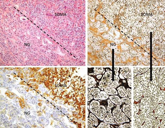
Figure 3b-2: Normal anterior gland (NG) and somatotroph adenoma (SOMA) interface. This figure illustrates histological principles of distinction of adenoma from normal gland, which may be difficult on routine HE stains (top left), but is greatly aided by a reticulin stain (top and bottom right). The normal adenohypophysis consists of very well demarcated cell nests separated by dense septa. Bottom left: Serial section to the top row images stained for GH. Dashed line: Border between normal gland and adenoma.