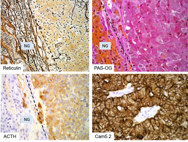
Figure 3b-12: Histology of a corticotroph microadenoma. These (often USP8-mutated) Cushing’s adenomas represent microscopic nodules well-demarcated from normal gland (NG). Reticulin stain is often essential for their detection (top left) and discrimination from corticotroph hyperplasia: a corticotroph adenoma results in completed destruction of the reticulin network as seen here (right side of the dashed line in top left image). Typical Cushing’s adenomas are deeply basophilic (top right) and show strong diffuse ACTH positivity (bottom left) and an intact keratin cytoskeleton (bottom right).