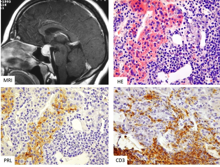
Figure 3b-26: MRI and histology of lymphocytic hypophysitis. The MRI appearances may be mistaken for a non-functioning adenoma; however, there are distinctive features, including homogeneous enhancement with extension posteriorly and rostrally via the infundibulum (‘infundibulohypophysitis’). There is a dense, destructive lymphocytic infiltrate, leaving islands of residual anterior gland (eosinophilic cells, top right) that in this instance are lactotrophs (bottom left; serial section to top right). The lymphocytes are predominantly T-cells (CD3-positive, bottom left).