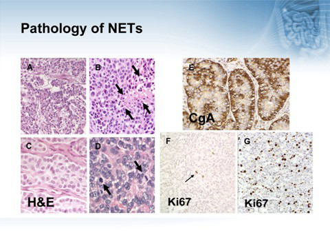
Figure 2 shows the histological findings supporting the neuroendocrine nature of the tumor with positive CGA
staining and the degree of dedifferentiation illustrated by the number of mitotic figures shown on H and E in
the top left and a blow up bottom right. The proliferative index is shown in F and G indicating a low level in F
and a high KI67 indexes in G.