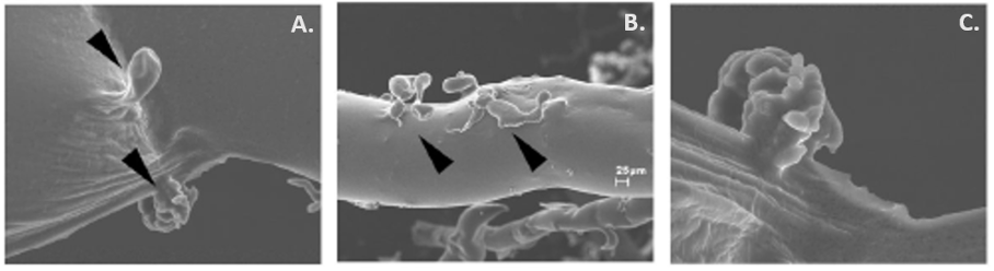
Figure 1. Visualization of the pancreatic duct glandular structures (PDGs) (arrowheads) in (A) large and (B) small ducts using scanning electron microscopy (SEM). PDGs can occur as single outpouches or form a complex of sac-like dilatations as illustrated in (C). This figure has been adapted from Gastroenterology, Strobel O., Rosow D. E., Rakhlin E. Y., et al., Pancreatic duct glands are distinct ductal compartments that react to chronic injury and mediate Shh-induced metaplasia, 138 (3): 1166-77 © 2010 (30).