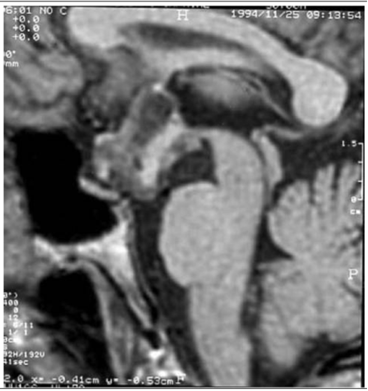
Figure 3. Resonance imaging T1-weighted sequences on sagittal plane before IV gadolinium chelate administration. Extra-axial craniopharyngioma in the intra and suprasellar space, with non-homogenous signal due to calcifications and cysts, in a 7- year-old boy presenting with reduced growth velocity, sleepiness, and visual loss. (Kindly provided by S. Cirillo, II University of Naples).