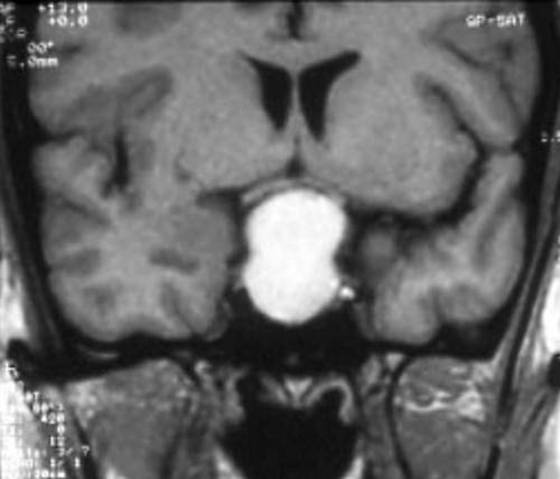
Figure 1a. Resonance Imaging T1-weighted sequences on coronal planes. Intra- and suprasellar craniopharyngioma in a 8 yr old boy presenting with reduced growth velocity and headache. This tumor has a total cystic component as shown by the hyper-intense spontaneous signal. (Kindly provided by S. Cirillo, II University of Naples).