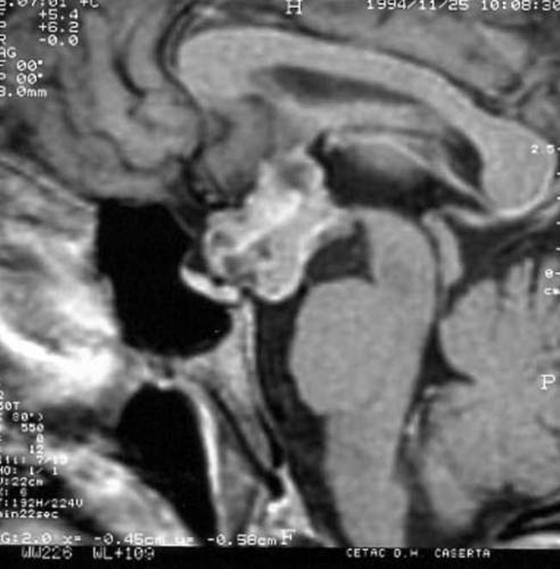
Figure 2b. Resonance Imaging T1-weighted sequences on sagittal plan after i.v. gadolinium chelate (diethylene-triamine pentacetate) administration. Extra-axial craniopharyngioma developing into the intra- and suprasellar space, with non-homogenous signal due to calcifications and cysts, in a 7 yr old boy presenting with reduced growth velocity, sleepiness and visual loss. After contrast medium (B) non-homogenous enhancement of the solid component. (Kindly provided by S. Cirillo, II University of Naples).