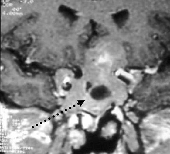
Figure 4b. Resonance Imaging T1-weighted sequences on sagittal plan after i.v. (B) gadolinium chelate (diethylene-triamine pentacetate) administration. Extra-axial macroadenoma developing into the intra- and suprasellar and parasellar space including carotid arteries (arrows), with non-homogenous signal due to calcifications and cysts. After contrast medium (B) non-homogenous enhancement of the solid and the cystic component (interrupted arrow). (Kindly provided by S. Cirillo, II University of Naples).