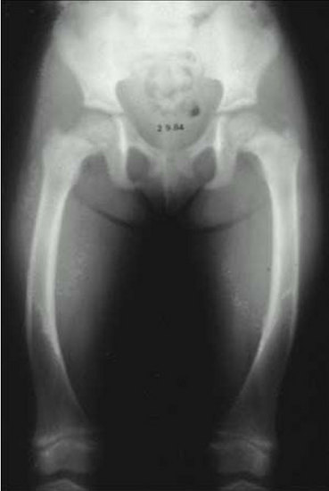
Figure 4.Radiograph of the lower extremeties in a patient with X-linked hypophosphatemia. Bowing of the femurs is evident bilaterally. The distal femoral metaphysis is cupped, frayed and widened, radiographic features of an expanded and disorganized growth plate.