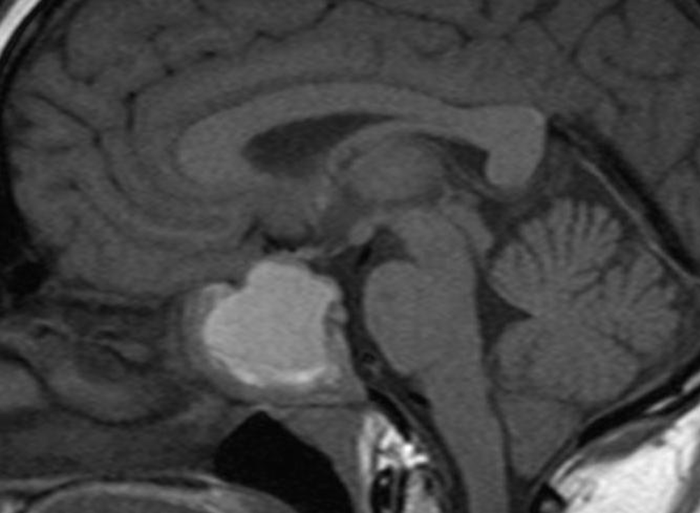
Figure 13. Sagittal T1 non-contrast image showing hemorrhage into an existing pituitary macroadenoma. The area of high signal represents the recent hemorrhage. There is a component of the tumor extending into the left cavernous sinus which does not show hemorrhage. The suprasellar extension is compressing the chiasm particularly on the right side.