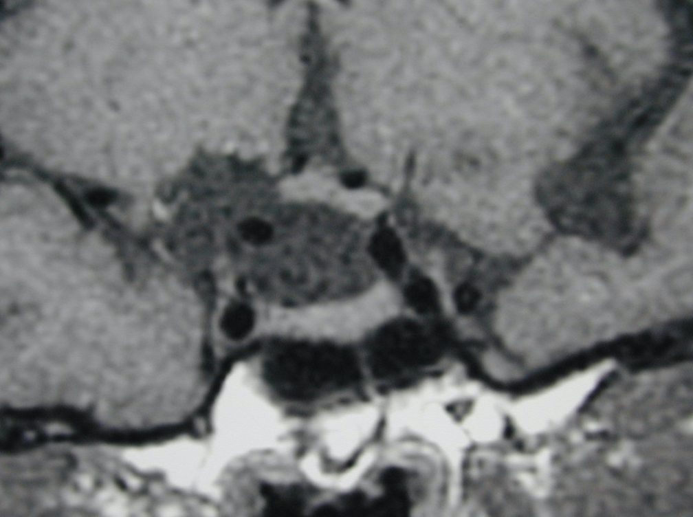
Figure 43. Coronal unenhanced T1 weighted images show an arachnoid cyst in the suprasellar region with elevation of the right side of the chiasm by the cyst. No cyst wall is evident and the pituitary tissue itself is normal.
Figure 43. Coronal unenhanced T1 weighted images show an arachnoid cyst in the suprasellar region with elevation of the right side of the chiasm by the cyst. No cyst wall is evident and the pituitary tissue itself is normal.