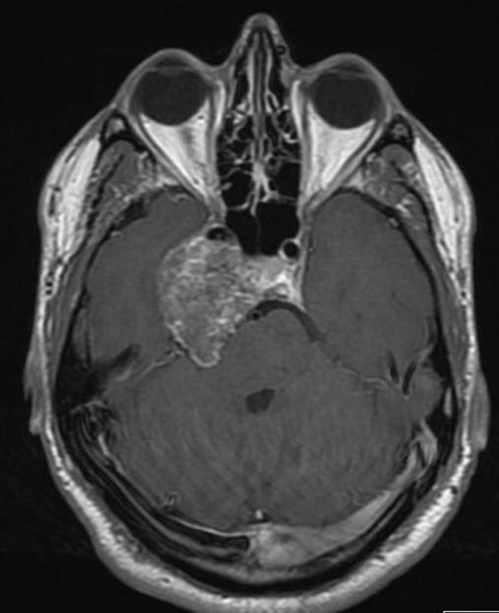
Figure 51. Axial-enhanced T1 weighted image of a chondrosarcoma. This shows the typical bubbly high T2 signal and involves the right side of the pituitary fossa, the right cavernous sinus and extends into the right middle cranial fossa.
Figure 51. Axial-enhanced T1 weighted image of a chondrosarcoma. This shows the typical bubbly high T2 signal and involves the right side of the pituitary fossa, the right cavernous sinus and extends into the right middle cranial fossa.