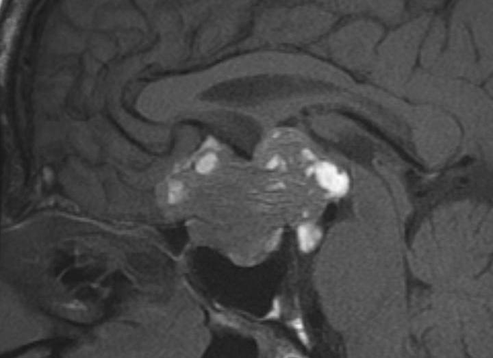
Figure 13A:Sagittal T1 weighted unenhanced (A) and coronal enhanced (B) images showing a partially cystic craniopharingioma. This is a large complex suprasellar mass which extends down into the pituitary fossa and up to deform the third ventricle. It is of mixed signal intensity and the solid components enhance after contrast. (B) There are patchy areas of high signal before contrast (A) which represent the cystic components with a high protein/ lipid content.