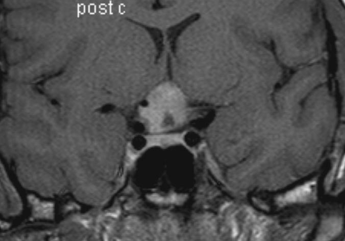
Figure 15B:Sagittal (A) T1 weighted and coronal (B) enhanced images show a partially cystic and partially solid hypothalamic glioma in the suprasellar region. The mass is centred on the region of the optic chiasm and is flattening the pituitary stalk and hypothalamus. The pituitary gland is normal.