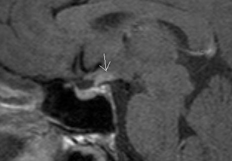
Figure 16B:Sagittal (A) and (B) T1 weighted enhanced images of two different germinomas. (A) demonstrates a large homogeneously enhancing soft tissue mass within the suprasellar region involving the upper aspect of the pituitary stalk and the hypothalamic region with a second separate mass visible in the region of the pineal gland -this is typical of a germinoma. (B) demonstrates a much smaller lesion in the region of the upper aspect of the stalk and hypothalamus (arrow).