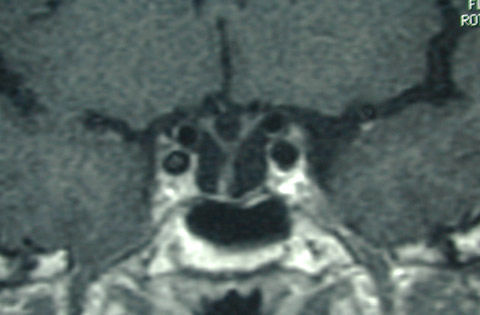
Figure 20B:Sagittal (A) and coronal (B) enhanced T1 weighted images show an empty sella. No pituitary tissue is visible and the stalk extends down to the floor of the sella. The optic chiasm has prolapsed inferiorly, a not uncommon appearance after a large mass has been removed.